Review
. 2022 Sep 6;26(1):42.
doi: 10.1186/s40824-022-00288-0.

Biomineralization of bone tissue: calcium phosphate-based inorganics in collagen fibrillar organic matrices

Affiliations
Review

Biomineralization of bone tissue: calcium phosphate-based inorganics in collagen fibrillar organic matrices

Min-Ho Hong et al. Biomater Res. .

Abstract

Background: Bone regeneration research is currently ongoing in the scientific community. Materials approved for clinical use, and applied to patients, have been developed and produced. However, rather than directly affecting bone regeneration, these materials support bone induction, which regenerates bone. Therefore, the research community is still researching bone tissue regeneration. In the papers published so far, it is hard to find an improvement in the theory of bone regeneration. This review discusses the relationship between the existing theories on hard tissue growth and regeneration and the biomaterials developed so far for this purpose and future research directions.

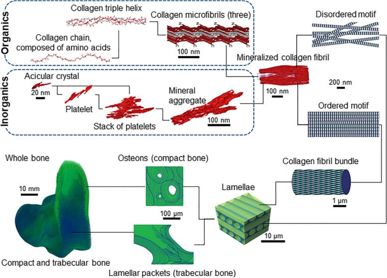
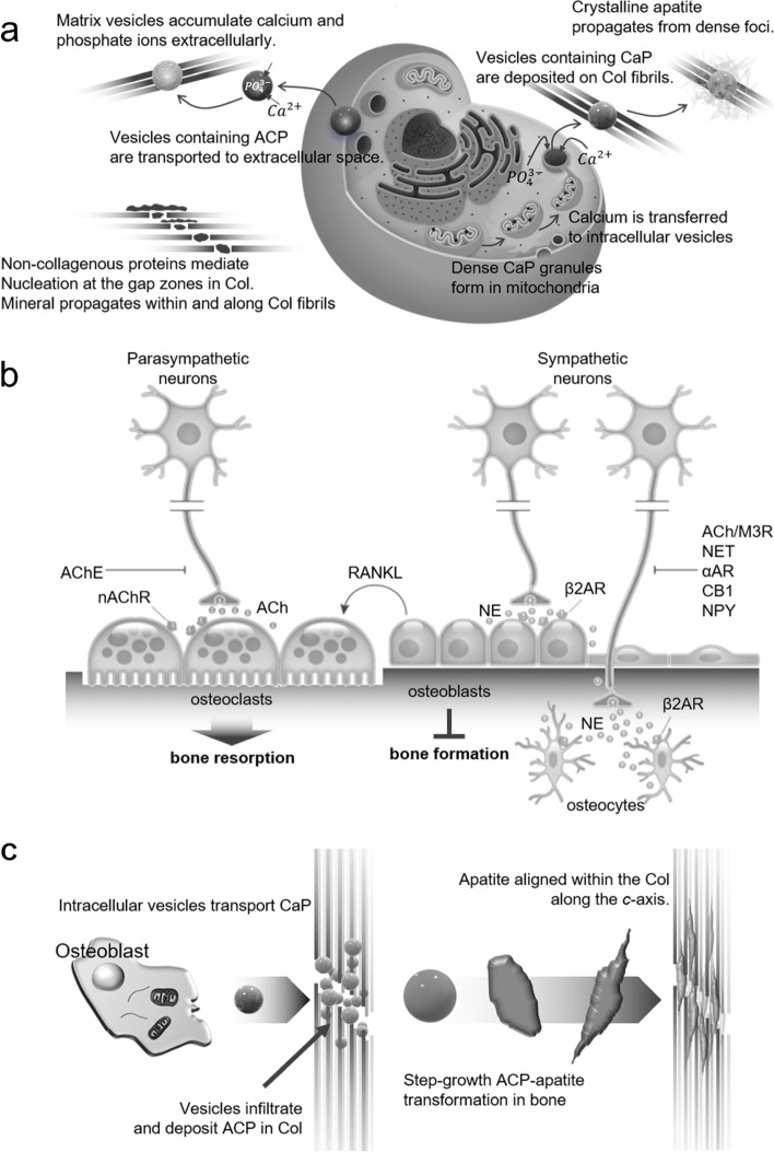
Mainbody: Highly complex nucleation and crystallization in hard tissue involves the coordinated action of ions and/or molecules that can produce different organic and inorganic composite biomaterials. In addition, the healing of bone defects is also affected by the dynamic conditions of ions and nutrients in the bone regeneration process. Inorganics in the human body, especially calcium- and/or phosphorus-based materials, play an important role in hard tissues. Inorganic crystal growth is important for treating or remodeling the bone matrix. Biomaterials used in bone tissue regeneration require expertise in various fields of the scientific community. Chemical knowledge is indispensable for interpreting the relationship between biological factors and their formation. In addition, sources of energy for the nucleation and crystallization processes of such chemical bonds and minerals that make up the bone tissue must be considered. However, the exact mechanism for this process has not yet been elucidated. Therefore, a convergence of broader scientific fields such as chemistry, materials, and biology is urgently needed to induce a distinct bone tissue regeneration mechanism.

Conclusion: This review provides an overview of calcium- and/or phosphorus-based inorganic properties and processes combined with organics that can be regarded as matrices of these minerals, namely collagen molecules and collagen fibrils. Furthermore, we discuss how this strategy can be applied to future bone tissue regenerative medicine in combination with other academic perspectives.

Keywords: Biomineralization; Bone growth; Bone regeneration; Collagen matrix; Hierarchical structure; Nucleation and crystallization.


Conflict of interest statement

The authors declare that they have no competing interests.

Figures

Fig. 1
Fig. 1
TreeMap Chart of publications for 15,943 results from Web of Science Core Collection between 2017 and 2021 (Data last updated: 2022–03-01)
Fig. 2
Fig. 2
A scheme of hierarchical structure of bone tissue composed of organic and inorganic materials [19], Copyright © 2018, The American Association for the Advancement of Science. The inorganics of the mineralized collagen fibrils themselves incorporate several nested structural motifs, listed as follows in decreasing order of complexity: mineral aggregates – stacks of platelets – platelets – acicular crystals. Collagen fibrils are composed of quasi-hexagonally packed microfibrils, each of which incorporates multiple staggered triple helices that in turn are formed from repetitive chains of amino acids. Ordered and disordered motifs of bone consist of mineralized collagen fibrils that are about 120 nm thick and build a continuous network
Fig. 3
Fig. 3
Summary and comparison of the size and size distribution of CaPs using various synthesis methods (solid-state method [27], mechanochemical method [38], precipitation method [39], sol–gel method [40], molecule direct crystallization method [–43], hydrothermal method [44, 45], hydrothermal decomposition of Ca-chelated complex method [46, 47], biomineralization method [–50], diffusion method [51, 52], gravity-assisted method [53], hydrolysis method [46], microemulsion method [54, 55], hydrothermal microemulsion method [55], sonochemistry assisted method [–58], microwave assisted method [59, 60], sonochemistry-assisted microwave method [57], precursor transformation method [–50, 61], combustion method [62, 63], pyrolysis method [64, 65], molten salt synthesis and flux technique method [66, 67], and spray-drying and flame-spray methods [64, 65].). TCP; tricalcium phosphate, DCPA; dicalcium phosphate anhydrous
Fig. 4
Fig. 4
Preparation of HAp nanoparticles via (a) conventional chemical precipitation, (b) hydrothermal condition, and (c) sol–gel process
Fig. 5
Fig. 5
a Transmission electron microscope (TEM) image of mineral particle aggregates (scale bars 200 nm) and (b) the corresponding SAED patterns: (b1) particles enclosed in the circle indicate amorphous scattering of the diffusion ring. (b2) the marked rectangular area has poor crystalline diffraction, and (b3) the particles in the area shown in the interpolated image produced a clear crystalline diffraction pattern [(002) and second order (004) showing well defined reflections of apatite planes.] (b4) the encircled area taken after being stored at room temperature for a week. The appearance of diffraction spots with (002) plane (arrowheads) spacing suggests a transition to the crystalline HAp phase [85]. c Cryogenic-scanning electron microscope (Cryo-SEM) image of the same area (scale bar 100 nm). d The amorphous (encircled area) and crystalline (rectangular area) portions of the electron selective backscatter (ESB) image shows no significant difference in signal strength (scale bar 100 nm)
Fig. 6
Fig. 6
a Pathways to crystallization by particle attachment [83]. b The mechanism, consisting of two steps, shows the formation of crystal nuclei inside them [82]. (i) The supersaturated solution. (ii) The dense liquid. (iii) The critical cluster at n2*. (iv) The nucleation cluster and ensuing growth of the crystal. (v) the thoroughly formed crystal. c The path to the free energy change ΔG according to the two feasible models of the two-phase nucleation mechanism [82]
Fig. 7
Fig. 7
Mineralization stages. a Two-dimensional projection images and (b) three-dimensional visualizations of tomograms. Stage 1 is in the absence of a monolayer, and stages 2–4 are that the inset SAED pattern in (a) shows that the spherical particles on the monolayer are amorphous phases. Finally, stage 5 is that the inset SAED pattern in (a) can be indexed as cHAp with a [110] zone axis. The yellow arrow in (b) indicates the preferred nucleation plane (110). The red arrows indicate the markers, and the blue shows the Au beads. Scale bars, 50 nm. c Surface-directed mineralization stages of CaP from SBF at 37 °C. Stage 1 represents the loose aggregation of the prenucleation cluster in equilibrium with the ions of the solution, and stage 2 shows that the nucleated cluster aggregates in the presence of a monolayer so that the loose aggregate still exists in the solution. Stages 3 and 4 show cohesion leading to densification near the monolayer, indicating the nucleation of amorphous spherical particles only on the monolayer surface, and the last 5 stage shows the development of crystallinity due to oriented nucleation directed by the monolayer [95], Copyright © 2010, Nature Publishing Group
Fig. 8
Fig. 8
ACC nanoparticle precursors and calcite crystal growth. a Calcite growth via classical and non-classical pathways. b Growth rates, R vs supersaturation, σcalcite. Inset show in situ AFM deflection images of spiral growth and birth-spread model, and an image of calcite growth through ACC attachment along stages according to the non-classical mechanism of particle-mediated growth from a colloidal dispersion at the interface between calcite and solution. c Nanoparticles on (10.4)calcite. ACC nanoparticle bulk diffusion (1), surface diffusion (2), and final diffusion/attachment to a corner (3) [108], Copyright © 2016, American Chemical Society
Fig. 9
Fig. 9
a Mechanism of mesocrystals; (1) alignments by the organic matrix and (2) physical forces, (3) crystalline bridges by epitaxial growth and secondary nucleation, (4) alignments by spatial constraints, (5) oriented attachment, and (6) face selective molecules [113]. b Cryo-TEM (1) image and (2) SAED pattern of the mineralized collagen fibril in the presence of pAsp [117]. c (1) TEM image of mineralized triple-helix protein molecule, (2) and (3) filtered and zoomed images of mineralized triple-helix protein molecules [123], Copyright © 2009 WILEY‐VCH Verlag GmbH & Co. KGaA, Weinheim
Fig. 10
Fig. 10
TEM images based on pAsp, is the most abundant amino acid group in NCPs and is used as a basic element in mineralization studies. a Cryo-TEM images according to pAsp for ACP precursor and Ca2+ concentration and time for crystal growth [130], Copyright © 2017, American Chemical Society. b Uranyl acetate staining TEM image of ACP mineralized reconstituted Col-I fibrils made anionic by binding with pAsp [132]. (1) The large and electrodense particles condensed on the fibril surface are ACP coacervates due to the electrostatic interaction between the polyvalent cation and the polyanionic electrolyte. (2) Adsorption of these electrodense particles slows the penetration of CaP. (3) In the maturation stage, the surface coacervate transforms into extrafibrillar CaP crystals, while mineralization intrafibrillar results in the formation of less heavy mineralized fibrils. c Mineralization of collagen fibrils in the presence of pAsp [134], © 2018 WILEY‐VCH Verlag GmbH & Co. KGaA, Weinheim. (1) Partially mineralized collagen fibers. (2) Enlarged image of mineralized collagen fibrils. (3) The SAED pattern in panel (2) is consistent with HAp and indicates oriented crystallization. (4) Elemental mapping of mineralized collagen fibrils
Fig. 11
Fig. 11
a Representation of model and mechanism for bone mineral formation [147]. b Representation diagram of mechanism by sympathetic nerves affecting bone formation inhibition and resorption promotion [148]. c Diagram describing a possible bone mineralization mechanism [149]. Col, collagen; AChE, acetylcholinesterase; nAChR, nicotinic acetylcholine receptor; Ach, acetylcholine; NE, norepinephrine; AR, adrenergic receptor; M3R, muscarinic 3 receptor; NET, norepinephrine transporter; CB, cannabinoid; NPY, anxiolytic neurotransmitter and cotransmitter
Fig. 12
Fig. 12
Schematic diagram of the hierarchical organization of bone (center; ordered material, green; disordered material, blue) [168]. Representative techniques for the assessment of the organization of bone tissue (both sides) [170]. GAG, glycosaminoglycan; QCT, quantitative computed tomography; NMR, nuclear magnetic resonance; HR-MRI, high resolution magnetic resonance imaging; HRpQCT, high-resolution peripheral quantitative computed tomography; FTIR, Fourier-transform infrared spectroscopy; CT, computed tomography; qBEI, quantitative backscattered electron image; XRD, X-ray diffraction; SAXS, small-angle X-ray scattering; HPLC, high performance liquid chromatography; TGA, thermal gravimetric analysis

References

    1. Madni AM. Disciplinary Convergence. Transdisciplinary Systems Engineering. Cham: Springer International Publishing; 2018. pp. 41–7.
    1. Convergence: Facilitating Transdisciplinary Integration of Life Sciences, Physical Sciences, Engineering, and Beyond. The National Academies Collection: Reports funded by National Institutes of Health. Washington (DC): National Academies Press (US); 2014. - PubMed
    1. Henkel J, Woodruff MA, Epari DR, Steck R, Glatt V, Dickinson IC, et al. Bone Regeneration Based on Tissue Engineering Conceptions - A 21st Century Perspective. Bone Res. 2013;1(3):216–248. doi: 10.4248/BR201303002. - DOI - PMC - PubMed
    1. Arola D, Murcia S, Stossel M, Pahuja R, Linley T, Devaraj A, et al. The limiting layer of fish scales: Structure and properties. Acta Biomater. 2018;67:319–330. doi: 10.1016/j.actbio.2017.12.011. - DOI - PubMed
    1. Kim RY, Oh JH, Lee BS, Seo YK, Hwang SJ, Kim IS. The effect of dose on rhBMP-2 signaling, delivered via collagen sponge, on osteoclast activation and in vivo bone resorption. Biomaterials. 2014;35(6):1869–1881. doi: 10.1016/j.biomaterials.2013.11.029. - DOI - PubMed