

Identification of HBEGF+ fibroblasts in the remission of rheumatoid arthritis by integrating single-cell RNA sequencing datasets and bulk RNA sequencing datasets
- PMID: 36068607
- PMCID: PMC9446562
- DOI: 10.1186/s13075-022-02902-x
Identification of HBEGF+ fibroblasts in the remission of rheumatoid arthritis by integrating single-cell RNA sequencing datasets and bulk RNA sequencing datasets
Abstract
Background: Fibroblasts are important structural cells in synovium and play key roles in maintaining the synovial homeostasis. By single-cell RNA sequencing (scRNA-seq), subpopulation of synovium-resident cells has been reported to protect intra-articular structures from chronic inflammation and promote tissue repair. However, a significant number of researchers have concentrated on the role of fibroblasts in the progress of rheumatoid arthritis (RA) while few reports had described the contribution of distinct fibroblast subsets in the RA remission. It is helpful to understand the role of fibroblast subpopulations in the RA process to provide predictive biomarkers and address RA remission mechanisms. Here, we found HBEGF+ fibroblasts that contributed to RA remission by integrating scRNA-seq datasets and bulk RNA sequencing (bulk RNA-seq) datasets.
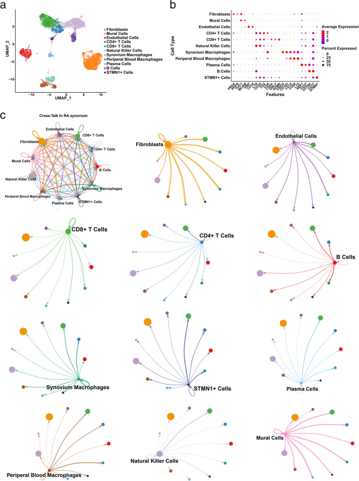
Method: Three single-cell RNA datasets of cells harvested from RA patients were processed and integrated by Seurat and Harmony R packages. After identifying cell types by classic marker genes, the integrated dataset was used to run CellChat for analysis of cell-cell communication. Specially, EGF signaling pathway was found and HBEGF+ fibroblasts were identified based on HBEGF expression. Differential expressed genes of HBEGF+ were shown in heatmap and volcano plot and used to run gene ontology (GO) enrichment analysis. Next, bulk RNA-seq datasets of synovium under different conditions (health, osteoarthritis (OA), rheumatoid arthritis, before and after classical treatment) were compared to show expression change of HBEGF and gene markers that are mainly expressed by HBEGF+ fibroblasts such as CLIC5, PDGFD, BDH2, and ENPP1. Finally, two single-cell RNA sequencing datasets of synovial cells from mice were integrated to identify Hbegf+ fibroblasts and calculate the population of Hbegf+ fibroblasts under different joint conditions (health, K/BxN serum transfer arthritis (STA), and remission of STA).
Result: After integrating three single-cell RNA sequencing datasets, we identified 11 clusters of synovial cells, such as fibroblasts, mural cells, endothelial cells, CD4+ T cells, CD8+ T cells, natural killer cells, synovium macrophage, peripheral blood macrophages, plasma cells, B cells, and STMN1+ cells. We found fibroblasts had an extensive communication network with other clusters and interacted with synovial macrophages through EGF signaling pathway via analysis of cell-cell communication between synovial cells. HBEGF, ligand to EGF signaling pathway, was highly expressed by a subset of fibroblasts and macrophages, and EGFR, receptor to EGF signaling pathway, was highly expressed by fibroblasts and meniscus cells. Moreover, HBEGF was downregulated under RA state and it had an increase after classical treatment. We then defined fibroblasts with high expression of HBEGF as HBEGF+ fibroblasts. In addition, we also found that HBEGF+ fibroblasts highly expressed CRTAC1, ITGB8, SCARA5, THBS4, and ITGBL1, genes relative to encoding extracellular matrix proteins and engaged in positive regulation of cell migration and motility, cellular component movement, and cell growth by GO enrichment analysis. We eventually identified HBEGF+ fibroblasts specially expressed CLIC5, PDGFD, BDH2, and ENPP1, which positively correlated with the expression of HBEGF. Moreover, the expression of CLIC5, PDGFD, BDH2, and ENPP1 was downregulated under RA state and elevated by classical therapy. On the contrary, the HBEGF+ macrophages specially expressed SLAMF8, GK, L1RN, and JAK2, which negatively correlated with the expression of HBEGF. The expression was upregulated in SLAMF8, GK, L1RN, and JAK2 under the RA state, whereas it was decreased after classical treatment. In mice, the number of Hbegf+ fibroblasts was reduced in the RA synovium but increased in the RA remitting synovium.
Conclusions: HBEGF+ fibroblasts play a role in the remission of rheumatoid arthritis, and HBEGF has potential to become a novel biomarker for prediction of RA progress.
© 2022. The Author(s).
Conflict of interest statement
The authors declare that they have no competing interests.
Figures


References
-
- Sokka T, Pincus T. Erythrocyte sedimentation rate, C-reactive protein, or rheumatoid factor are normal at presentation in 35%-45% of patients with rheumatoid arthritis seen between 1980 and 2004: analyses from Finland and the United States. J Rheumatol. 2009;36(7):1387–1390. doi: 10.3899/jrheum.080770. - DOI - PubMed
MeSH terms
Substances
LinkOut - more resources
Full Text Sources
Medical
Research Materials
Miscellaneous
