Review
. 2022 Nov;42(11):1112-1140.
doi: 10.1002/cac2.12345. Epub 2022 Sep 7.

Tumor-associated macrophages in liver cancer: From mechanisms to therapy

Affiliations
Review

Tumor-associated macrophages in liver cancer: From mechanisms to therapy

Kun Cheng et al. Cancer Commun (Lond). 2022 Nov.

Abstract

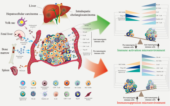
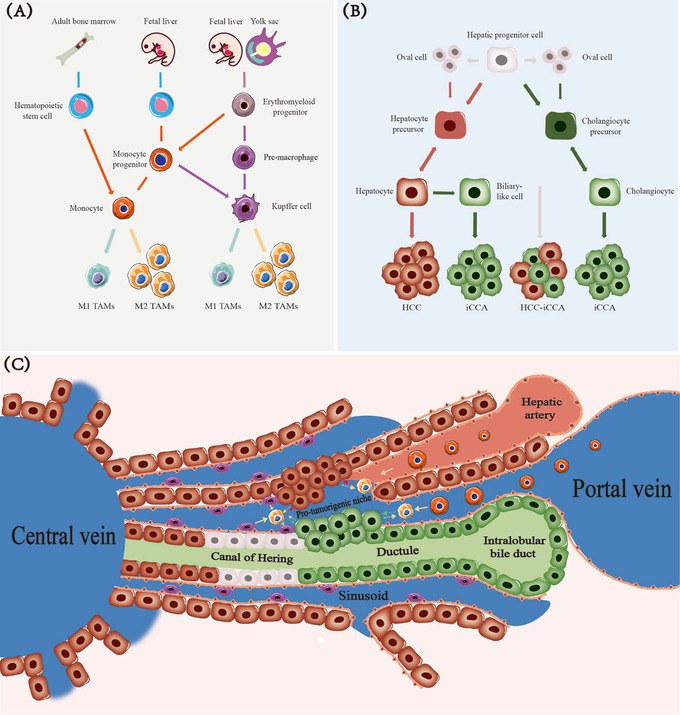
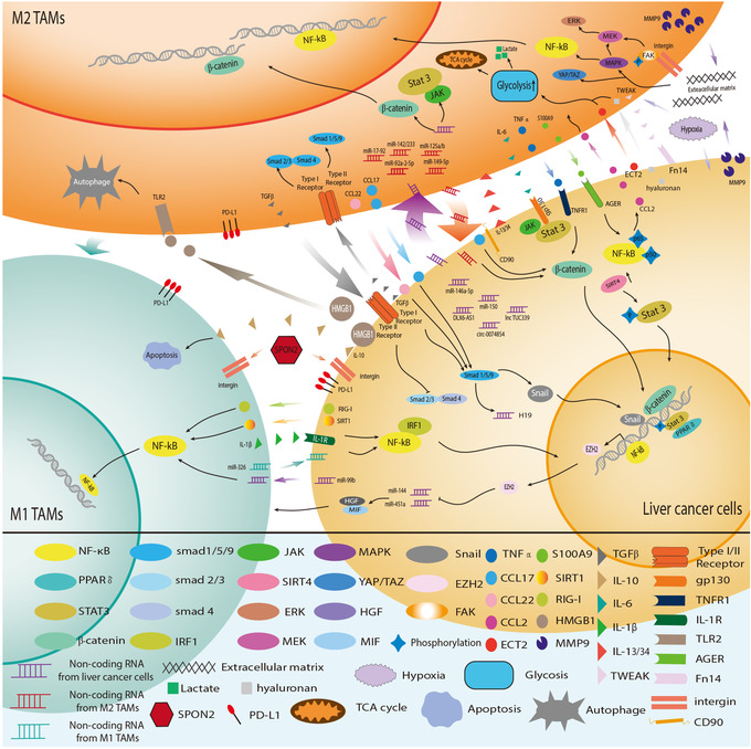
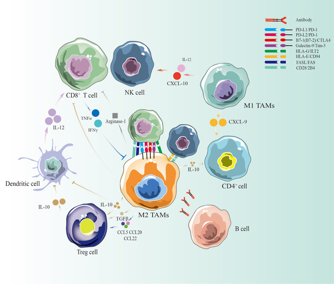
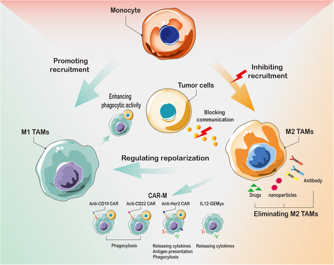
Multidimensional analyses have demonstrated the presence of a unique tumor microenvironment (TME) in liver cancer. Tumor-associated macrophages (TAMs) are among the most abundant immune cells infiltrating the TME and are present at all stages of liver cancer progression, and targeting TAMs has become one of the most favored immunotherapy strategies. In addition, macrophages and liver cancer cells have distinct origins. At the early stage of liver cancer, macrophages can provide a niche for the maintenance of liver cancer stem cells. In contrast, cancer stem cells (CSCs) or poorly differentiated tumor cells are key factors modulating macrophage activation. In the present review, we first propose the origin connection between precursor macrophages and liver cancer cells. Macrophages undergo dynamic phenotypic transition during carcinogenesis. In this course of such transition, it is critical to determine the appropriate timing for therapy and block specific markers to suppress pro-tumoral TAMs. The present review provides a more detailed discussion of transition trends of such surface markers than previous reviews. Complex crosstalk occurs between TAMs and liver cancer cells. TAMs play indispensable roles in tumor progression, angiogenesis, and autophagy due to their heterogeneity and robust plasticity. In addition, macrophages in the TME interact with other immune cells by directing cell-to-cell contact or secreting various effector molecules. Similarly, tumor cells combined with other immune cells can drive macrophage recruitment and polarization. Despite the latest achievements and the advancements in treatment strategies following TAMs studies, comprehensive discussions on the communication between macrophages and cancer cells or immune cells in liver cancer are currently lacking. In this review, we discussed the interactions between TAMs and liver cancer cells (from cell origin to maturation), the latest therapeutic strategies (including chimeric antigen receptor macrophages), and critical clinical trials for hepatocellular carcinoma (HCC) and intrahepatic cholangiocarcinoma (iCCA) to provide a rationale for further clinical investigation of TAMs as a potential target for treating patients with liver cancer.

Keywords: hepatocellular carcinoma; immunotherapy; intrahepatic cholangiocarcinoma; tumor-associated macrophages.


Figures

FIGURE 1
FIGURE 1
TAMs in the liver cancer immune microenvironment. TAMs constitute the core cell population of the tumor immune microenvironment in liver cancer. BM and embryos are the most critical sources of TAMs, followed by the spleen. The immunosuppressive cell population (based on TAMs) facilitates tumor progression and suppresses the immune response. In contrast, increasing infiltration of immune cell populations, including M1 and cytotoxic T lymphocytes, is associated with a better clinical prognosis. Abbreviations: TAMs, tumor‐associated macrophages; M1 TAMs, M1 type tumor‐associated macrophages; M2 TAMs, M2 type tumor‐associated macrophages; BM, bone marrow; NK cell, natural killer cell
FIGURE 2
FIGURE 2
Potential origin connection between TAMs and liver cancer cells. (A) Erythroid progenitor cells can differentiate into pMacs and monocyte progenitors. Hematopoietic stem cells are also a source of monocyte precursor cells. pMacs and monocyte progenitors differentiate into tissue‐resident macrophages (KCs in the liver) during growth in the body. Such cells maintain themselves by self‐renewal and can bind to peripheral blood monocytes to serve as two essential sources of TAMs when stimulated by certain cancer factors. (B) Liver progenitor cells have strong differentiation potential. Oval cells may be a subset of liver progenitor cells. Liver cancer and cholangiocarcinoma usually originate from mature hepatocytes and bile duct cells formed by precursor cells. Mixed cell carcinomas are primarily derived directly from progenitor cells. (C) The hepatic progenitor cells located in the canals of Hering have a subtle connection with TAM development during mature hepatocytes formations and subsequent transformation into hepatocarcinoma cells. TAMs can enable cancer cells to form a pre‐tumor niche. The development of cancer cells recruits a large number of mononuclear‐derived macrophages and activates tissue‐resident macrophages. Abbreviations: pMacs, pre‐macrophages; KCs, Kupffer cells; TAMs, tumor‐associated macrophages; M1 TAMs, M1 type tumor‐associated macrophages; M2 TAMs, M2 type tumor‐associated macrophages; HCC, hepatocellular carcinoma; iCCA, intrahepatic cholangiocarcinoma; HCC‐iCCA, hepatocellular carcinoma and intrahepatic cholangiocarcinoma
FIGURE 3
FIGURE 3
Dynamic phenotypic transition of TAMs during liver cancer progression. (A) KCs are unique macrophages in the liver and are part of the mononuclear phagocyte system. Those KCs in the liver sinusoids can phagocytize foreign antigens, antigen‐antibody complexes, and cell debris and also secrete cytokines, which is unfavorable for cancer initiation. DCs and HSCs present in the space of Disse cooperate with other immune cells such as KCs to maintain the liver immune microenvironment. (B) The immune microenvironment of liver cancer (HCC or iCCA) is constantly changing. The resident macrophages inside the tumor can alter their phenotype following stimulation. Furthermore, numerous circulating monocytes in the peripheral blood, including Tie+ monocytes, are recruited to the tumor milieu. Prior to converting pro‐tumoral TAMs, CD11b and Ly6C are highly expressed, while F4/80 is low. Nevertheless, TAMs, which suppresses the immune response, have higher F4/80 and lower CD11b and Ly6C levels of expression. HSCs in the space of Disse are also activated and may synergize with TAMs to promote liver cancer progression. Abbreviations: KCs, Kupffer cells; DCs, dendritic cells; HSCs, hepatic stellate cells; HCC, hepatocellular carcinoma; iCCA, intrahepatic cholangiocarcinoma; Clec4F, C‐type lectin domain family 4 member F; Tim‐4, T‐cell immunoglobulin and mucin domain‐containing 4; CX3CR, C‐X3‐C motif chemokine receptor; CSF1R, colony‐stimulating factor 1 receptor; CCR2, C‐C motif chemokine receptor 2; MHC‐II, major histocompatibility complex class II; GATA, glutamyl‐tRNA amidotransferase, subunit A; MoMFs, monocyte‐derived macrophages; MACRO, macrophage receptor with collagenous
FIGURE 4
FIGURE 4
Crosstalk between TAMs and liver cancer cells. An extensive and intricate network exists between tumor cells and TAMs. M1‐type macrophages exhibit anti‐tumor effects through various mechanisms, with abundant crosstalk between M2 macrophages and tumor cells, which is closely related to tumor progression. Tumor cells and HCC‐related macrophages coordinate to adapt to the immune environment of HCC. Abbreviations: TAMs, Tumor‐associated macrophages; M1 TAMs, M1 type Tumor‐associated macrophages; M2 TAMs, M2 type Tumor‐associated macrophages; HCC, hepatocellular carcinoma; PPAR, peroxisome proliferators‐activated receptors; IRF1, interferon regulatory factor 1; SIRT4, sirtuin 4; ERK, extracellular regulated protein kinase; MEK, mitogen‐activated protein kinase kinase; MAPK, mitogen‐activated protein kinase; YAP, yes‐associated protein 1; TAZ, tafazzin; HGF, hepatocyte growth factor; MIF, migration inhibitory factor; EZH2, enhancer of zeste 2 polycomb repressive complex 2 subunit; FAK, focal adhesion kinase; TNF‐α, tumor necrosis factor α; CCL2, C‐C motif chemokine ligand 2; CCL17, C‐C motif chemokine ligand 17; CCL22, C‐C motif chemokine ligand 22; ECT2, epithelial cell transforming 2; S100A9, S100 calcium binding protein A9; SIRT1, sirtuin 1; RIG‐I, retinotic acid‐inducible gene I; HMGB1, high mobility group box 1;MMP9, matrix metallopeptidase 9;TGF‐β, transforming growth factor‐β; IL‐6, interleukin 6;IL‐10, interleukin 10; IL‐1β, interleukin 1β; IL‐13/34, interleukin 13/34; TWEAK, tumor necrosis factor‐like weak inducer of apoptosis; TNFR1, tumor necrosis factor receptor 1; IL‐1R, interleukin 1 receptor; TLR2, toll‐like receptor 2; SPON2, matricellular protein spondin2; PD‐L1, programmed cell death protein 1 ligand 1; AGER, advanced glycosylation end product‐specific receptor; TCA, tricarboxylic acid cycle
FIGURE 5
FIGURE 5
TAMs coordinate with other immune cells in liver cancer. TAMs may be considered a double‐edged sword in the immune microenvironment of liver cancer. M2 macrophages and Tregs can secrete IL‐10 to inhibit DCs, T cells and NK cells. They can also secrete arginase and directly contact each other to inhibit the immune response. M1 macrophages can secrete IL‐12 and CXCL10 to recruit more T cells and NK cells, whereas CXCL9 derived from M1 macrophages can stimulate CD4+ T cells. B cells participate in anti‐tumor‐related macrophage activities by secreting antibodies. Abbreviations: TAMs, tumor‐associated macrophages; M1 TAMs, M1 type tumor‐associated macrophages; M2 TAMs, M2 type tumor‐associated macrophages; HCC, hepatocellular carcinoma; NK cell, natural killer cell; IL‐10, interleukin 10; IL‐12, interleukin 12; CCL5, C‐C motif chemokine ligand 5; CCL20, C‐C motif chemokine ligand 20; CCL22, C‐C motif chemokine ligand 22; TNF‐α, tumor necrosis factor α; TGF‐β, transforming growth factor‐β; IFNγ, interferon‐gamma; CXCL9, C‐X‐C motif chemokine ligand 9; CXCL10, C‐X‐C motif chemokine ligand 10; PD‐L1, programmed cell death protein 1 ligand 1; PD‐1, programmed cell death protein 1; CTLA‐4, cytotoxic T‐lymphocyte antigen 4; HLA‐E, human leukocyte antigen E; HLA‐G, human leukocyte antigen G; ILT2, Ig‐like transcript 2; FAS, factor‐related apoptosis; FASL, factor‐related apoptosis ligand
FIGURE 6
FIGURE 6
Therapeutic strategies with TAMs in liver cancer. Targeting TAMs can be carried out from three perspectives: 1. Cutting off the source and eliminating the production of M2 TAMs, including inhibiting the transition of monocytes to M2 TAMs and eliminating specific pro‐tumoral tissue‐resident macrophages in liver cancer. 2. Remodeling M2 TAMs to M1 TAMs and CAR‐M. 3. Blocking the communication between M2 TAMs and liver cancer cells. Abbreviations: TAMs, tumor‐associated macrophages; M1 TAMs, M1 type tumor‐associated macrophages; M2 TAMs, M2 type tumor‐associated macrophages. CAR‐M, chimeric antigen receptor macrophage; GEMys, genetically engineered myeloid cells. IL12, interleukin 12

References

    1. Hiam‐Galvez KJ, Allen BM, Spitzer MH. Systemic immunity in cancer. Nat Rev Cancer. 2021;21(6):345–59. - PMC - PubMed
    1. Asgharzadeh S, Salo JA, Ji L, Oberthuer A, Fischer M, Berthold F, et al. Clinical significance of tumor‐associated inflammatory cells in metastatic neuroblastoma. J Clin Oncol. 2012;30(28):3525–32. - PMC - PubMed
    1. Seton‐Rogers S. Taming TAMs in brain metastases. Nat Rev Cancer. 2022;22(1):2–3. - PubMed
    1. Sun L, Kees T, Almeida AS, Liu B, He XY, Ng D, et al. Activating a collaborative innate‐adaptive immune response to control metastasis. Cancer Cell. 2021;39(10):1361–74.e9. - PMC - PubMed
    1. Sunakawa Y, Stintzing S, Cao S, Heinemann V, Cremolini C, Falcone A, et al. Variations in genes regulating tumor‐associated macrophages (TAMs) to predict outcomes of bevacizumab‐based treatment in patients with metastatic colorectal cancer: results from TRIBE and FIRE3 trials. Ann Oncol. 2015;26(12):2450–6. - PMC - PubMed

Publication types

MeSH terms