. 2022 Oct 26;13(5):e0222122.
doi: 10.1128/mbio.02221-22. Epub 2022 Sep 7.

Treatment of Sindbis Virus-Infected Neurons with Antibody to E2 Alters Synthesis of Complete and nsP1-Expressing Defective Viral RNAs

Affiliations

Treatment of Sindbis Virus-Infected Neurons with Antibody to E2 Alters Synthesis of Complete and nsP1-Expressing Defective Viral RNAs

Jane X Yeh et al. mBio. .

Abstract

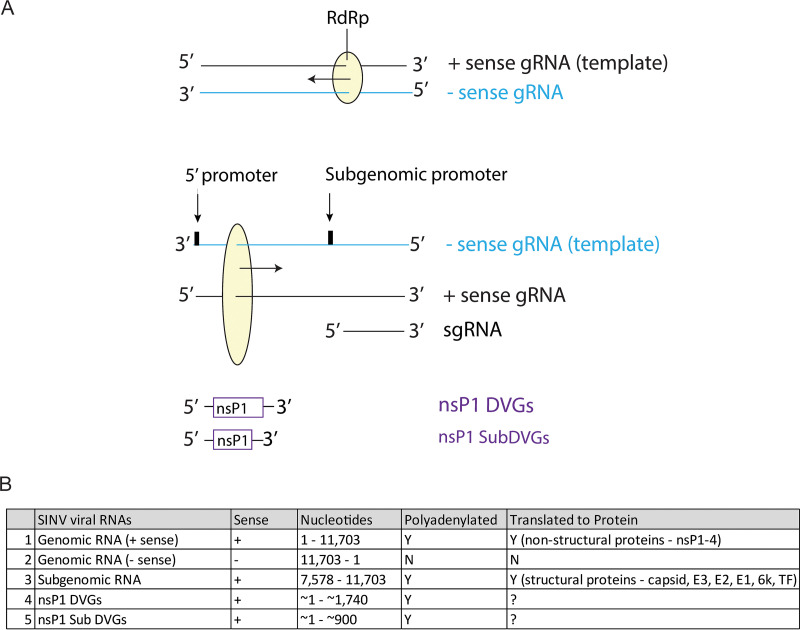
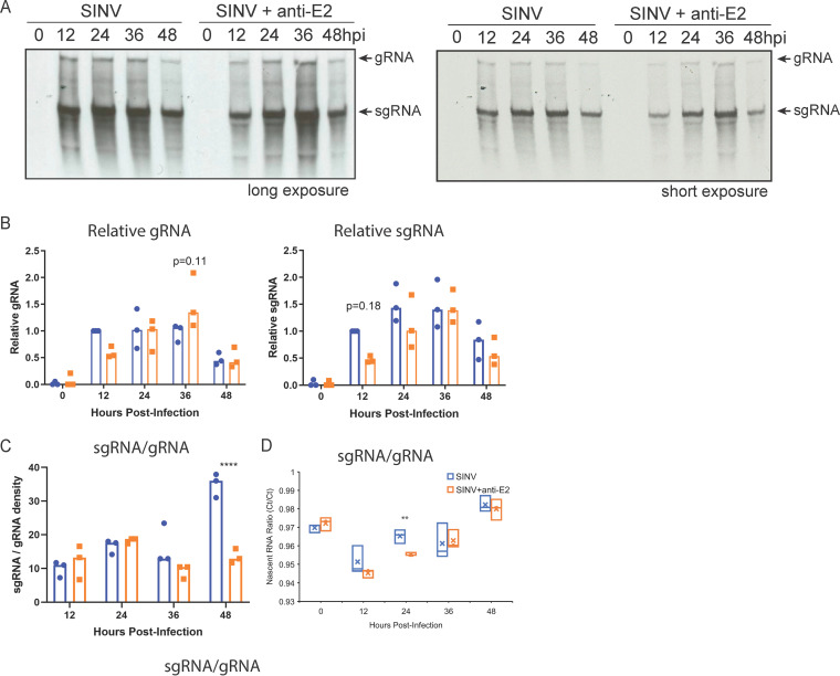
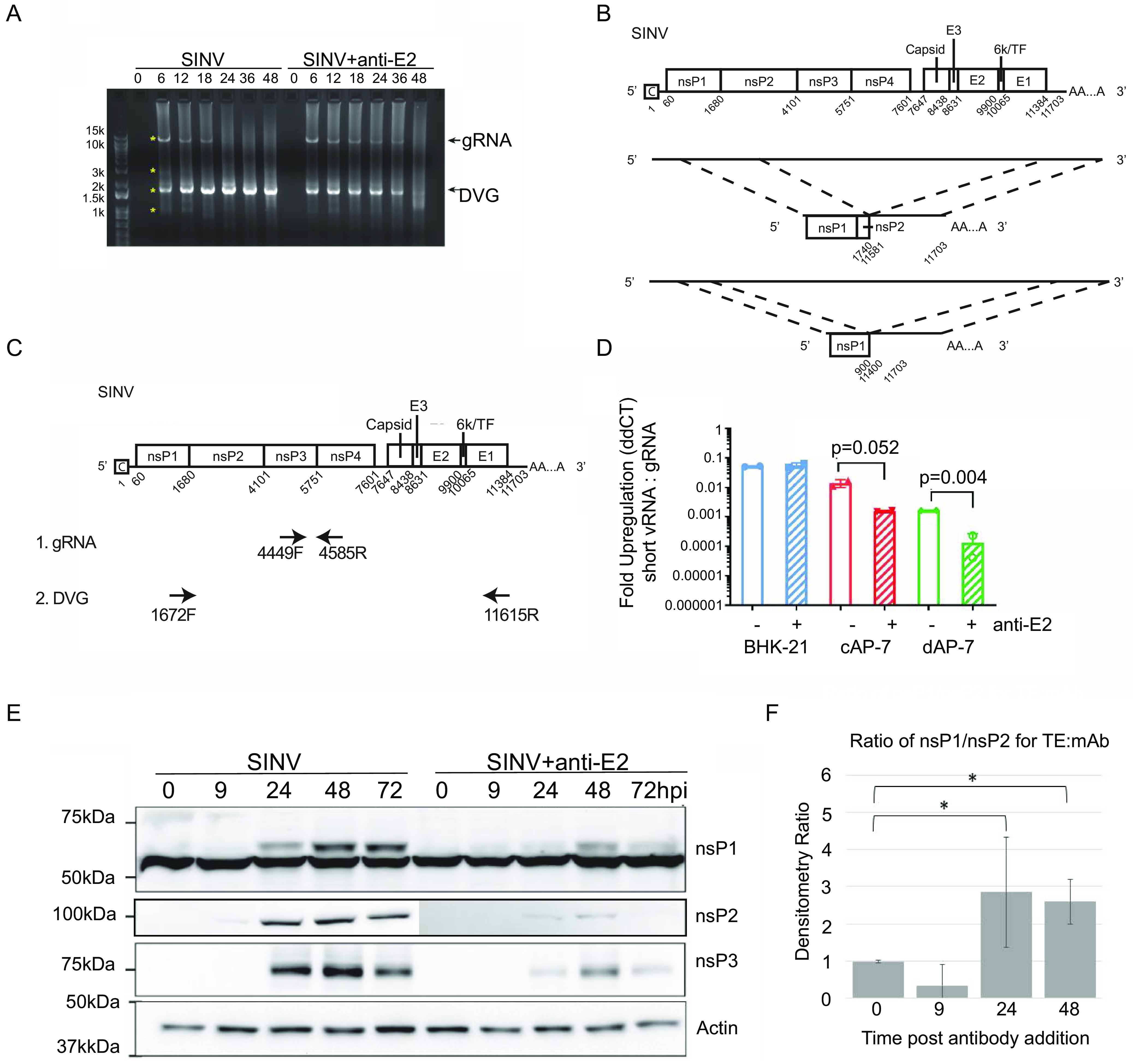
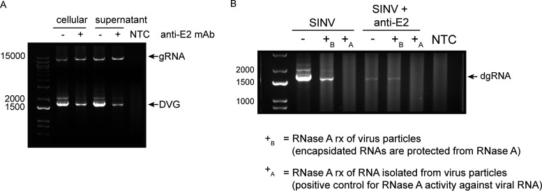
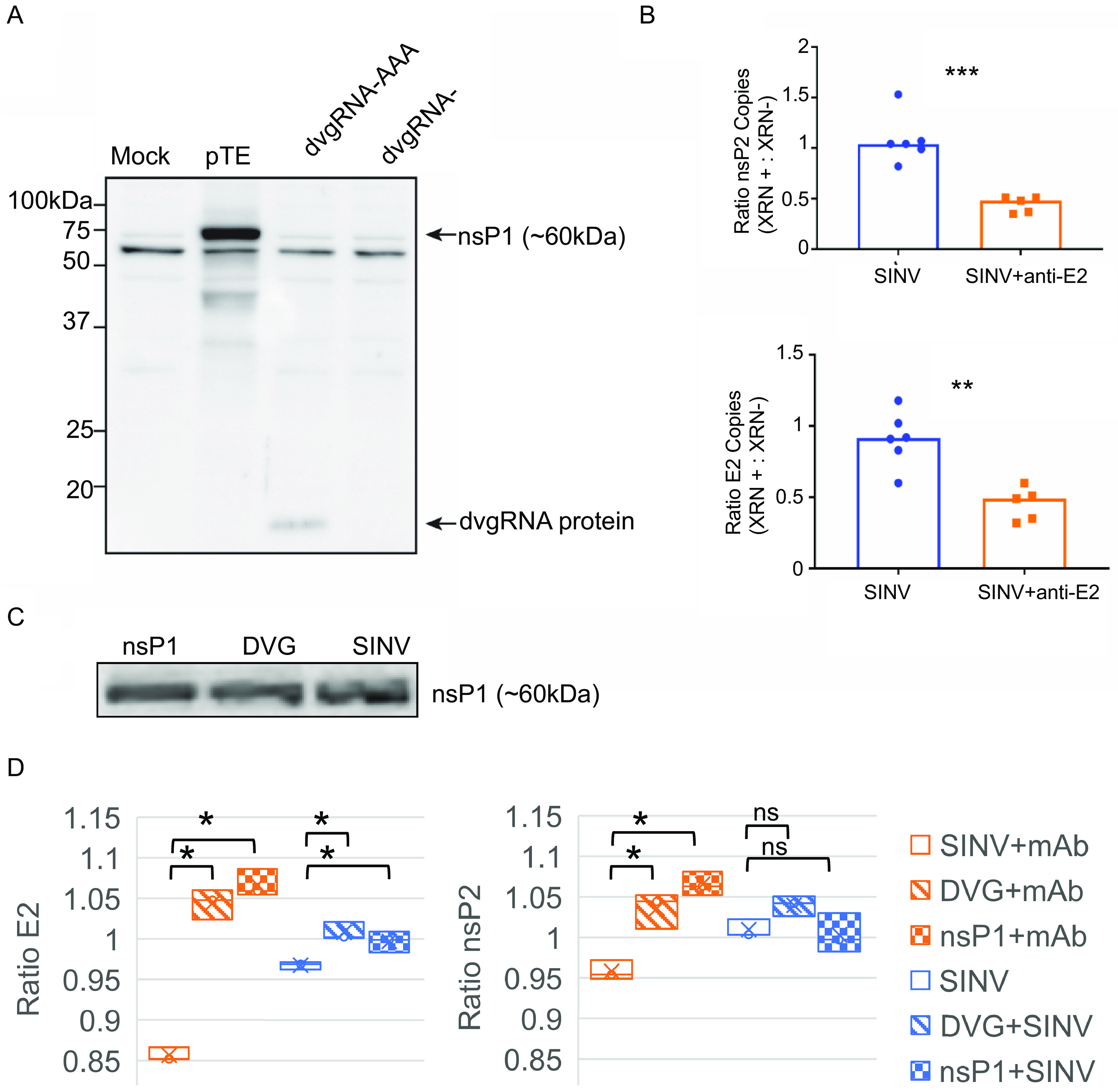
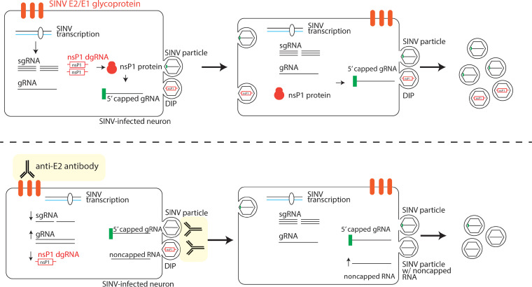
Alphaviruses are positive-sense RNA viruses that are important causes of viral encephalomyelitis. Sindbis virus (SINV), the prototype alphavirus, preferentially infects neurons in mice and is a model system for studying mechanisms of viral clearance from the nervous system. Antibody specific to the SINV E2 glycoprotein plays an important role in SINV clearance, and this effect is reproduced in cultures of infected mature neurons. To determine how anti-E2 antibody affects SINV RNA synthesis, Oxford Nanopore Technologies direct long-read RNA sequencing was used to sequence viral RNAs following antibody treatment of infected neurons. Differentiated AP-7 rat olfactory neuronal cells, an in vitro model for mature neurons, were infected with SINV and treated with anti-E2 antibody. Whole-cell RNA lysates were collected for sequencing of poly(A)-selected RNA 24, 48, and 72 h after infection. Three primary species of viral RNA were produced: genomic, subgenomic, and defective viral genomes (DVGs) encoding the RNA capping protein nsP1. Antibody treatment resulted in overall lower production of SINV RNA, decreased synthesis of subgenomic RNA relative to genomic RNA, and suppressed production of the nsP1 DVG. The nsP1 DVG was packaged into virus particles and could be translated. Because antibody-treated cells released a higher proportion of virions with noncapped genomes and transient transfection to express the nsP1 DVG improved viral RNA capping in antibody-treated cells, we postulate that one mechanism by which antibody inhibits SINV replication in neurons is to suppress DVG synthesis and thus decrease production of infectious virions containing capped genomes. IMPORTANCE Alphaviruses are important causes of viral encephalomyelitis without approved treatments or vaccines. Antibody to the Sindbis virus (SINV) E2 glycoprotein is required for immune-mediated noncytolytic virus clearance from neurons. We used direct RNA nanopore sequencing to evaluate how anti-E2 antibody affects SINV replication at the RNA level. Antibody altered the viral RNAs produced by decreasing the proportion of subgenomic relative to genomic RNA and suppressing production of a previously unrecognized defective viral genome (DVG) encoding nsP1, the viral RNA capping enzyme. Antibody-treated neurons released a lower proportion of SINV particles with capped genomes necessary for translation and infection. Decreased nsP1 DVG production in antibody-treated neurons led to lower expression of nsP1 protein, decreased genome capping efficiency, and release of fewer infectious virus particles. Capping was increased with exogenous expression of the nsP1 DVG. These studies identify a novel alphavirus DVG function and new mechanism for antibody-mediated control of virus replication.

Keywords: alphavirus; defective viral genomes; nanopore sequencing; neuron infection; viral encephalitis; virus clearance.


Conflict of interest statement

The authors declare a conflict of interest. D.E.G. is a member of advisory boards for GlaxoSmithKline, GreenLight Bioscience, Takeda Pharmaceuticals and Academia Sinica. J.X.Y. is an employee of Takeda Pharmaceuticals.

Figures

FIG 1
FIG 1
SINV RNA forms. (A) Schematic diagram of SINV RNA replication and the forms of SINV RNA made from each type of template strand. “gRNA” represents genomic RNA, “sgRNA” represents subgenomic RNA, and “DVG” represents the defective viral genome. (B) Table of the different SINV RNA forms indicating their sense, genome nucleotide composition, polyadenylation status, and translation to proteins.
FIG 2
FIG 2
Nanopore sequencing of SINV-infected differentiated AP-7 cells with or without anti-E2 antibody treatment. Differentiated AP-7 cells were infected with SINV (MOI of 10) and mock treated or treated with anti-E2 antibody (5 μg/mL) 4 h after infection. At 24, 48, and 72 h after infection, total intracellular RNA was collected in triplicate, enriched for poly(A), directly sequenced via nanopore sequencing, and aligned to the rat or SINV genome. (A) Proportion of RNA reads that align to the rat versus SINV genome. Proportions indicate the number of rat or SINV reads divided by the total number of RNA reads at each time point for mock-infected cells. The mock-infected 24-h sample is indicated in purple. (All mock time points had 0 SINV-aligned reads.) SINV-infected no-antibody-treatment samples are indicated in orange, and SINV-infected antibody-treated samples are indicated in green. Each dot indicates a different biological replicate. (B) Averaged sequence coverage of SINV-aligned RNA reads. Coverage plots indicate relative sequencing depth normalized to run yield across the SINV genome with (green) or without (orange) antibody treatment at 24, 48, and 72 h after infection. Coverage data represent the average of 3 biological replicates. (C) Reads were classified into genomic, subgenomic, DVG, or subDVG SINV RNA species using coverage differences at junction locations (see Materials and Methods). Abundance ratios for each species were calculated as a proportion of total SINV reads for each sample. Each point represents 1 biological replicate, and horizontal lines indicate the mean from 3 biological replicates. **, P < 0.01; ***, P < 0.001; ****, P < 0.0001.
FIG 3
FIG 3
Anti-E2 antibody increases production of SINV genomic RNA and decreases production of subgenomic RNA. Analysis of newly synthesized viral RNA. Differentiated AP-7 cells were infected with SINV (MOI of 10) and treated with anti-E2 antibody (5 μg/mL) at 4 h after infection. At the indicated time points, cells were labeled with 20 μCi/mL [3H]uridine in the presence of dactinomycin (1 μg/mL) for 2 h. (A) Representative assessment of radioactive viral RNA synthesis by agarose-formaldehyde gel and autoradiography. (Top) Long exposure; (bottom) short exposure of the same gel. (B) Densities of labeled genomic RNA (gRNA [left]) and subgenomic RNA (sgRNA [right]) normalized to SINV-infected, untreated cells at 12 h; (C) ratio of relative subgenomic RNA to genomic RNA density; (D) ratios of qRT-PCR-quantified biotin-captured 5-ethynyl uridine-labeled nascent subgenomic RNA to genomic RNA in SINV-infected dAP-7 cells with and without antibody treatment. Data from three independent experiments are presented as mean ± SD. **, P < 0.01; ****, P < 0.0001.
FIG 4
FIG 4
Antibody treatment increases SINV genomic RNA and decreases nsP1 defective viral genome RNA levels in dAP-7 cells. (A and B) Differentiated AP-7 cells were infected with SINV (MOI of 10) and treated with anti-E2 antibody (5 μg/mL) or medium (mock) at 4 h after infection. At the indicated time points, total cellular RNA lysates were collected. (A) Semiquantitative RT-PCR for SINV RNA using primers against the 5′ and 3′ ends of the SINV genome (SV-171F, SV-11655R). “gRNA” represents full-length SINV genomic RNA, and “DVG” represents the nsP1 defective viral genome. Asterisks indicate PCR bands excised for DNA extraction and Sanger sequencing. (B) SINV genome sequence alignment of the two defective viral genome sequences identified. (C and D) Differentiated AP-7 cells (dAP-7), undifferentiated cycling AP-7 cells (cAP-7), and BHK-21 cells were infected with SINV (MOI of 10) and treated with medium (mock) or anti-E2 antibody (5 μg/mL) at 4 h after infection. Twenty-four hours after infection, total cellular RNA was reverse transcribed to cDNA for qRT-PCR analysis of nsP1 defective viral genome levels. (C) Primer design used for qRT-PCR assay. Primers for the nsP1 defective viral genome span the deleted SINV region, while primers for the SINV genomic RNA are within the deleted region. (D) qRT-PCR analysis for nsP1 defective genome production. nsP1 RNA levels are expressed as fold regulation relative to SINV gRNA as calculated by ddCT. Data are presented as mean ± SD from two biological replicates. (E) Immunoblot of SINV nsP1, nsP2, and nsP3 expression from untreated and antibody-treated SINV-infected dAP-7 cells. (F) Densitometry was used to determine the ratios of nsP1 to nsP2 in immunoblots and to compare untreated (TE) to antibody-treated (MAb) SINV-infected dAP-7 cells. *, P < 0.05.
FIG 5
FIG 5
The nsP1 defective viral genome is packaged and released into viral particles. Differentiated AP-7 cells were infected with SINV (MOI of 10) and treated with medium (mock) or anti-E2 antibody (5 μg/mL) at 4 h after infection. Eighteen hours after infection, RNA from the cells and supernatant fluid was collected. (A) RNA was extracted from the cellular and supernatant samples. RT-PCR analysis for SINV genomic RNA (gRNA) and nsP1 defective viral genome RNA (DVG) was performed. “NTC” represents the no-template control. (B) RNase A protection assay. Culture supernatant was treated with RNase A (10 μg/mL) to digest all nonencapsidated RNAs (+B [before RNA extraction]) or untreated (−). Following RNase treatment, RNA was extracted from the supernatant and reverse transcribed for RT-PCR analysis of nsP1 defective genome RNA (dgRNA). As a positive control for RNase activity, extracted RNA was also treated with RNase A under the same conditions (+A [after RNA extraction]).
FIG 6
FIG 6
The nsP1 defective viral genome RNA can be translated, and expression is associated with increased SINV RNA capping efficiency. (A) The shorter nsP1 defective viral genome RNA (subDVG) was cloned into the pcDNA3.1 DNA expression vector and in vitro transcribed to RNA (dvgRNA). The RNA was capped and polyadenylated in vitro, purified, and transfected into uninfected undifferentiated AP-7 cells (dvgRNA-AAA). As controls, pTE (complete SINV plasmid) and unpolyadenylated defective viral genome RNA (dvgRNA−) were also transfected. Twenty-four hours after transfection, lysates were collected and assessed by immunoblot probing with a polyclonal antibody against SINV nsP1. (B) Proportion of capped genomes in virus particles released from untreated and antibody-treated SINV-infected dAP-7 cells. Differentiated AP-7 cells were infected with SINV (MOI of 10) and treated with medium (mock) or anti-E2 antibody (5 μg/mL) 4 h after infection. Twenty-four hours after infection, virus particles were purified from the supernatant fluid by ultracentrifugation and viral RNA was extracted and treated with XRN-1 exoribonuclease to degrade noncapped RNAs (XRN +) or left untreated (XRN −). Following treatment, the RNA was reverse transcribed to cDNA and assessed by qRT-PCR for SINV nsP2 and E2 transcripts. The y axis indicates the ratio of copy numbers of nsP2 or E2 transcripts in the XRN-treated versus untreated groups. (C) Immunoblot of nsP1 protein expression 24 h after SINV infection or transfection with pGenLenti vectors expressing nsP1 or nsP1 DVG; (D) proportion of capped genomes in virus particles released from untreated (blue) or antibody-treated (red) cells transiently transfected with lentivirus vector plasmids expressing either nsP1 or nsP1 DVG 24 h prior to infection with SINV. Virions were isolated with Viraffinity reagent, and RNA capping was analyzed using XRN degradation, as described above. Data are presented as mean ± SD and are from three biological replicates. *, P < 0.05; **, P < 0.01; ***, P < 0.001.
FIG 7
FIG 7
Model of nsP1 defective genome proviral activity (above) and interactions with antiviral anti-E2 antibody (below). nsP1 DVG expression results in increased production of SINV nsP1 protein and improved SINV RNA capping efficiency. The nsP1 DVG is packaged into defective interfering particles (DIPs) that can spread from cell to cell and replicate during coinfection with wild-type virus. Anti-E2 antibody suppresses viral RNA transcription and particularly affects SINV sgRNA and nsP1 DVG production. Anti-E2 antibody also inhibits viral and DIP budding by direct binding of surface E2. Inhibition of spread of the nsP1 DVG by inhibition of budding or neutralization contributes to overall decreased viral replication by decreasing production of nsP1 protein and decreasing SINV RNA capping efficiency during late infection.

References

    1. Suhrbier A, Jaffar-Bandjee MC, Gasque P. 2012. Arthritogenic alphaviruses—an overview. Nat Rev Rheumatol 8:420–429. doi:10.1038/nrrheum.2012.64. - DOI - PubMed
    1. Griffin D, Levine B, Tyor W, Ubol S, Despres P. 1997. The role of antibody in recovery from alphavirus encephalitis. Immunol Rev 159:155–161. doi:10.1111/j.1600-065x.1997.tb01013.x. - DOI - PubMed
    1. Jackson AC, Moench TR, Trapp BD, Griffin DE. 1988. Basis of neurovirulence in Sindbis virus encephalomyelitis of mice. Lab Invest 58:503–509. - PubMed
    1. Griffin DE. 2016. Alphavirus encephalomyelitis: mechanisms and approaches to prevention of neuronal damage. Neurotherapeutics 13:455–460. doi:10.1007/s13311-016-0434-6. - DOI - PMC - PubMed
    1. Lustig S, Jackson AC, Hahn CS, Griffin DE, Strauss EG, Strauss JH. 1988. Molecular basis of Sindbis virus neurovirulence in mice. J Virol 62:2329–2336. doi:10.1128/JVI.62.7.2329-2336.1988. - DOI - PMC - PubMed

Publication types

LinkOut - more resources