. 2022 Jul-Sep;13(3):19476035221121792.
doi: 10.1177/19476035221121792.

Sexual Dimorphism in the Extracellular and Pericellular Matrix of Articular Cartilage

Affiliations

Sexual Dimorphism in the Extracellular and Pericellular Matrix of Articular Cartilage

Paula A Hernandez et al. Cartilage. 2022 Jul-Sep.

Abstract

Objective: Women have a higher prevalence and burden of joint injuries and pathologies involving articular cartilage than men. Although knee injuries affecting young women are on the rise, most studies related to sexual dimorphism target postmenopausal women. We hypothesize that sexual dimorphism in cartilage structure and mechanics is present before menopause, which can contribute to sex disparities in cartilage pathologies.

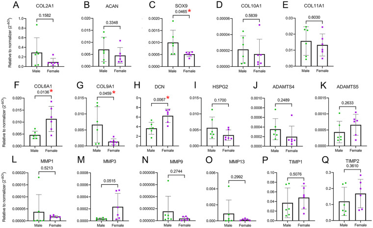
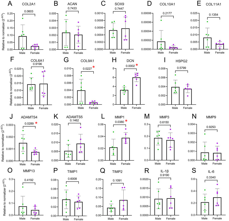
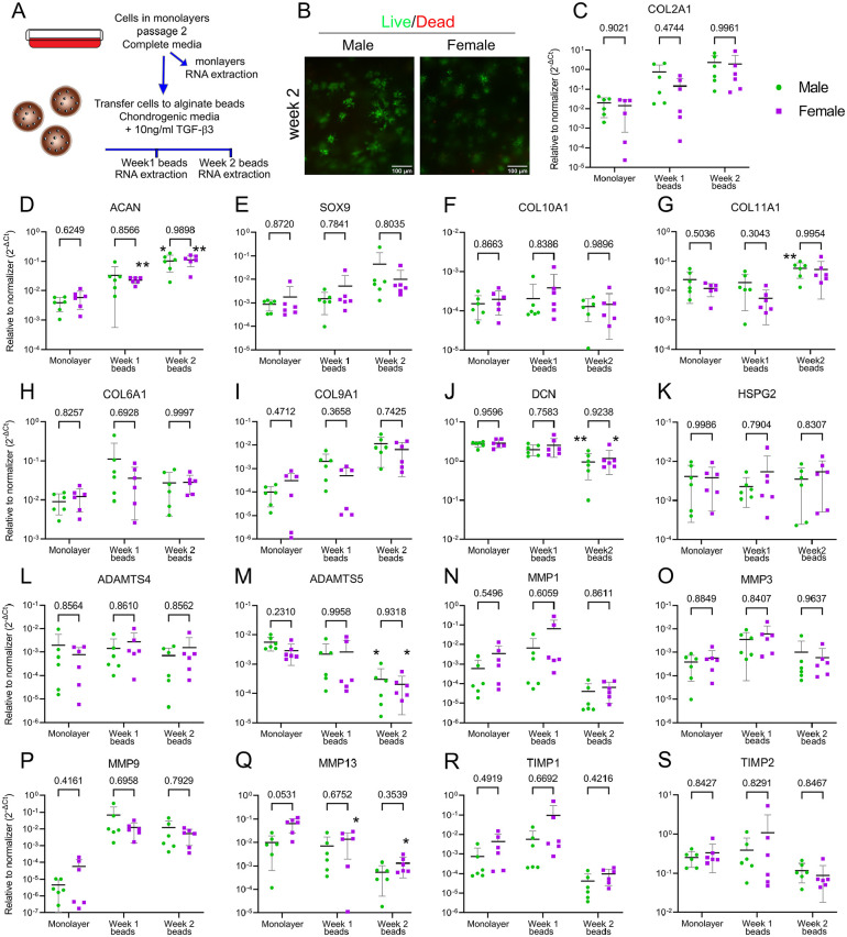
Design: Bovine knee was used as a model to study healthy adult cartilage. We compared elastic moduli under compression, abundances of extracellular and pericellular matrix (PCM) proteins using proteomics, and PCM constituency with tissue immunofluorescence. The gene expression of matrix-related genes under basal, anabolic, and catabolic conditions was assessed by quantitative polymerase chain reaction (qPCR).

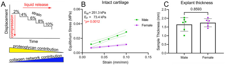
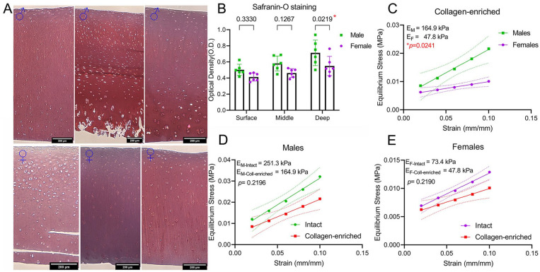
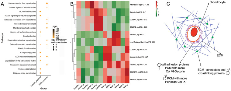
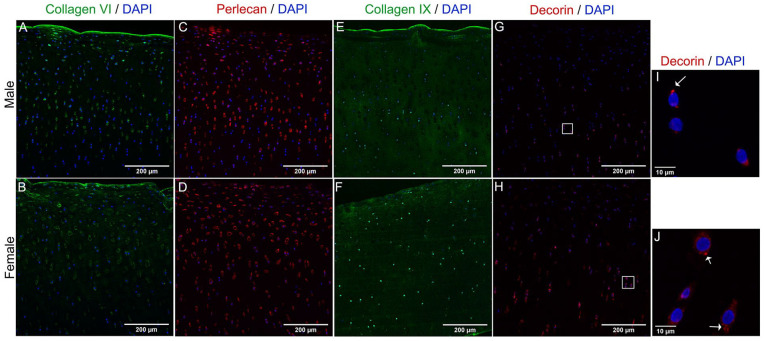
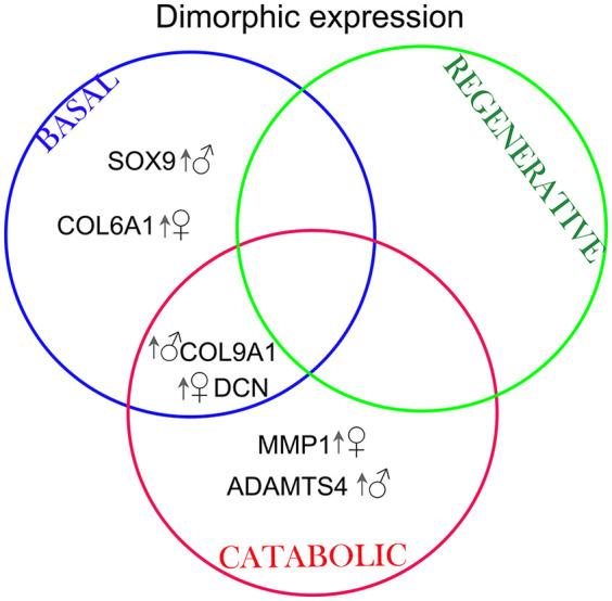
Results: The equilibrium modulus was higher in male cartilage compared with female cartilage. Proteoglycans were not associated with this biomechanical dimorphism. Proteomic and pathway analyses of tissue showed dimorphic enriched pathways in extracellular matrix (ECM)-related proteins in which male cartilage was enriched in matrix interconnectors and crosslinkers that strengthen the ECM network. Moreover, male and female tissue differed in enriched PCM components. Females had more abundance of collagen type VI and decorin, suggesting different PCM mechanics. Furthermore, the activation of regenerative and catabolic function in chondrocytes triggered sex-dependent signatures in gene expression, indicating dimorphic genetic regulation that is dependent on stimulation.

Conclusions: We provide evidence for sexual dimorphism in cartilage before menopause. Some differences are intrinsic to chondrocytes' gene expression defined by their XX versus XY chromosomal constituency.

Keywords: articular cartilage; extracellular matrix; pericellular matrix; sex differences.


Conflict of interest statement

Declaration of Conflicting Interests: The author(s) declared no potential conflicts of interest with respect to the research, authorship, and/or publication of this article.

Figures

Figure 1.
Figure 1.
Dimorphism in cartilage network mechanics. (A) Schematics of the stress relaxation test used. The expected relative contribution of proteoglycans and collagen networks to equilibrium modulus are indicated at the bottom of the graph. (B) Equilibrium modulus calculated from the linear portion of the stress-strain curve in intact cartilage. (C) Sample thickness of intact cartilage samples. Data in B and C are presented as mean ± 95% CI (dotted lines in B); n = 6♂ and 6♀, with each measured in duplicate. CI = confidence interval.
Figure 2.
Figure 2.
Proteoglycans are not responsible for mechanical dimorphism. (A) Representative images of safranin-O staining of male and female cartilage. Fast green was omitted. Scale bars are 200 µm. (B) Quantification of optical density from safranin-O staining in surface, middle, and deep layers; n = 6♂ and 6♀, mean ± SD. (C) Stress-strain curve showing calculated equilibrium moduli for male and female tissue. Proteoglycans were digested with chondroitinase ABC. (D-E) Stress-strain slopes and equilibrium modulus of males and females comparing intact with proteoglycan-depleted cartilage. Data in C-E are presented as mean ± 95% CI (dotted lines in C-E); n = 6♂ and 6♀, with each measured in duplicate.
Figure 3.
Figure 3.
Dimorphic enrichment of ECM proteins in male and female cartilage. (A) Pathway analysis of protein abundances in male and female cartilage. (B) Heat map with ECM proteins showing a significant difference in male and female cartilage. Upregulation is shown in red and downregulation is shown in green. Fold change of male compared with female is indicated in parenthesis; n = 6♂ + 6♀. (C) Summary schematics of main protein functions enriched in female and male cartilage. Male cartilage was enriched in ECM connectors and crosslink proteins, whereas female cartilage was enriched in PCM proteins and cell matrix adhesion regulators. A-B: Empirical Bayes moderated t test implemented in limma. ECM = extracellular matrix; FDR = False Discovery Rate; PCM = pericellular matrix; NCAM1 = neural cell adhesion molecule 1.
Figure 4.
Figure 4.
Dimorphic abundance of PCM proteins in male and female cartilage. Representative images of Z-project with maximum intensity of collagen type VI (A, B), perlecan (C, D), collagen type IX (E, F), and decorin (G, H) in cartilage sections. DAPI was used for nuclear counterstain (blue). Scale bars are 200 µm. A higher magnification of chondrocytes stained for decorin (I, J) reveals an accumulation of this protein inside the cell, as indicated by white arrows. Scale bars are 10 µm. Brightness was adjusted equally in males and females for display purposes only. DAPI = 4′,6-diamidino-2-phenylindole; PCM = pericellular matrix.
Figure 5.
Figure 5.
Chondrocytes show sexual dimorphism in gene expression of PCM proteins. (A-B) No difference between male and female was detected in the expression of COL2A1 and ACAN. (C) SOX9 was higher in male chondrocytes. (D-E) COL10A1 and COL11A1 were not dimorphic. (F-H) The PCM genes COL6A1, COL9A1, and DCN were expressed in a sex-dependent manner, although HSPG2 (I) was not dimorphic. (J-K) No difference was observed in ADAMTS4 and ADAMTS5 expression. (L-O) MMP1, MMP9, and MMP13 were similar between sexes, but MMP3 showed a trend for higher expression in females. (P-Q) None of the TIMPs tested were differentially expressed depending on the sex of the cell. Data are presented as mean ± SD of relative quantity to normalizer (geometric mean of 18S and GAPDH). Cells from 6♂ + 6♀ were used in passage 0. PCM = pericellular matrix; MMP = metalloprotease; ADAMTS = a disintegrin and metalloproteinase with thrombospondin motifs; DCN = decorin; TIMP = tissue inhibitor of metalloproteases; ACAN = agreccan; COL = collagen; SOX9 = SRY (sex determining region Y)-Box 9; HSPG2 = heparan sulfate proteoglycan 2 (perlecan); GAPDH = glyceraldehyde-3-phosphate dehydrogenase.
Figure 6.
Figure 6.
Catabolic function elicits a dimorphic response. Chondrocytes were treated with 10 ng/ml IL-1β for 24 hours to provoke an inflammatory response and activate the catabolic function. No differences were detected in the expression of COL2A1, ACAN, SOX9 (A-C), COL10A1, and COL11A (D-E). COL6A1 decreased similarly for males and females (F), but COL9A1 and DCN decreased, resulting in the same trend observed for basal conditions (G-H). No dimorphism was found for HSPG2 (I). All proteases tested increased with treatment (J-O), but dimorphism was observed only in ADAMTS4 and MMP1. No significant differences were detected in TIMP1 and TIMP2 expression (P-Q). The pro-inflammatory cytokines IL-1β and IL-6 increased with treatment; however, no dimorphism was detected (R-S). Data are presented as mean ± SD of relative quantity to normalizer (geometric mean of 18S and GAPDH). Cells from 6♂ + 6♀ were used in passage 0. MMP = metalloprotease; ADAMTS = a disintegrin and metalloproteinase with thrombospondin motifs; DCN = decorin; TIMP = tissue inhibitor of metalloproteases; ACAN = agreccan; COL = collagen; SOX9 = SRY (sex determining region Y)-Box 9; HSPG2 = heparan sulfate proteoglycan 2 (perlecan); GAPDH = glyceraldehyde-3-phosphate dehydrogenase.
Figure 7.
Figure 7.
No dimorphism was detected in chondrocyte regenerative function. (A) Schematics of the experimental design. (B) Representative images showing high viability of cells kept for up to 2 weeks in alginate beads in the presence of 10 ng/ml of TGF-β3. Live cells are in green (calcein) and dead are in red (ethidium homodimer-1; scale bar, 100 µm). (C-S) A qPCR analysis was done on the expression of COL2A1, ACAN, SOX9, COL6A1, COL9A1, COL10A1, COL11A1, MMP1, MMP3, MMP9, MMP13, ADAMTS4, ADAMTS5, TIMP1, and TIMP2. Data are shown as mean ± SD of relative quantity to normalizer (18S and SDHA were used as normalizers). Males are shown as green circles and females as purple squares. Cells originate from 6♂ + 6♀ in passage 2. Shown P values indicate comparison between sexes per time point (two-way ANOVA with Sidak’s test for multiple comparisons). The * indicates P < 0.05 and ** indicates P < 0.001 comparing relative quantities of Week 1 to monolayer and Week 2 to monolayer per sex (repeated measures two-way ANOVA with Dunnett’s test for multiple comparisons). TGF-β3 = transforming growth factor β3; qPCR = quantitative polymerase chain reaction; MMP = metalloprotease; ADAMTS = a disintegrin and metalloproteinase with thrombospondin motifs; ACAN = agreccan; COL = collagen; SOX9 = SRY (sex determining region Y)-Box 9; HSPG2 = heparan sulfate proteoglycan 2 (perlecan); SDHA = succinate dehydrogenase complex flavoprotein subunit A; TIMP = tissue inhibitor of metalloproteases; ANOVA = analysis of variance.
Figure 8.
Figure 8.
Venn diagram summarizing the targets that were differentially regulated in male and female chondrocytes depending on the particular stimulation. Arrow and symbol indicate which biological sex had an upregulated gene expression. MMP = metalloprotease; ADAMTS = a disintegrin and metalloproteinase with thrombospondin motifs; DCN = decorin; COL = collagen; SOX9 = SRY (sex determining region Y)-Box 9.

References

    1. Hewett TE, Myer GD, Ford KR. Anterior cruciate ligament injuries in female athletes: Part 1, mechanisms and risk factors. Am J Sports Med. 2006;34(2):299-311. doi:10.1177/0363546505284183. - DOI - PubMed
    1. Hosseinzadeh S, Kiapour AM. Sex differences in anatomic features linked to anterior cruciate ligament injuries during skeletal growth and maturation. Am J Sports Med. 2020;48(9):2205-12. doi:10.1177/0363546520931831. - DOI - PMC - PubMed
    1. Racine J, Aaron RK. Post-traumatic osteoarthritis after ACL injury. R I Med J. 2014;97(11):25-8. - PubMed
    1. Boyan BD, Tosi L, Coutts R, Enoka R, Hart DA, Nicolella DP, et al.. Sex differences in osteoarthritis of the knee. J Am Acad Orthop Surg. 2012;20(10):668-9. doi:10.5435/jaaos-20-10-668. - DOI - PMC - PubMed
    1. Hame SL, Alexander RA. Knee osteoarthritis in women. Curr Rev Musculoskelet Med. 2013;6(2):182-7. doi:10.1007/s12178-013-9164-0. - DOI - PMC - PubMed

Publication types