. 2022 Oct 11;69(4):T25-T36.
doi: 10.1530/JME-22-0113. Print 2022 Nov 1.

Retinoic acid receptor structures: the journey from single domains to full-length complex

Affiliations

Retinoic acid receptor structures: the journey from single domains to full-length complex

Fraydoon Rastinejad. J Mol Endocrinol. .

Abstract

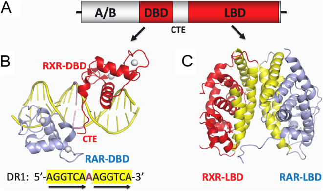
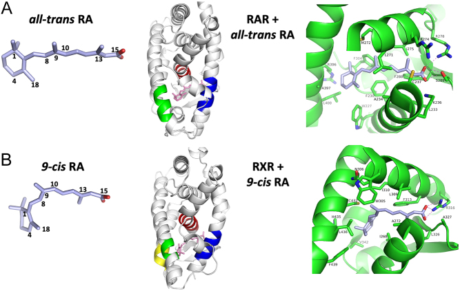
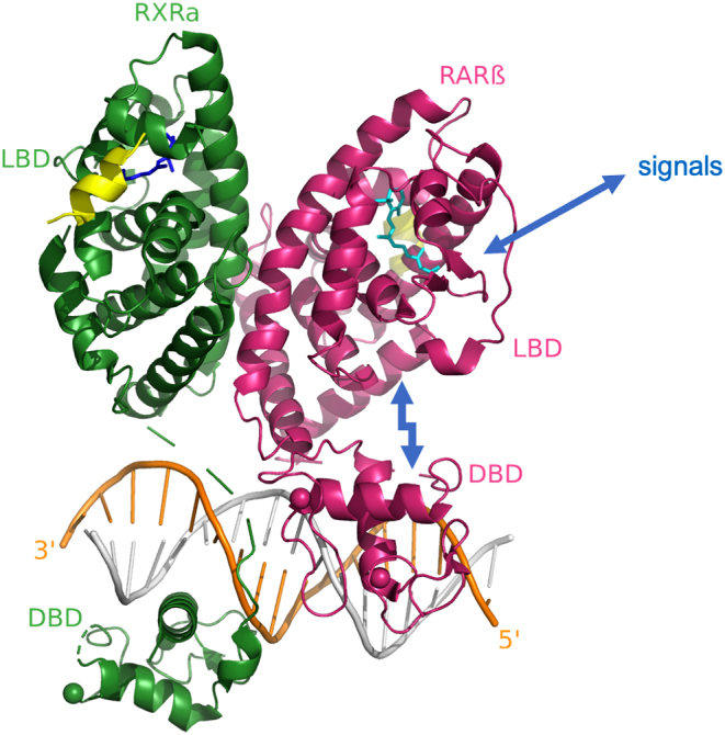
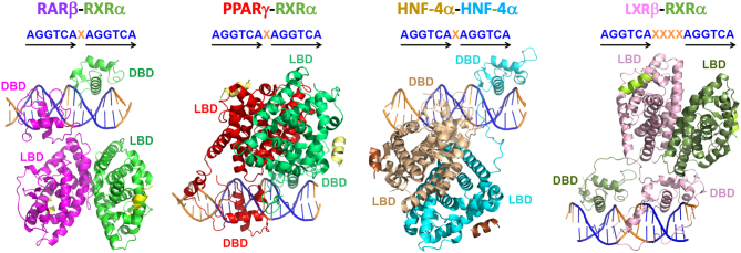
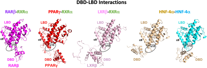
The retinoic acid receptors (RARα, β, and γ) are multi-domain polypeptides that heterodimerize with retinoid X receptors (RXRα, β, and γ) to form functional transcription factors. Understanding the three-dimensional molecular organization of these nuclear receptors (NRs) began with RAR and RXR DNA-binding domains (DBDs), and were followed with studies on isolated ligand-binding domains (LBDs). The more complete picture emerged in 2017 with the multi-domain crystal structure of RXRα-RARβ on its response element with retinoic acid molecules and coactivator segments on both proteins. The analysis of that structure and its complementary studies have clarified the direct communication pathways within RXR-RAR polypeptides, through which DNA binding, protein-ligand, and protein-protein interactions are integrated for overall functional responses. Understanding the molecular connections in the RXR-RAR complex has benefited from direct observations of the multi-domain structures of RXRα-PPARγ, RXRα-LXRβ, HNF-4α homodimer, and androgen receptor homodimer, each bound to its response element. These comprehensive NR structures show unique quaternary architectures, yet all have DBD-DBD, LBD-LBD, and DBD-LBD domain-domain contacts within them. These convergence zones allow signals from discrete domains of their polypeptides to be propagated and integrated across their entire complex, shaping their overall responses in an allosteric fashion.

Keywords: hormone receptors; receptors; retinoic acid; structure.


Conflict of interest statement

The author is a founder and consultant for Flare Therapeutics.

Figures

Figure 1
Figure 1
Individual domains of RXR–RAR polypeptides and their abilities to form DBD–DBD and LBD–LBD interactions. (A) The generalized polypeptide organization of NRs showing domain locations. (B) The first crystal structure for RXR–RAR, observed on a complex with its DR1 response element (Rastinejad et al. 2000) PDB 1DSZ. RARα binds to the 5’-AGGTCA half-site and RXRα binds to the 3’ half-site of the DR1 element. Each DBD uses its recognition helix to recognize half-sites at their major grooves. In addition, there is an observed DBD–DBD interaction that is DNA dependent, forming at the one base-pair spacer minor groove of the DR1 element. (C) The crystal structure of the RXRα–RARβ LBD–LBD dimer (Pogenberg et al. 2005) PDB 1XDK. The dimerization interface that joins two LBDs is DNA independent, and much larger in size than that formed between the two DBDs, in terms of buried solvent exposed surfaces. The LBD–LBD interface involves helices H7, H9 and H10, and loops L8–9 and L9–10 of each subunit (shown in yellow) which face each other.
Figure 2
Figure 2
Molecular structures of RA molecules and their binding sites within RAR and RXR LBDs. (A) 3D structure of all-trans RA and its binding location and interactions with RARγ LBD (from PDB 2LBD). (B) 3D structure of 9-cis RA and its binding location and interactions with RXRα LBD (from PDB 1FBY). Each ligand becomes lodged in a cavity formed mainly through helix-3 (blue), helix-5 (red) and helix-10/11 (green).
Figure 3
Figure 3
The multi-domain crystal structure of the RXRα–RARβ heterodimer with DNA, ligands, and coregulator peptides (Chandra et al. 2017) PDB 5UAN. All of the previously known LBD–LBD, DBD–DBD, and DBD–DNA interactions (Fig. 1) were preserved within this multi-domain complex. But there were new findings including the LBD–DBD interface within the RARβ protein. Biochemical and cell-based functional studies indicate that signals detected at the LBD (different ligands) or DBD (different response elements) can be allosterically transmitted in a bidirectional manner (Chandra et al. 2017).
Figure 4
Figure 4
Uniqueness of NR quaternary structures. Here a comparison of the multi-domain quaternary crystal structures of RXRα–RARβ (Chandra et al. 2017) PDB 5UAN, RXRα–PPARγ (Chandra et al. 2008) PDB 3DZY, 3E00, 3DZU, HNF-4α homodimer (Chandra et al. 2013) PDB 4IQR, and RXRα–LXRβ–DR4 heterodimer (Lou et al. 2014) PDB 4NQA is shown. All four complexes are aligned the same way, with their DBDs and half-sites held in identical positions for direct comparison of quaternary structures.
Figure 5
Figure 5
DBD–LBD interactions in all four NR–DNA complexes. The DBD–LBD interface is established between a loop in the DBD (preceding the DBD’s α1 helix) and the region of the LBD located between α9 and α10 helices The PDB IDs are indicated in Fig. 3.

Similar articles

Cited by

References

    1. Benbrook D, Lernhardt E, Pfahl M.1988A new retinoic acid receptor identified from a hepatocellular carcinoma. Nature 333669–672. (10.1038/333669a0) - DOI - PubMed
    1. Black BE, Holaska JM, Rastinejad F, Paschal BM.2001DNA binding domains in diverse nuclear receptors function as nuclear export signals. Current Biology 111749–1758. (10.1016/s0960-9822(0100537-1) - DOI - PubMed
    1. Bourguet W, Ruff M, Chambon P, Gronemeyer H, Moras D.1995Crystal structure of the ligand-binding domain of the human nuclear receptor RXR-alpha. Nature 375377–382. (10.1038/375377a0) - DOI - PubMed
    1. Bourguet W, Vivat V, Wurtz JM, Chambon P, Gronemeyer H, Moras D.2000Crystal structure of a heterodimeric complex of RAR and RXR ligand-binding domains. Molecular Cell 5289–298. (10.1016/s1097-2765(0080424-4) - DOI - PubMed
    1. Brand N, Petkovich M, Krust A, Chambon P, de The H, Marchio A, Tiollais P, Dejean A.1988Identification of a second human retinoic acid receptor. Nature 332850–853. (10.1038/332850a0) - DOI - PubMed

LinkOut - more resources