. 2022 Dec 7;70(1):e220120.
doi: 10.1530/JME-22-0120. Print 2023 Jan 1.

Structure of full-length TSH receptor in complex with antibody K1-70™

Affiliations

Structure of full-length TSH receptor in complex with antibody K1-70™

Ricardo Núñez Miguel et al. J Mol Endocrinol. .

Abstract

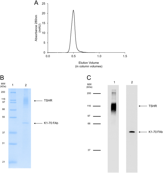
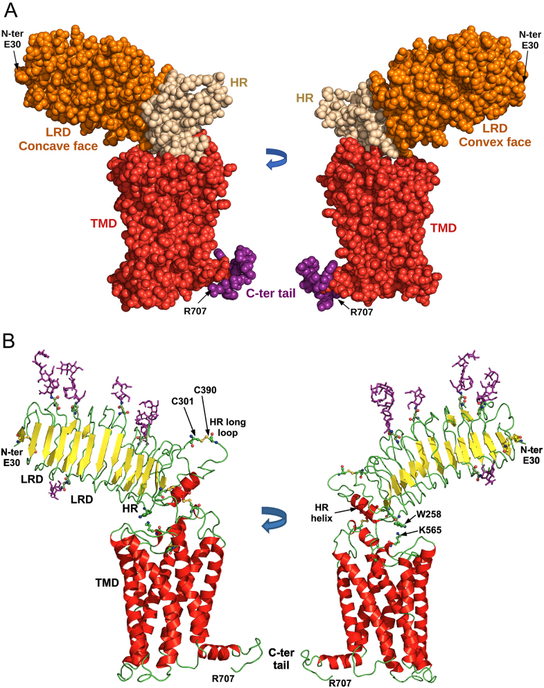
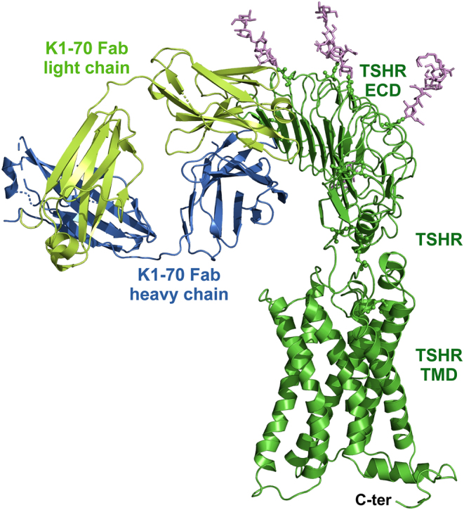
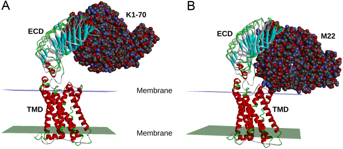
Determination of the full-length thyroid-stimulating hormone receptor (TSHR) structure by cryo-electron microscopy (cryo-EM) is described. The TSHR complexed with human monoclonal TSHR autoantibody K1-70™ (a powerful inhibitor of TSH action) was detergent solubilised, purified to homogeneity and analysed by cryo-EM. The structure (global resolution 3.3 Å) is a monomer with all three domains visible: leucine-rich domain (LRD), hinge region (HR) and transmembrane domain (TMD). The TSHR extracellular domain (ECD, composed of the LRD and HR) is positioned on top of the TMD extracellular surface. Extensive interactions between the TMD and ECD are observed in the structure, and their analysis provides an explanation of the effects of various TSHR mutations on TSHR constitutive activity and on ligand-induced activation. K1-70™ is seen to be well clear of the lipid bilayer. However, superimposition of M22™ (a human monoclonal TSHR autoantibody which is a powerful stimulator of the TSHR) on the cryo-EM structure shows that it would clash with the bilayer unless the TSHR HR rotates upwards as part of the M22™ binding process. This rotation could have an important role in TSHR stimulation by M22™ and as such provides an explanation as to why K1-70™ blocks the binding of TSH and M22™ without activating the receptor itself.

Keywords: TSHR; autoantibodies; autoimmunity; cryo-EM; structure.


Figures

Figure 1
Figure 1
(A) Analysis of purified TSHR–K1-70™ complex by size exclusion chromatography (Superdex 200 XK 16-100 column run in 50 mmol/L Tris–HCl pH 8.0, 150 mmol/L NaCl, 0.02% LMNG and 0.002% CHS). (B) Analysis of purified TSHR–K1-70™ complex by SDS-PAGE (12% acrylamide gel) under non-reducing conditions. The positions of the molecular weight standards are shown and the positions of the TSHR and the K1-70™ Fab are marked (lane 1: molecular weight standards; lane 2: purified TSHR–K1-70™ complex). (C) Western blotting analysis of purified TSHR–K1-70™ complex. Lane 1: blotting with mouse monoclonal TSHR antibody 8E2 labelled with horseradish peroxidase; lane 2: blotting with mouse anti-human IgG (Fab-specific) MAB labelled with horseradish peroxidase. See text for experimental details.
Figure 2
Figure 2
Structural features of the full-length TSHR. (A) TSHR structure (amino acids E30 to R707) in two views rotated 180o. The structure is a space fill representation with the leucine-rich-repeat domain (LRD) in orange, the hinge region (HR) in beige and the transmembrane domain (TMD) in red with the intracellular C-terminus in purple. (B) TSHR structure in two views rotated 180o. The structure is in cartoon representation with the disulphide bonds and glycosylation sites in ball and stick representation with oxygen in red, nitrogen in blue and sulphur in yellow. The glycans are shown in purple. The LRD, HR and TMD are marked. Cys301 forms a disulphide bond with Cys390 and the residues in between (amino acids 302–389) are missing from the structure.
Figure 3
Figure 3
Structural features of the full-length TSHR in complex with the blocking monoclonal autoantibody K1-70™. The TSHR–K1-70™ Fab structure is in cartoon representation with the glycans shown in purple sticks. The K1-70™ heavy chain is shown in blue, the K1-70™ light chain in light green and the TSHR in green. The TSHR, extracellular domain (ECD), transmembrane domain (TMD) and intracellular C-terminus are marked.
Figure 4
Figure 4
Proposed mechanism of TSHR activation by M22™. The cryo-EM structure of the TSHR bound to the blocking autoantibody K1-70™ was superimposed with the crystal structure of the TSHR leucine-rich domain (LRD) in complex with K1-70™ (A) and with the crystal structure of the TSHR LRD in complex with the stimulating monoclonal autoantibody M22™ (B). K1-70™ does not contact the membrane while the stimulating antibody M22™ is seen to clash with the membrane. This clash between M22™ and the membrane would inhibit M22™ binding to the TSHR unless the receptor’s ECD rotates upwards as part of the M22™ binding process. This rotation could have an important role in TSHR stimulation by M22™.

References

    1. Bahn RS.2010Graves’ ophthalmopathy. New England Journal of Medicine 362726–738. (10.1056/NEJMra0905750) - DOI - PMC - PubMed
    1. Ballesteros JA, Weinstein H.1995Integrated methods for the construction of three dimensional models and computational probing of structure function relations in G protein-coupled receptors. Methods in Neurosciences 25366–428. (10.1016/S1043-9471(0580049-7) - DOI
    1. BIOVIA Dassault Systèmes 2021Discovery Studio 2021. San Diego, CA, USA: BIOVIA Dassault Systèmes.
    1. Birk HW, Koepsell H.1987Reaction of monoclonal antibodies with plasma membrane proteins after binding on nitrocellulose: renaturation of antigenic sites and reduction of non-specific antibody binding. Analytical Biochemistry 16412–22. (10.1016/0003-2697(8790360-5) - DOI - PubMed
    1. Boutin A, Krieger CC, Marcus-Samuels B, Klubo-Gwiezdzinska J, Neumann S, Gershengorn MC.2020TSH receptor homodimerization in regulation of cAMP production in human thyrocytes in vitro. Frontiers in Endocrinology 11 276. (10.3389/fendo.2020.00276) - DOI - PMC - PubMed

Publication types

LinkOut - more resources