Review
. 2022 Sep 28;122(18):14842-14880.
doi: 10.1021/acs.chemrev.2c00212. Epub 2022 Sep 7.

Microfluidics for Neuronal Cell and Circuit Engineering

Affiliations
Review

Microfluidics for Neuronal Cell and Circuit Engineering

Rouhollah Habibey et al. Chem Rev. .

Abstract

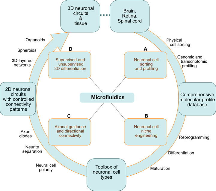
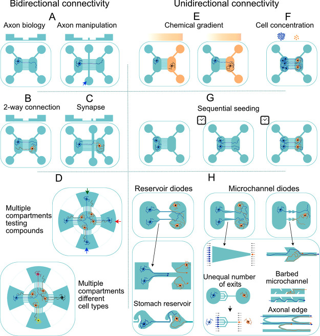
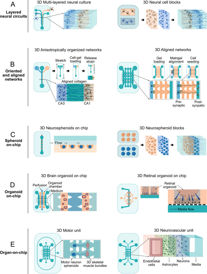
The widespread adoption of microfluidic devices among the neuroscience and neurobiology communities has enabled addressing a broad range of questions at the molecular, cellular, circuit, and system levels. Here, we review biomedical engineering approaches that harness the power of microfluidics for bottom-up generation of neuronal cell types and for the assembly and analysis of neural circuits. Microfluidics-based approaches are instrumental to generate the knowledge necessary for the derivation of diverse neuronal cell types from human pluripotent stem cells, as they enable the isolation and subsequent examination of individual neurons of interest. Moreover, microfluidic devices allow to engineer neural circuits with specific orientations and directionality by providing control over neuronal cell polarity and permitting the isolation of axons in individual microchannels. Similarly, the use of microfluidic chips enables the construction not only of 2D but also of 3D brain, retinal, and peripheral nervous system model circuits. Such brain-on-a-chip and organoid-on-a-chip technologies are promising platforms for studying these organs as they closely recapitulate some aspects of in vivo biological processes. Microfluidic 3D neuronal models, together with 2D in vitro systems, are widely used in many applications ranging from drug development and toxicology studies to neurological disease modeling and personalized medicine. Altogether, microfluidics provide researchers with powerful systems that complement and partially replace animal models.


Conflict of interest statement

The authors declare no competing financial interest.

Figures

Figure 1
Figure 1
Diverse applications of microfluidic platforms: from molecular characterization of cells in the central nervous system to engineering neuronal cell types and neural circuits in vitro. (A) Neuronal cells extracted from native brain tissue are sorted based on their physical properties or surface markers and are classified based on their genomic or transcriptomic profile (qRT-PCR and single-cell RNA-Seq). (B) The information gathered on the molecular identity of the diverse neurons in the brain, retina, and spinal cord is useful for devising strategies to reprogram and differentiate hiPSCs into specific neuronal cell types. (C) HiPSC-derived neurons can be used to engineer 2D neural circuits or (D) be incorporated in physiologically relevant systems as 3D layered networks and organoids.
Figure 2
Figure 2
Different microfluidic cell-sorting strategies. (A) Cell separation using viscoelastically tuned hydrodynamic spreading. Depending on the viscosity of the elution flow and on cell size, specific cells can be separated. (B) Inertial separation of neurons and glia in a serpentine microchannel. Large cells (neurons) tend to migrate to the center of the microchannel, while small glial cells that experience stronger inertial forces stay close to the sidewalls. (C) Isolating single cells in neurospheres using inertial microfluidics. The curvature of the spiral microfluidic channel induces Dean’s forces that push small particles and single cells toward the inner wall. Larger particles, as cell clusters, move toward the center. (D) As whole cell membrane capacitance is a biomarker of stem cell fate potential and, conversely, of ongoing differentiation processes, label-free dielectrophoresis-assisted continuous sorters exploit this electrophysiological property of the plasma membrane for sorting more (e.g., neuron- or astrocyte-forming cells) or less differentiated cells (e.g., stem cells)., (E) Acoustophoresis-based separation of live neuroblastoma and human ESCs from apoptotic cells. A first piezoceramic transducer aligns the cells close to the wall, while a second one deflects their trajectory based on their acoustic properties and morphology. (F) Real-time deformability cytometry enables on-the-fly analysis of cells deforming as they pass through narrow microchannels without exposing them to shear stresses or pressure gradients. (G) Low-cost and simple microfluidic FACS (μFACS). Label-based neuronal cell sorting can be performed in μFACS at a reduced cost. (H) Characterizing the differentiation state of neuronal stem cells based on specific membrane capacitance and cytoplasm conductivity. Cells are continuously aspirated into a constriction channel to measure these properties. (I) Sorting cells based on their dynamic response to a chemical stimulus. Cells are introduced to the sorting device through a flow line (depicted in green), and their movement and positions are adjusted by control lines (depicted in yellow). After trapping the cells, a stimulus is delivered through the appropriate flow line, and the cell response is measured based on calcium influx. As proof of principle, this method has been applied to separate olfactory sensory neurons that respond to specific odor cues.
Figure 3
Figure 3
Contribution of microfluidics-based concepts to scRNA sequencing. Cells obtained either from primary neuronal tissues or from models engineered in vitro are dissociated and sorted by FACS. Purified cells are processed using either low-throughput RNA-Sequencing tools like Smart-Seq and CEL-Seq, or high-throughput microfluidic systems. In general, three main microfluidic approaches are used for single-cell analysis: valve-based (e.g., Fluidigm 1), droplet-based (Drop-Seq, inDrop, 10× Chromium, and Quartz-Seq), and microwell-based (Seq-well) systems. In all cases, trapped single cells are lysed, their RNA is hybridized and reverse transcribed (RT), and cDNA is then amplified either by PCR or linear isothermal amplification by T7-based in vitro transcription (IVT). Thereafter, the cDNA libraries generated in these steps are sequenced, and the data are demultiplexed, aligned to a reference transcriptome, and interpreted for classification of neuronal cell subpopulations. STAMP: single-cell transcriptomes attached to microparticles.
Figure 4
Figure 4
Engineering the neuronal cell niche using microfluidic gradient generators. (A) Microfluidic channels and microwells are used to deposit solid or surface-bound cues. (B,C) Surface-bound binary or gradient patterns have been generated by microchannel devices to probe neuronal cell polarization and axonal growth in response to attractant or repellent factors (also shown in E).,,,, (D) Similarly, chemical gradients integrated with topographical gradients or cues have been deployed to guide neurites. (E) Schematic axonal growth cone response to attractant (upper panel) and repellent (lower panel) cue gradients. (F) Two basic diffusive gradient generators are Y-junction and T-junction configurations. (G) Osmotic pump-derived ultraslow flow rate generates continuous and overlapping chemical gradients to induce a common stem cell population to differentiate into neurons and Schwann cells. The lower panel shows a device with asymmetric peripheral channels whereby gradually changing gradients of soluble Netrin-1 are created. In such a device, the axon growth response can be subsequently measured. (H) Christmas tree microfluidic channel networks have been used to create 1D or 2D gradients of neuronal growth factors to differentiate NSCs into neurons, of Shh and Netrin-1 to guide axons, and of Wnt to model neural tube development. (I) 3D gradient of neurotrophic factors and axon guiding factors has also been generated in scaffold-based neuronal cultures embedded in microfluidic devices.,,
Figure 5
Figure 5
Main approaches for engineering neuronal circuits in microfluidic devices. (A) Two-compartment microfluidic devices can be used for axonal isolation. In such devices, a third compartment is often added to manipulate isolated axonal branches.,, (B) Seeding neurons in both compartments of such two-compartment devices allows for the generation of bidirectionally connected networks.,,,,, (C) Inserting a third compartment to two-compartment devices close to one neuronal population is a commonly used strategy to study synapses., (D) Multicompartment devices connecting different neuronal populations bidirectionally enables the engineering of complex circuits and the testing of chemical compounds in specific populations.,,, (E–H) Unidirectional neuronal circuits are constructed in compartmentalized devices connected by straight microchannels or by diode-style configurations. In straight microchannels, the probability of axonal growth in one direction is manipulated by (E) the use of axonal attractant or repellent gradients,, (F) the seeding of different cell densities in the two connected compartments,, and/or (G) the sequential seeding of cells in each compartment., (H) The axonal diode configuration of microfluidic devices is one of the most widely used approaches to direct axonal growth., Diode structures can be embedded in reservoirs or, alternatively, entire reservoirs can be designed in a particular shape, e.g., stomach, to facilitate unidirectional axonal growth., Microchannel diodes can also be designed by simply narrowing them on one side,, to create arrow-like structures,, by including barbs that prompt axons to grow in one direction, or by connecting adjacent channels with arches that allow growing axons to turn backward. Overall, axonal diode layouts are designed to increase the probability of axons growing in a particular direction.
Figure 6
Figure 6
Construction of 3D neuronal circuits in microfluidic devices. (A) Layered neuronal circuits: Different neuronal types are embedded in hydrogel and pushed through microchannels in microfluidic devices to engineer 3D layers. Hydrogel-embedded neuronal cell blocks can be generated using PDMS devices and placed next to each other afterward. (B) Oriented and aligned networks: 3D hydrogel scaffolds supporting neuronal cells are aligned either by stretching the microfluidic device during the polymerization process or by applying hydrostatic pressure. Aligning collagen or Matrigel fibers enables the axons to be better guided from presynaptic to postsynaptic compartments and to thereby form unidirectional networks. (C) Spheroid-on-a-chip: High-throughput scaffold-free neurospheroid cultures are generated in perfused microwell microfluidic systems. Spheroid blocks from different neuronal cell types can be engineered and subsequently placed next to each other. (D) Organoid-on-a-chip: Growing brain and retinal organoids in microfluidic devices with improved diffusion extends their lifespan. (E) Organ-on-a-chip: By integrating additional cell types such as myocytes or endothelial cells, functional units like the motor unit or the blood–brain barrier (BBB) can be replicated in microfluidic platforms.

Similar articles

Cited by

References

    1. Huang Z. J.; Paul A. The Diversity of GABAergic Neurons and Neural Communication Elements. Nat. Rev. Neurosci. 2019, 20, 563–572. 10.1038/s41583-019-0195-4. - DOI - PMC - PubMed
    1. Poulin J. F.; Tasic B.; Hjerling-Leffler J.; Trimarchi J. M.; Awatramani R. Disentangling Neural Cell Diversity Using Single-Cell Transcriptomics. Nat. Neurosci. 2016, 19, 1131–1141. 10.1038/nn.4366. - DOI - PubMed
    1. Mu Q.; Chen Y.; Wang J. Deciphering Brain Complexity Using Single-Cell Sequencing. Genomics, Proteomics Bioinforma. 2019, 17, 344–366. 10.1016/j.gpb.2018.07.007. - DOI - PMC - PubMed
    1. Bassett D. S.; Gazzaniga M. S. Understanding Complexity in the Human Brain. Trends Cogn. Sci. 2011, 15, 200–209. 10.1016/j.tics.2011.03.006. - DOI - PMC - PubMed
    1. Pastrana E. Focus on Mapping the Brain. Nat. Methods 2013, 10, 481–481. 10.1038/nmeth.2509. - DOI - PubMed

Publication types