. 2022 Sep 7;12(1):15192.
doi: 10.1038/s41598-022-19469-x.

Construction of a culture protocol for functional bile canaliculi formation to apply human iPS cell-derived hepatocytes for cholestasis evaluation

Affiliations

Construction of a culture protocol for functional bile canaliculi formation to apply human iPS cell-derived hepatocytes for cholestasis evaluation

Shinichiro Horiuchi et al. Sci Rep. .

Abstract

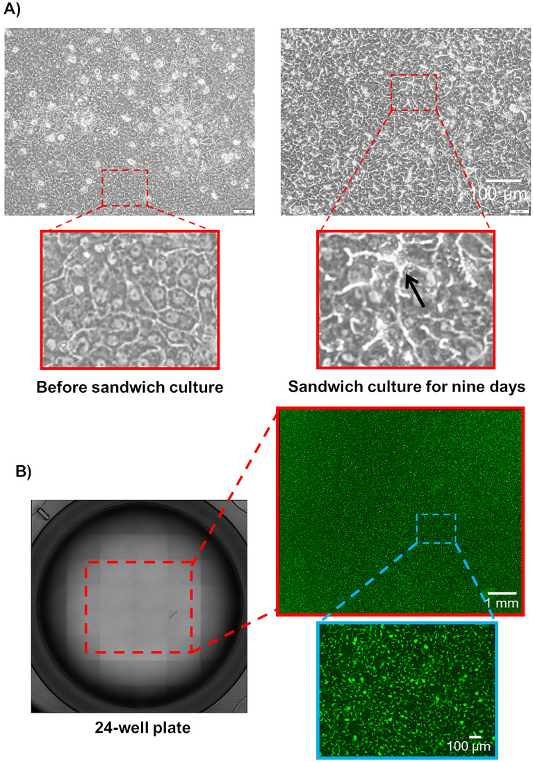
Cholestatic toxicity causes the failure of pharmaceutical agents during drug development and, thus, should be identified at an early stage of drug discovery and development. The formation of functional bile canaliculi in human hepatocytes is required for in vitro cholestasis toxicity tests conducted during the early stage of drug development. In this study, we investigated the culture conditions required for the formation of bile canaliculi using human-induced pluripotent stem cell-derived hepatocytes (hiPSC-Heps). When hiPSC-Heps were sandwich-cultured under the condition we established, extended bile canaliculi were formed on the whole well surfaces. Biliary efflux transporters were localized in the formed bile canaliculi structures which had junctional complexes. After the model substrates of the biliary efflux transporters were taken up into cells, their subsequent excretion into the bile canaliculi was observed and was found to be impeded by each inhibitor of the biliary efflux transporter. These findings suggest that bile canaliculi have transporter-specific bile excretion abilities. We will continue to study the application of this culture protocol to cell-based cholestasis assay system. As a result, the culture protocol could lead to a highly predictable, robust cell-based cholestasis assay system because it forms functional bile canaliculi reproducibly and efficiently.


Conflict of interest statement

The authors declare no competing interests.

Figures

Figure 1
Figure 1
Accumulation of model fluorescein substrates into bile canaliculi when the culture conditions for the formation of extended bile canaliculi were examined. Human-induced pluripotent stem cell-derived hepatocytes were cultured in a long-term maintenance medium for 14 days, 21 days, and 28 days and then, they were sandwich-cultured in a maintenance medium for 9 days. Following the sandwich culture, the excretion of model fluorescein substrates (fluorescein diacetate and tauro-nor-THCA-24-DBD) into bile canaliculi was observed. Fluorescence images show (A) Fluorescein and (B) Tauro-nor-THCA-24-DBD, which were accumulated into bile canaliculi.
Figure 2
Figure 2
Bile canaliculi formed in suitable culture conditions. Human-induced pluripotent stem cell-derived hepatocytes were cultured in a long-term maintenance medium for 28 days and then, they were sandwich-cultured in a maintenance medium for 9 days. (A) The cell morphology that was observed using a phase-contrast microscope before and after the sandwich culture. The black arrow shows the extended bile canaliculi. After sandwich culture, the excretion of tauro-nor-THCA-24-DBD into a bile canaliculus was observed in a wide area within the well (B: Red dotted line). Fluorescence images show (B) Tauro-nor-THCA-24-DBD, which was accumulated into bile canaliculi.
Figure 3
Figure 3
Expression of the genes which relate to the maturity of hepatocytes while cells were cultured in a long-term maintenance medium. The expression levels of ALB and AFP were measured using quantitative polymerase chain reaction over time during culture in a long-term maintenance medium for 28 days. The bar shows the relative expression levels of ALB or AFP. Pooled human liver RNA was used for the standard curve, and the expression level was set as one. The relative expression level was calculated using the equation of the line for the standard curve. Data are presented as means ± S.D. (n = 3). ** Shows that expression significantly increased compared with that on day 4 (P < 0.01).
Figure 4
Figure 4
Gene expressions and the localization of biliary efflux transporters when the bile canaliculi were formed. Human-induced pluripotent stem cell-derived hepatocytes were cultured in a long-term maintenance medium for 28 days and then, they were sandwich-cultured in a maintenance medium for 9 days. (A) Expression levels of the biliary efflux transporters, multidrug resistance protein 2 (MRP2) and bile salt export pump (BSEP), were measured using quantitative polymerase chain reaction before and after sandwich culture. The bar shows the relative expression levels of MRP2 or BSEP. Pooled RNA from human liver was used for the standard curve, and the expression level was set as one. The relative expression level was calculated using the equation of the line for the standard curve. Data are presented as means ± S.D. (n = 3). ** Indicates that the expression significantly increased compared with that prior to the sandwich culture (P < 0.01). (B) Immunostaining images of MRP2 and BSEP after sandwich culture. Actin filaments were stained with phalloidin as an index of bile canaliculi.
Figure 5
Figure 5
Effect of a biliary efflux transporter inhibitor on the excretion of the model substrate into bile canaliculi. Human-induced pluripotent stem cell-derived hepatocytes were cultured in a long-term maintenance medium for 28 days and then, they were sandwich-cultured in a maintenance medium for 9 days. After that, the effect of a biliary efflux transporter inhibitor on the excretion of the model substrate into bile canaliculi was examined. Fluorescence images show (A) Fluorescein or (B) Tauro-nor-THCA-24-DBD, when cells were treated with the biliary efflux transporter inhibitor (MK571 or cyclosporine A) or left untreated.
Figure 6
Figure 6
Accumulation of model fluorescein substrates into bile canaliculi when the stability of the canalicular efflux ability was examined. The term of sandwich culture in a maintenance medium following culture in an LTM medium for 28 days was examined to determine how long the assay is possible. Fluorescence images show fluorescein or tauro-nor-THCA-24-DBD, which accumulated in bile canaliculi when biliary efflux assay was performed on the 7th to 14th day of the sandwich culture.
Figure 7
Figure 7
(A) Microstructure of bile canaliculi was observed using transmission electron microscopy. There are bile canaliculi in the dotted line in the left electron microscopy image. The right electron microscopy image is an enlarged view of the dotted line in the left that shows the microstructure of bile canaliculi. hiPSC-Heps were cultured in the LTM medium for 28 days and then sandwich-cultured in a maintenance medium for 9 days. (B) Accumulation of model fluorescein substrates into bile canaliculi under maintenance or disruption conditions and the microstructure of bile canaliculi. Human-induced pluripotent stem cell-derived hepatocytes (hiPSC-Heps) were cultured in a long-term maintenance (LTM) medium for 28 days and then, they were sandwich-cultured in a maintenance medium for 9 days. Fluorescence images reveal the presence of 5-(and-6)-carboxy-2′,7′-dichloro-fluorescein (CDF), which accumulated into bile canaliculi under maintenance or disruption conditions. hiPSC-Heps were cultured in the LTM medium for 28 days and then sandwich-cultured in a maintenance medium for 9 days.
Figure 8
Figure 8
Gene expression of cytochrome P450 when the bile canaliculi were formed. Human-induced pluripotent stem cell-derived hepatocytes were cultured in a long-term maintenance medium for 28 days and then, they were sandwich-cultured in a maintenance medium for 9 days. The expression levels of major CYPs were measured using quantitative polymerase chain reaction before and after sandwich culture. The bar shows the relative expression levels of CYPs. Pooled RNA from human liver was used for the standard curve, and the expression level was set as one. The relative expression level was calculated using the equation of the line for the standard curve. The red line shows the maximum value of expression in 22 lots of human cryopreserved hepatocytes. The blue line shows the minimum value of expression in 22 lots of human cryopreserved hepatocytes. Data are presented as means ± SD. (n = 3). ** Shows that expression significantly increased compared with that before sandwich culture (P < 0.01).

References

    1. Onakpoya IJ, Heneghan CJ, Aronson JK. Post-marketing withdrawal of 462 medicinal products because of adverse drug reactions: A systematic review of the world literature. BMC Med. 2016;14:10. doi: 10.1186/s12916-016-0553-2. - DOI - PMC - PubMed
    1. Takikawa H, Murata Y, Horiike N, Fukui H, Onji M. Drug-induced liver injury in Japan: An analysis of 1676 cases between 1997 and 2006. Hepatol. Res. 2009;39(5):427–431. doi: 10.1111/j.1872-034X.2008.00486.x. - DOI - PubMed
    1. Padda MS, Sanchez M, Akhtar AJ, Boyer JL. Drug-induced cholestasis. Hepatology. 2011;53(4):1377–1387. doi: 10.1002/hep.24229. - DOI - PMC - PubMed
    1. Kolarić TO, Ninčević V, Smolić R, Smolić M, Wu GY. Mechanisms of hepatic cholestatic drug injury. J. Clin. Transl. Hepatol. 2019;7(1):86–92. - PMC - PubMed
    1. Morales M, Vélez L, Muñoz M. Hepatotoxicity: A drug-induced cholestatic pattern. Rev. Colomb. Gastroenterol. 2016;31(1):36–47. doi: 10.22516/25007440.71. - DOI

Publication types

Substances