. 2022 Jun;15(6):1296-1308.
doi: 10.1038/s41385-022-00563-2. Epub 2022 Sep 7.

Enteric glial cells favor accumulation of anti-inflammatory macrophages during the resolution of muscularis inflammation

Affiliations

Enteric glial cells favor accumulation of anti-inflammatory macrophages during the resolution of muscularis inflammation

Michelle Stakenborg et al. Mucosal Immunol. 2022 Jun.

Abstract

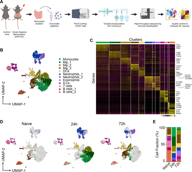
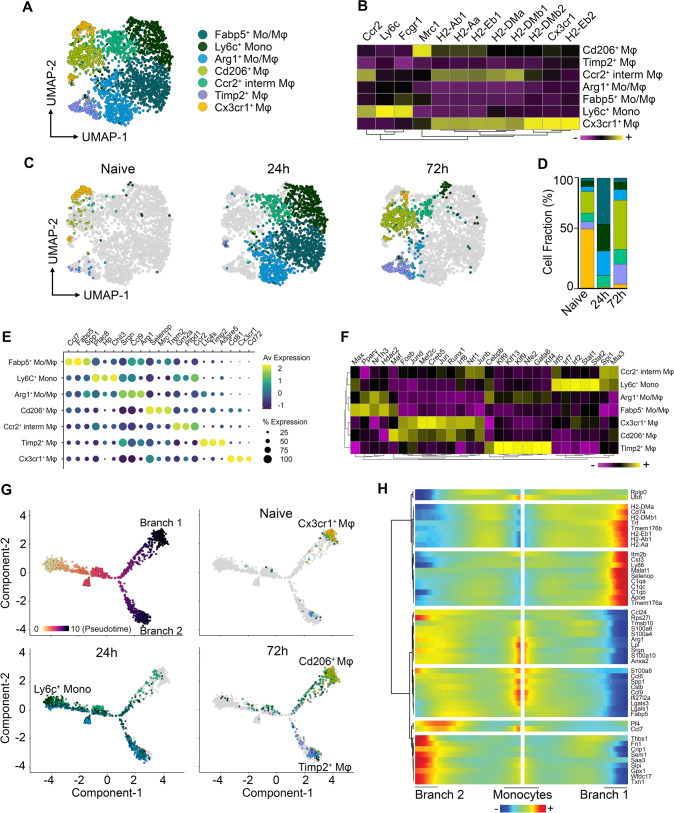
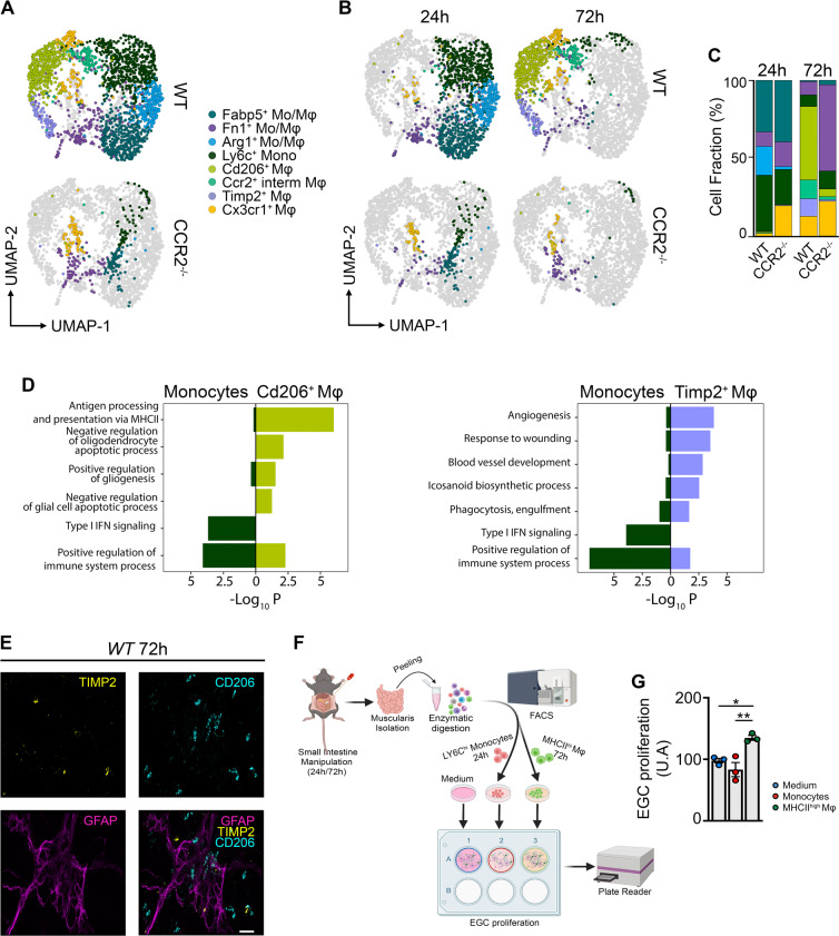
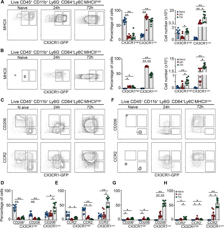
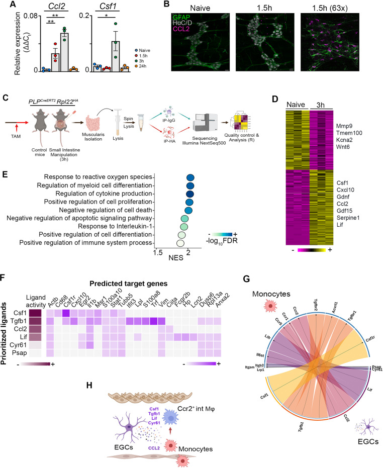
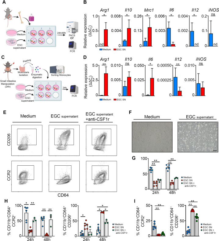
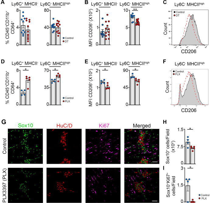
Monocyte-derived macrophages (Mφs) are crucial regulators during muscularis inflammation. However, it is unclear which micro-environmental factors are responsible for monocyte recruitment and anti-inflammatory Mφ differentiation in this paradigm. Here, we investigate Mφ heterogeneity at different stages of muscularis inflammation and determine how environmental cues can attract and activate tissue-protective Mφs. Results showed that muscularis inflammation induced marked alterations in mononuclear phagocyte populations associated with a rapid infiltration of Ly6c+ monocytes that locally acquired unique transcriptional states. Trajectory inference analysis revealed two main pro-resolving Mφ subpopulations during the resolution of muscularis inflammation, i.e. Cd206+ MhcIIhi and Timp2+ MhcIIlo Mφs. Interestingly, we found that damage to the micro-environment upon muscularis inflammation resulted in EGC activation, which in turn stimulated monocyte infiltration and the consequent differentiation in anti-inflammatory CD206+ Mφs via CCL2 and CSF1, respectively. In addition, CSF1-CSF1R signaling was shown to be essential for the differentiation of monocytes into CD206+ Mφs and EGC proliferation during muscularis inflammation. Our study provides a comprehensive insight into pro-resolving Mφ differentiation and their regulators during muscularis inflammation. We deepened our understanding in the interaction between EGCs and Mφs, thereby highlighting pro-resolving Mφ differentiation as a potential novel therapeutic strategy for the treatment of intestinal inflammation.


Conflict of interest statement

The authors declare no competing interests.

Figures

Fig. 1
Fig. 1. Identification of CD45+ immune cell populations by unsupervised scRNA-seq clustering in the healthy and inflamed muscularis.
a Experimental pipeline of scRNA-seq experiment. b UMAP of sorted CD45+ immune cells from the healthy muscularis, 24 h and 72 h post-injury from WT mice. Each sample was pooled from 3–4 mice. c Heatmap of the 50 most differentially expressed genes in each cluster. d UMAPs of time-dependent infiltration of immune cells upon muscularis inflammation. Data of Fig. 1b has been split based on different time points. e Cell fraction of each cluster relative to the total number of CD45+ immune cells at different time points after muscularis inflammation.
Fig. 2
Fig. 2. Two main anti-inflammatory Mφ subpopulations with a unique transcriptional state are present during the resolution of muscularis inflammation.
a UMAP of reclustered monocyte/Mφ subpopulations from Fig. 1a from the healthy muscularis, 24 h and 72 h post-injury from WT mice. b Heatmap of typical monocyte and Mφ markers including MHCII genes. c UMAPs of time-dependent infiltration of monocyte and Mφ subsets upon muscularis inflammation. d Cell fraction of each subcluster relative to the total number of monocytes/Mφs at different time points after muscularis inflammation. e Dotplot showing expression of selected differentially expressed genes in each subcluster. f Heatmap of regulon activity per cluster according to SCENIC analysis. g Pseudotime analysis of monocytes/Mφs at different time points after muscularis inflammation. h Heatmap of gene expression showing the top 50 genes of different branches of the pseudotime trajectory tree.
Fig. 3
Fig. 3. Two distinct Mφ subpopulations during the resolution of muscularis inflammation are derived from CCR2+ monocytes.
a UMAP of monocyte and Mφ subclusters from the muscularis of WT and CCR2−/− mice at 24 h and 72 h after induction of muscularis inflammation. Each sample was pooled from 3–4 mice. b UMAPs of myeloid cells at different time points post-injury. c Cell fraction of each subcluster relative to the total number of myeloid cells at different time points after muscularis inflammation. d GO analysis of monocytes versus Cd206+ Mφs (left) or Timp2+ Mφs (right) showing negative Log10(p-value). e Immunofluorescent images of muscularis whole-mount preparations 3 days after the induction of muscularis inflammation stained for GFAP (purple), TIMP2 (yellow) and CD206 (light blue). Scale bar 15 µm. f Experimental outline of in vitro EGC proliferation by stimulation with supernatant of monocytes or Mφs from different time points post-injury. g Fold induction of EGCs stimulated with the supernatant of LY6Chi monocytes from 24 h post-injury or MHCIIhi Mφs from 72 h post-injury relative to control medium. Every data point is an independent sorting and culture experiment One-way ANOVA; test *p < 0.05; **p < 0.01; ns not significant.
Fig. 4
Fig. 4. Flow cytometry validates unique Mφ subpopulations during muscularis inflammation.
a–h from naïve CX3CR1GFP/+ mice, 24 h and 72 h after muscularis inflammation. a Contour plots representing CX3CR1 expression in Ly6C MHCIIhi Mφs (left). Percentages and absolute numbers of CX3CR1hi and CX3CR1lo cells from Ly6C MHCIIhi Mφs are shown as mean ± SEM (right). b Contour plots representing CX3CR1 expression in Ly6C MHCIIlo Mφs (left). Percentages and absolute numbers of CX3CR1hi and CX3CR1lo cells from Ly6C MHCIIlo Mφs are shown as mean ± SEM (right). ch CD206 and CCR2 expression in Ly6C MHCIIhi and MHCIIlo Mφs. Contour plots representing CD206 or CCR2 expression in Ly6C MHCIIhi (c) and Ly6C MHCIIlo Mφs (f). Percentages of CD206+ CX3CR1hi, CD206 CX3CR1hi and CD206+ CX3CR1lo cells in the LY6C MHCIIhi Mφs (d) and MHCIIlo Mφs (g). Percentages of CCR2+ CX3CR1hi, CCR2 CX3CR1hi and CCR2+ CX3CR1lo cells in the LY6C MHCIIhi Mφs (e) and MHCIIlo Mφs (h). One-way ANOVA; test *p < 0.05; **p < 0.01; ns not significant.
Fig. 5
Fig. 5. EGCs produce factors essential for the recruitment and differentiation of monocytes during inflammation.
a Relative mRNA levels for Ccl2 and Csf1 normalized to the housekeeping gene rpl32 from ganglia isolated from the muscularis of the small intestine from naïve wild-type mice and 1.5 h, 3 h and 24 h after muscularis inflammation. One-way ANOVA; test *p < 0.05; **p < 0.01; ns not significant. b Immunofluorescent images of muscularis whole-mount preparations at homeostasis and 1.5 h after the induction of muscularis inflammation stained for GFAP (green), HuC/D (gray) and CCL2 (purple). Scale bar (25x) 25 µm, (63x) 15 µm. c Experimental overview of experiments in PLP-CreERT2 Rpl22HA mice. d Heatmap of HA-enriched differentially expressed genes between immunoprecipitated samples from naïve PLP-CreERT2 Rpl22HA mice and 3 h after intestinal manipulation. e Selected significant GO terms enriched (GSEA) in PLP1+ EGCs 3 h post-injury compared to naive PLP1+ EGCs. f Heat map of ligand-target pairs showing regulatory potential scores between top positively correlated prioritized ligands and their target genes among the differentially expressed genes between Ly6c+ monocytes and Ccr2+ int Mφs. g Circos plot showing top NicheNet ligand-receptor pairs between EGCs and Ly6c+ monocytes corresponding to the prioritized ligands in Fig. 5f. h Schematic overview of interactions between EGCs and infiltrating Ly6c+ monocytes.
Fig. 6
Fig. 6. EGCs stimulate the differentiation of monocytes into anti-inflammatory CD206+ Mφs in part via CSF-1 in vitro.
a Experimental outline of in vitro primary bone marrow monocytes stimulated with supernatant of EGCs. b Bone marrow-derived monocytes were stimulated for 24 h with/without supernatant of EGCs. Relative mRNA levels for pro- and anti-inflammatory cytokines normalized to the housekeeping gene rpl32 in bone marrow monocytes cultured with/without EGC supernatant. c Experimental outline of in vitro experiment using sorted Ly6C+ MHCII monocytes stimulated with/without EGC supernatant for 24 h. d Ly6C+ MHCII monocytes were sorted from the muscularis of WT mice 24 h after the induction of muscularis inflammation and were stimulated for 24 h with/without supernatant of EGCs. Relative mRNA levels of pro- and anti-inflammatory mediators normalized to the housekeeping gene rpl32 in sorted Ly6C+ MHCII monocytes stimulated with/without EGC supernatant. eh Bone marrow monocytes were cultured for 24–48 h with/without EGC supernatant and supplemented with anti-CSF1r antibody. e Contour plots of bone marrow monocytes showing expression of CD206 (top) or CCR2 (bottom) upon culture for 48 h with/without EGC supernatant and supplemented with anti-CSF1r antibody. f Brightfield images of monocytes upon culture for 24 h with/without EGC supernatant. g Quantification of 7-AAD+ cells in bone marrow monocytes cultured for 24 h or 48 h with/without EGC supernatant and supplemented with anti-CSF1r antibody. h Percentages of CCR2+ and CD206+ cells in live CD45+ CD11b+ Ly6G CD64+ population. i Ly6C+ MHCII monocytes were sorted from the muscularis of WT mice 24 h after the induction of muscularis inflammation and were cultured with/without EGC supernatant and supplemented with anti-CSF1r antibody for 48 h. Percentages of CCR2+ and CD206+ cells in live CD45+ CD11b+ Ly6G CD64+ population. ad T-test. gi one-way ANOVA. *p < 0.05; **p < 0.01.
Fig. 7
Fig. 7. EGCs are crucial for monocyte differentiation into anti-inflammatory CD206+ Mφs in part via CSF-1 in vivo.
ac A small portion of the small intestine of PLPCreERT2 iDTR mice was exposed to saline or DT after intestinal manipulation and mice were sacrificed 72 h after the induction of muscularis inflammation. a Percentages of Ly6C+ MHCII monocytes and Ly6C MHCIIhi Mφs in live CD45+ CD11b+ Ly6G CD64+ population. b Mean fluorescent intensity (MFI) of CD206 in Ly6C+ MHCII monocytes and Ly6C MHCIIhi Mφs. c Histogram of CD206 expression in Ly6C MHCIIhi Mφs in control and DT exposed PLPCreERT2 iDTR mice 72 h after muscularis inflammation. dh Wild-type mice were gavaged daily with vehicle or PLX-3397 (50 mg/kg) starting from the day of the manipulation and sacrificed 72 h after the induction of muscularis inflammation. D Percentages of Ly6C+ MHCII monocytes and Ly6C MHCIIhi Mφs in live CD45+ CD11b+ Ly6G CD64+ population. e Mean fluorescent intensity (MFI) of CD206 in Ly6C+ MHCII monocytes and Ly6C MHCIIhi Mφs. f Histogram of CD206 expression in Ly6C MHCIIhi Mφs in control and PLX treated mice 72 h after muscularis inflammation. g Immunofluorescent images of muscularis whole-mounts in control and PLX treated mice 72 h after muscularis inflammation stained for SOX10 (green), HuC/D (red) and Ki-67 (purple). Scale bar 25 µm. h Quantification of the number of SOX10+ cells per field in control and PLX treated mice 72 h after muscularis inflammation (average of 4–5 pictures/mouse). i Quantification of the number of Ki-67+ SOX10+ cells per field in control and PLX treated mice 72 h after muscularis inflammation (N = 4–5 pictures/mouse). a, b; d, e; h, i. T-test. *p < 0.05; **p < 0.01.

References

    1. Gabanyi I, et al. Neuro-immune interactions drive tissue programming in intestinal macrophages. Cell. 2016;164:378–391. doi: 10.1016/j.cell.2015.12.023. - DOI - PMC - PubMed
    1. Asano K, et al. Intestinal CD169(+) macrophages initiate mucosal inflammation by secreting CCL8 that recruits inflammatory monocytes. Nat. Commun. 2015;6:7802. doi: 10.1038/ncomms8802. - DOI - PMC - PubMed
    1. Hadis U, et al. Intestinal tolerance requires gut homing and expansion of FoxP3+ regulatory T cells in the lamina propria. Immunity. 2011;34:237–246. doi: 10.1016/j.immuni.2011.01.016. - DOI - PubMed
    1. Mowat AM. Publisher Correction: To respond or not to respond - a personal perspective of intestinal tolerance. Nat. Rev. Immunol. 2018;18:536. doi: 10.1038/s41577-018-0033-3. - DOI - PubMed
    1. Zigmond E, et al. Macrophage-restricted interleukin-10 receptor deficiency, but not IL-10 deficiency, causes severe spontaneous colitis. Immunity. 2014;40:720–733. doi: 10.1016/j.immuni.2014.03.012. - DOI - PubMed

Publication types

Substances