. 2022 Sep;609(7927):547-551.
doi: 10.1038/s41586-022-05160-8. Epub 2022 Sep 7.

Surgical amputation of a limb 31,000 years ago in Borneo

Affiliations

Surgical amputation of a limb 31,000 years ago in Borneo

Tim Ryan Maloney et al. Nature. 2022 Sep.

Abstract

The prevailing view regarding the evolution of medicine is that the emergence of settled agricultural societies around 10,000 years ago (the Neolithic Revolution) gave rise to a host of health problems that had previously been unknown among non-sedentary foraging populations, stimulating the first major innovations in prehistoric medical practices1,2. Such changes included the development of more advanced surgical procedures, with the oldest known indication of an 'operation' formerly thought to have consisted of the skeletal remains of a European Neolithic farmer (found in Buthiers-Boulancourt, France) whose left forearm had been surgically removed and then partially healed3. Dating to around 7,000 years ago, this accepted case of amputation would have required comprehensive knowledge of human anatomy and considerable technical skill, and has thus been viewed as the earliest evidence of a complex medical act3. Here, however, we report the discovery of skeletal remains of a young individual from Borneo who had the distal third of their left lower leg surgically amputated, probably as a child, at least 31,000 years ago. The individual survived the procedure and lived for another 6-9 years, before their remains were intentionally buried in Liang Tebo cave, which is located in East Kalimantan, Indonesian Borneo, in a limestone karst area that contains some of the world's earliest dated rock art4. This unexpectedly early evidence of a successful limb amputation suggests that at least some modern human foraging groups in tropical Asia had developed sophisticated medical knowledge and skills long before the Neolithic farming transition.


Conflict of interest statement

The authors declare no competing interests.

Figures

Fig. 1
Fig. 1. Location of Liang Tebo.
a, Sunda, the continental shelf region encompassing the present-day island of Borneo during periods of lowered sea levels, is situated to the west of Wallacea and northwest of the Pleistocene low-sea-level landmass of Sahul (Australia and New Guinea). The Sangkulirang–Mangkalihat Peninsula (SMP) is adjacent to the easternmost edge of Sunda. The area shown in b is highlighted. b, Liang Tebo and surrounding archaeological sites, including those with dated Late Pleistocene rock art (shown in red). Map source, Shuttle Radar Topography Mission 1 Arc-Second Global by NASA/NGS/USGS; GEBCO_2014 Grid, version 20150318 (http://gebco.net). Base maps generated using ArcGIS by M. Kottermair and A. Jalandoni. Scale bars, 500 km (a) and 10 km (b). ‘ka equals thousands of year’.
Fig. 2
Fig. 2. Liang Tebo burial feature.
a, A single adult inhumation (TB1). The skull is to the right of the scale bar, as shown by the exposure of the supraorbital ridge. A flexed burial position with the right knee brought to the chest and a complete right foot, and the left knee flexed below the pelvis, with the tibia and fibula underneath the femur. b, In situ nodule of red ochre (a natural earth pigment) next to the mandible. c, Maxilla and mandible. Scale bar, 5 cm.
Fig. 3
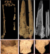
Fig. 3. Surgically amputated site of the left tibia and fibula.
a, TB1 left and right legs with pelvic girdle, demonstrating the complete absence of the distal third of the left lower leg. b, Left tibia and fibula showing the amputation surface, atrophy and necrosis. The bone surface is more porous because lysis occurred to remove the dead bone (necrosis). c, Radiograph of the left tibia and fibula. df, Remodelled bone covering the amputation surfaces, demonstrating healing after the amputation. d, Left tibia medial aspect. e, Left tibia medial aspect. f, left fibula anterior aspect. Images in df taken using an Olympus DSX1000 digital microscope. Scale bars, 5 cm (a), 5 mm (b and c) and 2 mm (df).
Extended Data Fig. 1
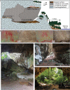
Extended Data Fig. 1. Liang Tebo cave topography and site plan.
a, Liang Tebo is a cathedral-sized (~160 m3) limestone cave with three large chambers. Following GPR and ERT survey, a 2 x 2 m excavation was positioned over an area of likely deep deposit in the centre of the caves’ lower and largest chamber. This area was excavated to a depth of 1.5 m before the project was forced to cease, following the COVID19 pandemic. The human burial (TB1) was excavated in its entirety before the field season was terminated. b, The uppermost chamber of the cave contains pigment rock art, including negative hand stencils, shown modified using D-Stretch. c, View of excavation square in lower chamber looking south towards upper chamber 1. d, View from upper chamber 1 looking northeast into lower chamber. e, Liang Tebo north cave mouth showing rainforest vegetation outside of the dripline.
Extended Data Fig. 2
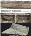
Extended Data Fig. 2. Liang Tebo stratigraphic profile.
a, Nine major stratigraphic units (SU) with mostly horizontal bedding and strongly to moderately interpretable boundaries shown across west and north profiles. Sediments were assessed and described using Munsell colour; compaction (cemented, weakly cemented, moderate, loose to very loose); and sorting grades of well (<0.5 mm), moderate (~1 mm), poor (>1.5 mm), and very poor (>2 mm) particles. Calibrated radiocarbon ages and laboratory codes are presented within respective SUs represented in stratigraphic profile. b, Squares C and D, west wall profile showing burial feature cut that continues into the profile. c, Squares D and A, north wall profile. d, 3D laser scan depicting thorax of TB1 skeleton, with stratigraphic units above the feature (SU5,6,7) depicted on the western wall, and a boundary line of the underlying strata SU9.
Extended Data Fig. 3
Extended Data Fig. 3. TB1 burial feature.
Figure viewed from left to right, upper to lower. The TB1 burial feature was carefully excavated in 32 episodic stages (R1 to R32). Upper left, large limestone burial markers positioned above the skull and arms post-burial. Centre, skull (left red bounding box) and left femur (right red bounding box) under which amputated tibia and fibula lie. Middle right, TB1 amputated tibia shown in situ (white arrow). Lower right, burial feature post-removal demonstrating distinct grave cut margins.
Extended Data Fig. 4
Extended Data Fig. 4. TB1 amputation.
a, Comparison of left and right tibiae and fibulae. b, Left tibia showing healed amputation surfaces of amputation site. c, Left fibula showing healed amputation surfaces of amputation site. d, Cortex thickness of the left and right tibia are presented for comparison demonstrating atrophy of the lower left limb in association with the amputation.
Extended Data Fig. 5
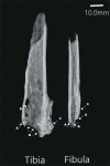
Extended Data Fig. 5. Radiograph of TB1 amputated distal left limb.
White arrows demonstrate (A) Clear cutting margins indicating use of a sharp instrument. (B) Resorption of the bone due to lack of proper vascularisation of the bone tissue. (C) Heterogenic ossification of the interosseus membrane. (D) Post-mortem insect damage. Note: fibula is placed postero-anteriorly.
Extended Data Fig. 6
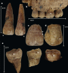
Extended Data Fig. 6. Dental pathology of TB1.
a, notches (arrows) on the left maxillary central and lateral incisors. b, dental calculus (arrows) and periodontal disease (dashed line) of the left maxillary 1st premolar to 2nd molar. c, Linear Enamel Hypoplasia of the right maxillary central incisor. Two defects are observable. d, Pitted hypoplasia (arrow) of the right maxillary canine. e, Carious lesions (arrows) on the buccal surfaces of right mandibular 3rd molar, right mandibular 2nd molar and left mandibular 3rd molar (left to right).
Extended Data Fig. 7
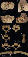
Extended Data Fig. 7. Other trauma.
a, Osteolysis of the right proximal tibia (superior view). b, Osteolysis of the right distal femur (posteroinferior view). c, Healed fracture of the C2 right pars interarticularis (superior and inferior views). d, DJD of the C3 (superior view) and C4 (superior and inferior view) due to malalignment of C2 fracture. e, Potential fracture or new bone deposit on the left 4th interproximal digital phalanx (dorsal view). f, Unilateral muscle activity of the clavicles (inferior view). The right clavicle is associated with a deep costoclavicular sulcus indicating muscle strain associated with repetitive rotary motion of the right shoulder.
Extended Data Fig. 8
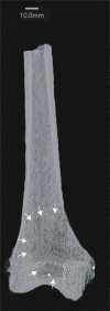
Extended Data Fig. 8. Radiograph of osteomyelitis of the distal right femur.
Note: the distinct radiolucent region that extends to the margins of the lysis that is macroscopically visible on the external bone indicated by asterisk (*). The margins of the lesion (white arrows) are irregular consistent with an active and fast spreading infection. There is too much post-mortem damage to the underlying cancellous bone of the proximal tibia and the same lysis cannot be clearly observed as with the femur.
Extended Data Fig. 9
Extended Data Fig. 9. Antemortem fracture of the C2 denoted by white arrows.
(A) is not macroscopically visible and more consistent with an infracture. (B) is macroscopically visible and of great consequence to deformity of the original bone shape than (A).
Extended Data Fig. 10
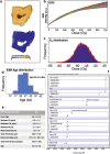
Extended Data Fig. 10. Direct US-ESR dating of mandibular left third molar (M3) and dating sequence.
a, Tooth section and polished Uranium-series analyses (red rectangles indicate location of laser rasters), with Trace elemental map: distribution of uranium in the M3 dental tissues. b, Dose response curve (DRC) obtained using McDoseE 2.0 program60 with an iteration of 100,000. c, Dose equivalent De frequency distribution; d, Age distribution frequency for the third molar (M3). e, Table summarising parameters and data used for modelling the ESR age (2-sigma). f, Results of the Bayesian model age sequence.

Comment in

  • Reply to: Common orthopaedic trauma may explain 31,000-year-old remains.
    Vlok M, Maloney T, Dilkes-Hall IE, Oktaviana AA, Setiawan P, Priyatno AAD, Ririmasse M, Geria IM, Effendy MAR, Istiawan B, Atmoko FT, Adhityatama S, Moffat I, Joannes-Boyau R, Brumm A, Aubert M. Vlok M, et al. Nature. 2023 Mar;615(7952):E15-E18. doi: 10.1038/s41586-023-05757-7. Nature. 2023. PMID: 36922613 Free PMC article. No abstract available.
  • Common orthopaedic trauma may explain 31,000-year-old remains.
    Murphy NJ, Davis JS, Tarrant SM, Balogh ZJ. Murphy NJ, et al. Nature. 2023 Mar;615(7952):E13-E14. doi: 10.1038/s41586-023-05756-8. Epub 2023 Mar 15. Nature. 2023. PMID: 36922615 Free PMC article. No abstract available.

References

    1. Roberts, C. A. in The Archaeology of Medicine (ed. Arnott, R.) Vol. 1046, 1–20 (Archaeopress, 2002).
    1. Richards MP. A brief review of the archaeological evidence for Palaeolithic and Neolithic subsistence. Euro. J. Clin. Nutrition. 2002;56:1270–1278. doi: 10.1038/sj.ejcn.1601646. - DOI - PubMed
    1. Buquet-Marcon, C., Philippe, C. & Anaick, S. The oldest amputation on a Neolithic human skeleton in France. Nat. Prec.10.1038/npre.2007.1278.1 (2007).
    1. Aubert M, et al. Palaeolithic cave art in Borneo. Nature. 2018;564:254–257. doi: 10.1038/s41586-018-0679-9. - DOI - PubMed
    1. Brumm A, et al. Skeletal remains of a Pleistocene modern human (Homo sapiens) from Sulawesi. PLoS ONE. 2021;9:e0257273. doi: 10.1371/journal.pone.0257273. - DOI - PMC - PubMed

Publication types

Substances