. 2022 Sep 8;5(1):927.
doi: 10.1038/s42003-022-03852-5.

Engineered marble-like bovine fat tissue for cultured meat

Affiliations

Engineered marble-like bovine fat tissue for cultured meat

Yedidya Zagury et al. Commun Biol. .

Abstract

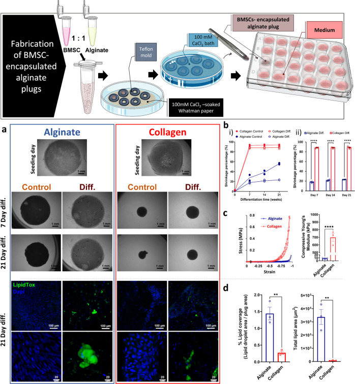
Cultured meat can provide a sustainable and more ethical alternative to conventional meat. Most of the research in this field has been focused on developing muscle tissue, as it is the main component of meat products, while very few studies address cultured fat tissue, an essential component in the human diet and determinant of meat quality, flavor, juiciness, and tenderness. Here, we engineered bovine fat tissue for cultured meat and incorporated it within engineered bovine muscle tissue. Mesenchymal stem cells (MSCs) were derived from bovine adipose tissue and exhibited the typical phenotypic profile of adipose-derived MSCs. MSC adipogenic differentiation and maturation within alginate-based three-dimensional constructs were optimized to yield a fat-rich edible engineered tissue. Subsequently, a marble-like construct, composed of engineered bovine adipose and muscle tissues, was fabricated, mimicking inter- and intra-muscular fat structures.


Conflict of interest statement

This research was sponsored by Aleph Farms. Prof. Shulamit Levenberg is the chief scientific advisor and Dr. Neta Lavon is the chief technology officer and vice president of R&D of Aleph Farms. The remaining authors declare no competing interests.

Figures

Fig. 1
Fig. 1. Adipogenic differentiation of isolated BMSCs within 3D construct.
Top panel: BMSCs were seeded in 2D culture plates, and then differentiated into adipocytes by application of differentiation medium for 12 days. Image credits: this figure was created using elements from Servier Medical Art (https://smart.servier.com). Representative bright-field and ORO staining images of b, c, e, f post-differentiated BMSCs and a, d non-induced BMSCs at passages 2 and 5; scale bar 50 µm. Bottom panel: BMSCs were loaded into 0.5% alginate hydrogel beads and cultured in differentiation medium (illustration of the process in the bottom panel). Image credits: this figure was created using elements from Servier Medical Art (https://smart.servier.com). ORO-stained sections of BMSC-loaded beads cultured in g standard or h, i differentiation medium for 19 days. PPARγ-stained sections of BMSC-loaded beads cultured in j standard or k, l differentiation medium for 19 days (red, PPARγ; blue, DAPI); scale bar 20 µm. m Relative fold-change of LPL and PPARγ mRNA expression, measured using real-time qPCR, in differentiated BMSCs of each isolation (BMSC-I, BMSC-II, and BMSC-III). Fold-change in expression was calculated using the ΔΔCt method after normalization with endogenous reference 18s or GAPDH. Differences between gene expression levels of BMSCs loaded into beads cultured in differentiation versus standard medium (control) were evaluated using Student’s t-test and reported as the mean ± SE of triplicates, *p < 0.05, **p < 0.01.
Fig. 2
Fig. 2. Influence of matrix material on engineered adipose tissue formation.
Top panel: Illustration of BMSC-loaded alginate plug fabrication. Image credits: this figure was created using elements from Servier Medical Art (https://smart.servier.com). a (top panel) Bright-field images of BMSC-loaded alginate and BMSC-loaded collagen constructs cultured in differentiation medium (diff.) or standard medium (control), as captured on seeding day, and after 7, 14, and 21 days of differentiation, scale bar 1 mm. b Shrinkage percentage of the constructs (relative to their original size) (i) All samples: differentiation or control in alginate or in collagen over 21 days of differentiation, (ii) BMSC-loaded alginate vs. BMSC-loaded collagen constructs after 7, 14, and 21 days in differentiation medium n =4. c Compressive stress–strain curves and calculated Young’s modulus of BMSC-loaded alginate vs. BMSC-loaded collagen constructs after 21 days in differentiation medium, n = 3, Student’s t-test, error bars show SE, ****p < 0.0001. a (bottom panel) Confocal laser scanning microscopy images (green, LipidTox; blue, DAPI) of BMSC-loaded alginate and BMSC-loaded collagen constructs cultured in differentiation medium or in standard medium, for 21 days, scale bars: 100 µm (top) and 20 µm (bottom). d Quantified lipid droplet coverage and total lipid area of LipidTox-stained differentiated alginate vs. collagen constructs, n = 3, Student’s t-test, error bars show SE, **p < 0.01.
Fig. 3
Fig. 3. Optimization of adipogenic differentiation and maturation periods of BMSC-loaded alginate 3D constructs.
a Scheme of the 6-week experimental timeline. Image credits: this figure was created using elements from Servier Medical Art (https://smart.servier.com). b Percentage of lipid droplet coverage, c differentiation percentage, and d lipid droplet content per differentiated cell, in BMSCs subjected to 2 weeks of differentiation followed by 1–4 weeks of maturation (blue circles) or to 3 weeks of differentiation followed by 2–3 weeks of maturation (purple squares); n = 4. Multiple unpaired t-test, Holm-Sidak post hoc, error bars show standard error (SE), ns: non-significant, *p < 0.05, **p < 0.01. e Whole-mount C/EBPα and lipid staining (red, C/EBPα; green, LipidTox; blue, DAPI), lipid droplet size distribution and mean diameter after 2 weeks of differentiation followed by 2 weeks of maturation (blue, ‘2w diff. + 2w mat.’) and after 3 weeks of differentiation followed by 1 week of maturation (purple, ‘3w diff. + 1w mat.’); Student’s t-test, error bars show SE, ****p < 0.0001. f Scheme of the 2-week experimental timeline, each color line represents a different experimental timeline. g Whole-mount lipid staining (green, LipidTox; blue, DAPI) and h lipid droplet coverage percentage of lipid-stained constructs subjected to different differentiation/maturation schedules, n = 4, one-way ANOVA followed by Tukey’s post hoc test, **p < 0.01, ***p < 0.001, ****p < 0.0001, error bars show SE. i Mean (±SE) differentiation percentage and lipid droplet content per cell in lipid-stained BMSC-loaded constructs after 5 days in differentiation medium followed by 9 days in maturation medium, n = 3. Each color represents unique combination of differentiation and maturation periods as specified in the figure; d = days; diff. = differentiation medium; mat. = maturation medium.
Fig. 4
Fig. 4. Engineered bovine adipose-muscle marbled-like construct.
a Engineered mold-cast marble-like constructs composed of bovine fat and muscle in a (i) ring and a (ii) semicircular configuration. The outer ring/left semi-circle is a lyophilized casted alginate-RGD plug onto which bovine satellite cells were seeded and myogenically differentiated into myotubes. The inner ring/right semi-circle is an alginate plug loaded with differentiated BMSCs. The integrated constructs were incubated in adipogenic maturation medium for 1 week. From left to right: Illustration of the attachment process, bright field images in low and high magnifications, and confocal images of lipid and desmin staining of constructs (green, LipidTox; red, Desmin; blue, DAPI) in low and high magnifications. b Marbled-like construct on a 3D-printed scaffold composed of extracted mature adipocytes (differentiated BMSCs) within bovine engineered muscle tissue. From left to right: illustration of the construct fabrication process, bright field image, and confocal images of lipid and desmin staining of constructs (green, LipidTox; red, Desmin; blue, DAPI) at different magnifications. Image credits: this figure was created using elements from Servier Medical Art (https://smart.servier.com).
Fig. 5
Fig. 5. Comparative analysis of mature adipocytes within marble-like constructs.
a Confocal images of lipid staining of the ring-shape integrated constructs before the integration procedure versus after integration and 1-week co-culture, for 2 types of adipose constructs; 2-week differentiation followed by 3-week maturation (‘2w diff. + 3w mat.’) and 3-weeks differentiation followed by 2-week maturation (‘3w diff. + 2w mat.’). The images were taken from the inner ring region of the integrated constructs (green, LipidTox; blue, DAPI), scale bar 200 µm. b Lipid droplet coverage percentage of the Lipid-stained constructs before the integration procedure ‘Before attachment’, after the integration procedure and 1-week co-culture in maturation medium (‘Co-culture’), and of constructs that contained only adipocytes (adipose construct that were integrated with non-cell scaffold) after the integration procedure and 1-week co-culture in maturation medium (‘Control’) for the ‘2w diff. + 3w mat.’ and ‘3w diff. + 2w mat.’ adipose constructs. n = 4, one-way ANOVA followed by Tukey’s post hoc test, ns: non-significant, error bars show standard error.

References

    1. Bhat ZF, Fayaz H. Prospectus of cultured meat—advancing meat alternatives. J. Food Sci. Technol. 2011;48:125–140.
    1. Post MJ. Cultured meat from stem cells: challenges and prospects. Meat Sci. 2012;92:297–301. - PubMed
    1. Zhang G, et al. Challenges and possibilities for bio-manufacturing cultured meat. Trends Food Sci. Technol. 2020;97:443–450.
    1. Wood JD, et al. Effects of fatty acids on meat quality: a review. Meat Sci. 2004;66:21–32. - PubMed
    1. Ng S, Kurisawa M. Integrating biomaterials and food biopolymers for cultured meat production. Acta Biomater. 2021;124:108–129. - PubMed

Substances