

Brain regions involved in fractional amplitude of low-frequency fluctuation in cluster headache patients: a resting-state functional MRI study
- PMID: 36071405
- PMCID: PMC9450424
- DOI: 10.1186/s12883-022-02863-3
Brain regions involved in fractional amplitude of low-frequency fluctuation in cluster headache patients: a resting-state functional MRI study
Abstract
Background: We used resting-state functional magnetic resonance imaging (RS-fMRI) to assess the possible pathogenic role of fALFF in CH. A limited number of studies have reported on fractional amplitude of low-frequency fluctuation (fALFF) in cluster headache (CH).
Methods: RS-fMRI scans of 23 patients with CH were obtained (11with left-sided headache and 12 with right-sided headache), along with scans of 23 age- and sex-matched normal controls. The RS-fMRI data were analyzed to explore abnormal brain activity in the left CH and right CH patients during the non-painful state in one cluster period. fALFF was compared between patients and controls, and correlation analysis between the regional mean fALFF values and clinical characteristics was performed.
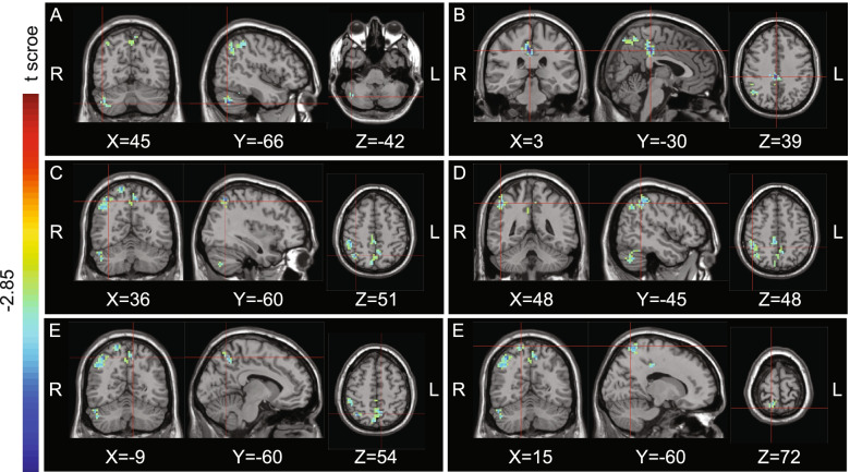
Results: A decrease in fALFF was detected in the left cerebellum, left lentiform nucleus, left frontal lobe, left anterior cingulate, and right postcentral gyrus in the left CH group compared to the controls, while a decrease of fALFF was detected in the right cerebellum, right cingulate gyrus, right superior parietal lobule, right inferior parietal lobule, right postcentral gyrus, and left precuneus in the right CH group. No patient had a region with increased fALFF. A moderate correlation was observed between some regional mean fALFF values and the clinical characteristics.
Conclusions: We deduced that dysfunction in multiple brain areas is involved in the non-painful state of CH during a cluster period.
Keywords: Brain activity; Cluster headache; Fractional amplitude of low-frequency fluctuation; Resting-state functional magnetic resonance imaging.
© 2022. The Author(s).
Conflict of interest statement
The authors declare that they have no competing interests
Figures


Similar articles
-
Amplitude of low-frequency oscillations in Parkinson's disease: a 2-year longitudinal resting-state functional magnetic resonance imaging study.Chin Med J (Engl). 2015 Mar 5;128(5):593-601. doi: 10.4103/0366-6999.151652. Chin Med J (Engl). 2015. PMID: 25698189 Free PMC article.
-
Spontaneous brain activity in chronic smokers revealed by fractional amplitude of low frequency fluctuation analysis: a resting state functional magnetic resonance imaging study.Chin Med J (Engl). 2014;127(8):1504-9. Chin Med J (Engl). 2014. PMID: 24762597
-
Alterations of spontaneous brain activity in systematic lupus erythematosus patients without neuropsychiatric symptoms: A resting-functional MRI study.Lupus. 2021 Oct;30(11):1781-1789. doi: 10.1177/09612033211033984. Epub 2021 Oct 8. Lupus. 2021. PMID: 34620007
-
Functional magnetic resonance imaging studies in bipolar disorder in resting state: A coordinates-based meta-analysis.Psychiatry Res Neuroimaging. 2024 Oct;344:111869. doi: 10.1016/j.pscychresns.2024.111869. Epub 2024 Aug 10. Psychiatry Res Neuroimaging. 2024. PMID: 39146823 Review.
-
Alterations of amplitude of low-frequency fluctuations and fractional amplitude of low-frequency fluctuations in end-stage renal disease on maintenance dialysis: An activation likelihood estimation meta-analysis.Front Hum Neurosci. 2022 Dec 1;16:1040553. doi: 10.3389/fnhum.2022.1040553. eCollection 2022. Front Hum Neurosci. 2022. PMID: 36530199 Free PMC article.
Cited by
-
Pain Lateralization in Cluster Headache and Associated Clinical Factors.J Clin Neurol. 2025 May;21(3):220-229. doi: 10.3988/jcn.2024.0457. J Clin Neurol. 2025. PMID: 40308017 Free PMC article.
-
Intrinsic Brain Functional Activity Abnormalities in Episodic Tension-Type Headache.Neural Plast. 2023 May 24;2023:6560298. doi: 10.1155/2023/6560298. eCollection 2023. Neural Plast. 2023. PMID: 37266410 Free PMC article.
-
MRI-based analysis of the microstructure of the thalamus and hypothalamus and functional connectivity between cortical networks in episodic cluster headache.J Headache Pain. 2025 Jan 15;26(1):12. doi: 10.1186/s10194-024-01920-1. J Headache Pain. 2025. PMID: 39815195 Free PMC article.
-
Cluster headache: understandings of current knowledge and directions for whole process management.Front Neurol. 2024 Aug 21;15:1456517. doi: 10.3389/fneur.2024.1456517. eCollection 2024. Front Neurol. 2024. PMID: 39233684 Free PMC article. Review.
-
A state-of-the-art review of functional magnetic resonance imaging technique integrated with advanced statistical modeling and machine learning for primary headache diagnosis.Front Hum Neurosci. 2023 Sep 1;17:1256415. doi: 10.3389/fnhum.2023.1256415. eCollection 2023. Front Hum Neurosci. 2023. PMID: 37746052 Free PMC article. Review.
References
-
- Headache Classification Committee of the International Headache Society (IHS) The International Classification of Headache Disorders, 3rd edition. Cephalalgia: Int J Headache. 2018;38(1):1–211. 10.1177/0333102417738202. - PubMed
MeSH terms
LinkOut - more resources
Full Text Sources
