. 2022 Jul 16;9(Pt 5):551-561.
doi: 10.1107/S2052252522006820. eCollection 2022 Sep 1.

Organism-specific differences in the binding of ketoprofen to serum albumin

Affiliations

Organism-specific differences in the binding of ketoprofen to serum albumin

Mateusz P Czub et al. IUCrJ. .

Abstract

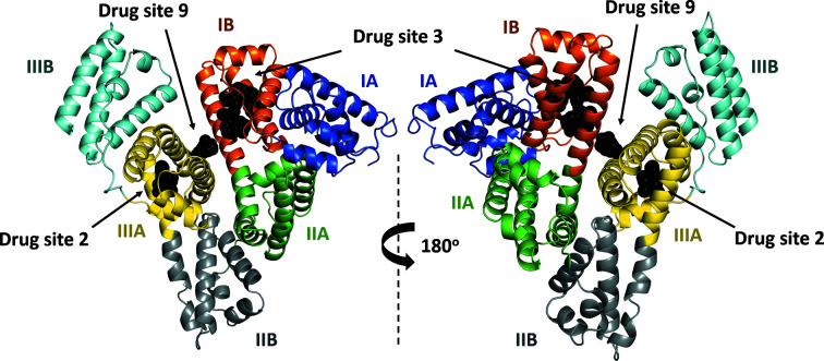
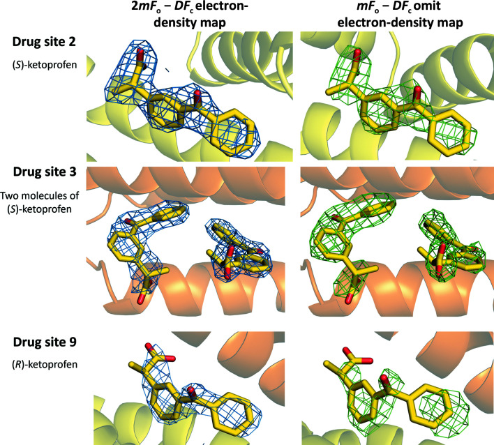
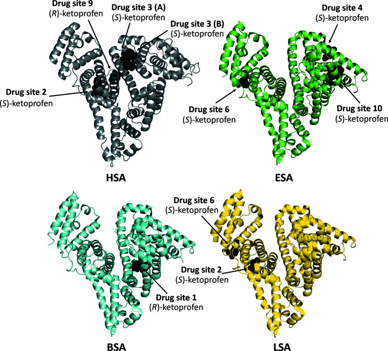
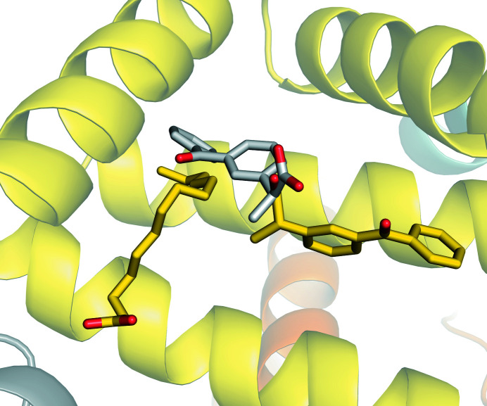
Serum albumin is a circulatory transport protein that has a highly conserved sequence and structure across mammalian organisms. Its ligand-binding properties are of importance as albumin regulates the pharmacokinetics of many drugs. Due to the high degree of structural conservation between mammalian albumins, nonhuman albumins such as bovine serum albumin or animal models are often used to understand human albumin-drug interactions. Ketoprofen is a popular nonsteroidal anti-inflammatory drug that is transported by albumin. Here, it is revealed that ketoprofen exhibits different binding-site preferences when interacting with human serum albumin compared with other mammalian albumins, despite the conservation of binding sites across species. The reasons for the observed differences were explored, including identifying ketoprofen binding determinants at specific sites and the influence of fatty acids and other ligands on drug binding. The presented results reveal that the drug-binding properties of albumins cannot easily be predicted based only on a complex of albumin from another organism and the conservation of drug sites between species. This work shows that understanding organism-dependent differences is essential for assessing the suitability of particular albumins for structural or biochemical studies.

Keywords: NSAIDs; anti-inflammatory drugs; drug interactions; drug transport; human serum albumin; ketoprofen; organism-dependent studies.


Figures

Figure 1
Figure 1
Chemical structure of ketoprofen; the chiral center is labeled with an asterisk.
Figure 2
Figure 2
The overall structure of the complex of HSA with ketoprofen. Albumin subdomains are each shown in a different color. Roman numerals (I, II, III) are associated with domains and letters (for example IB) with subdomains. Ketoprofen molecules are shown with atoms in black spheres.
Figure 3
Figure 3
Ketoprofen binding sites in HSA (PDB entry 7jwn). The 2mF oDF c electron-density map (r.m.s.d. of 1.0 Å) is presented in blue and the mF oDF c omit electron-density map (map calculated after ten REFMAC refinement cycles without the drug in the model, r.m.s.d. of 2.5 Å) is presented in green. Ketoprofen molecules are shown in stick representation with O atoms in red and C atoms in yellow. The colors of the helices correspond to the colors used in Fig. 2 ▸. The electron density and the model can be inspected interactively at https://molstack.bioreproducibility.org/project/view/VW8s7hb1Z9mnCLbg3NBU/.
Figure 4
Figure 4
Ketoprofen binding sites in mammalian serum albumins. Structures of ketoprofen complexes with HSA (PDB entry 7jwn), ESA (Czub et al., 2020; PDB entry 6u4r), BSA (Castagna et al., 2019; PDB entry 6qs9) and LSA (Zielinski et al., 2020; PDB entry 6ock).
Figure 5
Figure 5
Superposition of ketoprofen binding sites in BSA (a) (PDB entry 6qs9) and LSA (b, c) (PDB entry 6ock) with analogous sites in ligand-free HSA (PDB entry 4k2c). C atoms in BSA, LSA and HSA are shown in cyan, yellow and gray, respectively. Residue numbers correspond to positions in HSA. Residues labeled in black are conserved between BSA or LSA and HSA, while those labeled in red differ. The naming scheme for differing residues is as follows: residue in BSA or LSA, residue number, corresponding residue in HSA.
Figure 6
Figure 6
Comparison of (S)-ketoprofen binding to drug site 2 in HSA (PDB entry 7jwn) and LSA (PDB entry 6ock). The (S)-ketoprofen molecule and a molecule of a fatty acid bound to HSA are shown in stick representation with O atoms in red and C atoms in yellow, while a molecule of (S)-ketoprofen bound to LSA is shown in stick representation with O atoms in red and C atoms in gray. The colors of the helices correspond to the colors used in Fig. 2 ▸.

Similar articles

Cited by

References

    1. Acharya, M. R., Sparreboom, A., Sausville, E. A., Conley, B. A., Doroshow, J. H., Venitz, J. & Figg, W. D. (2006). Cancer Chemother. Pharmacol. 57, 275–281. - PubMed
    1. Anguizola, J., Matsuda, R., Barnaby, O. S., Hoy, K. S., Wa, C., DeBolt, E., Koke, M. & Hage, D. S. (2013). Clin. Chim. Acta, 425, 64–76. - PMC - PubMed
    1. Bi, S., Yan, L., Sun, Y. & Zhang, H. (2011). Spectrochim. Acta A Mol. Biomol. Spectrosc. 78, 410–414. - PubMed
    1. Boden, G. (2011). Curr. Opin. Endocrinol. Diab. Obes. 18, 139–143. - PMC - PubMed
    1. Bohnert, T. & Gan, L.-S. (2013). J. Pharm. Sci. 102, 2953–2994. - PubMed