. 2022 Sep 4:10:tkac030.
doi: 10.1093/burnst/tkac030. eCollection 2022.

Co-culture of Schwann cells and endothelial cells for synergistically regulating dorsal root ganglion behavior on chitosan-based anisotropic topology for peripheral nerve regeneration

Affiliations

Co-culture of Schwann cells and endothelial cells for synergistically regulating dorsal root ganglion behavior on chitosan-based anisotropic topology for peripheral nerve regeneration

Tiantian Zheng et al. Burns Trauma. .

Abstract

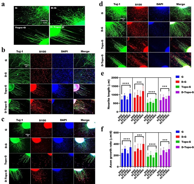
Background: Anisotropic topologies are known to regulate cell-oriented growth and induce cell differentiation, which is conducive to accelerating nerve regeneration, while co-culture of endothelial cells (ECs) and Schwann cells (SCs) can significantly promote the axon growth of dorsal root ganglion (DRG). However, the synergistic regulation of EC and SC co-culture of DRG behavior on anisotropic topologies is still rarely reported. The study aims to investigate the effect of anisotropic topology co-cultured with Schwann cells and endothelial cells on dorsal root ganglion behavior for promoting peripheral nerve regeneration.

Methods: Chitosan/artemisia sphaerocephala (CS/AS) scaffolds with anisotropic topology were first prepared using micro-molding technology, and then the surface was modified with dopamine to facilitate cell adhesion and growth. The physical and chemical properties of the scaffolds were characterized through morphology, wettability, surface roughness and component variation. SCs and ECs were co-cultured with DRG cells on anisotropic topology scaffolds to evaluate the axon growth behavior.

Results: Dopamine-modified topological CS/AS scaffolds had good hydrophilicity and provided an appropriate environment for cell growth. Cellular immunofluorescence showed that in contrast to DRG growth alone, co-culture of SCs and ECs could not only promote the growth of DRG axons, but also offered a stronger guidance for orientation growth of neurons, which could effectively prevent axons from tangling and knotting, and thus may significantly inhibit neurofibroma formation. Moreover, the co-culture of SCs and ECs could promote the release of nerve growth factor and vascular endothelial growth factor, and up-regulate genes relevant to cell proliferation, myelination and skeletal development via the PI3K-Akt, MAPK and cytokine and receptor chemokine pathways.

Conclusions: The co-culture of SCs and ECs significantly improved the growth behavior of DRG on anisotropic topological scaffolds, which may provide an important basis for the development of nerve grafts in peripheral nerve regeneration.

Keywords: Anisotropic topology; Co-culture; Dorsal root ganglion behavior; Endothelial cells; Nerve, Regeneration; Regulation mechanism; Schwann cells.


Figures

Figure 1.
Figure 1.
Schematic diagram of the preparation of patterned CS/AS scaffolds for cell co-culture. CS chitosan, AS artemisia sphaerocephala, PDMS polydimethylsiloxane, DRG dorsal root ganglion, EC endothelial cell, SC Schwann cell
Figure 2.
Figure 2.
Morphological characteristics of the patterned CS/AS scaffolds. (a) Surface topography taken by optical microscope and scanning electron microscope. (b) Quantitative analysis of the particle diameter of surface dopamine, n = 30. (c) Quantitative analysis of concave and convex width of topological structure. Data are shown as means ± SD. Statistical analysis: ***p < 0.001, ****p < 0.0001. CS chitosan, AS artemisia sphaerocephala, SD standard deviation, OM the optical microscopy, SEM scanning electron microscopy, G glass group, D-G the pure slide (glass group) treated with dopamine, Topo-G the pure slide treated with topological structure, D-Topo-G the pure slide was treated with topological structure with dopamine coating
Figure 3.
Figure 3.
Atomic force microscopy of patterned CS/AS scaffolds. (a) Dopamine particles. (b) Topological structure. (c) Topological structure with dopamine coating. (d) 3D structure of samples. (e) Quantitative analysis of surface roughness, n = 5. Data are shown as means ± SD. One-way analysis of variance was used. Statistical analysis: *p < 0.05, **p < 0.01, ns no significance. CS chitosan, AS artemisia sphaerocephala, SD standard deviation, D-G the pure slide was treated with dopamine, Topo-G the pure slide was treated with topological structure, D-Topo-G the pure slide was treated with topological structure with dopamine coating
Figure 4.
Figure 4.
Infrared spectrum of the patterned CS/AS scaffolds. CS chitosan, AS artemisia sphaerocephala, G glass group, D-G the pure slide was treated with dopamine, Topo-G the pure slide was treated with topological structure, D-Topo-G the pure slide was treated with topological structure with dopamine coating
Figure 5.
Figure 5.
Wettability of the CS/AS scaffolds. (a) Physical image of the contact angle. (b) Quantitative analysis of the contact angle, n = 20. Data are shown as means ± SD. Statistical analysis: ****p < 0.0001, ns no significant difference. CS chitosan, AS artemisia sphaerocephala, SD standard deviation, G glass group, D-G the pure slide was treated with dopamine, Topo-G the pure slide was treated with topological structure, D-Topo-G the pure slide was treated with topological structure with dopamine coating
Figure 6.
Figure 6.
Cell co-culture system promotes the growth of DRG axons. Immunofluorescence staining of (a) DRG cultured alone; (b) SCs and DRG co-culture; (c) ECs and DRG co-culture; (d) SCs, ECs and DRG co-culture (scale bar, 100 μm). (e) Quantitative analysis of the mean axon outgrowth length of DRG, n = 22. (f) Quantitative analysis of the mean axon outgrowth rate of DRG, n = 22. Data are shown as means ± SD. One-way analysis of variance was used. Statistical analysis: ***p < 0.001, ****p < 0.0001. CS chitosan, AS artemisia sphaerocephala, SD standard deviation, G glass group, D-G the pure slide was treated with dopamine, Topo-G the pure slide was treated with topological structure, D-Topo-G the pure slide was treated with topological structure with dopamine coating, DRG dorsal root ganglion, EC endothelial cell, SC Schwann cell
Figure 7.
Figure 7.
Cell-oriented growth on topological structure. (a) Quantitative analysis of unknotted axon outgrowth length in SC-DRG, EC-DRG and SC-EC-DRG co-culture systems, n = 30. (b) Top panels: the cell growth status under low magnification when SCs and DRG are co-cultured on the glass group (scale bar, 100 μm). Left image: SCs around neurons were observed to grow along the axon poles, while they grew in circles at the end of the axons. However, SCs grew in multipolarity when they were far away from the DRG soma (right image). Below: cell entanglement under magnification when EC-DRG and SC-EC-DRG are co-cultured on the glass group (scale bar, 50 μm). The red arrows indicate the axon outgrowth orientation. (c) Quantification of cell orientation angle in the SC-EC-DRG co-culture system on the samples of the four groups. Data are shown as means ± SD. One-way analysis of variance is used. Statistical analysis: ***p < 0.001, ****p < 0.0001. SD standard deviation, G glass group, D-G the pure slide was treated with dopamine, Topo-G the pure slide was treated with topological structure, D-Topo-G the pure slide was treated with topological structure with dopamine coating, DRG dorsal root ganglion, EC endothelial cell, SC Schwann cell
Figure 8.
Figure 8.
Determination of NGF and VEGF concentration in cell culture medium by enzyme-linked immunosorbent assay, n = 3. The method of one-way analysis of variance is used. Data are shown as means ± SD. Statistical analysis: *p < 0.05, **p < 0.01, ns no significant difference. SD standard deviation, EC endothelial cell, SC Schwann cell, NGF nerve growth factor, VEGF vascular endothelial growth factor
Figure 9.
Figure 9.
Gene expression of cells co-cultured on patterned CS/AS scaffolds. (a) Transcriptome sequence. Upper panel: up-regulated/down-regulated genes in cells. Lower panels: heat map of differentially expressed genes in different groups. (b) Volcano map of differentially expressed genes; red dots indicate up-regulation and blue dots indicate down-regulation. (c) Gene expression detected by quantitative real-time PCR. One-way analysis of variance is used. Data are shown as means ± SD. Statistical analysis: *p < 0.05, **p < 0.01, ***p < 0.001, ****p < 0.0001, ns no significant difference. CS chitosan, AS artemisia sphaerocephala, SD standard deviation, R RSC96 rat Schwann cell, E endothelial cell, Con control group, Exp experiment group, D dorsal root gGanglion, G glass, T topological structure, YAP yes-associated protein, MPZ myelin protein zero, Sox10 SRY-box transcription factor 10, EGR2 early growth response 2
Figure 10.
Figure 10.
Summary of the possible influence mechanism of anisotropic topological structure and cell co-culture on RSC96 cells. NGF nerve growth factor, VEGF vascular endothelial growth factor, Akt protein kinase B, NF-kB nuclear factor kappa-B, EGFR epidermal growth factor receptor, PI3K the phosphoinositide 3-kinase, IKK inhibitor of nuclear factor kappa-B kinase, MAPK mitogen-activated protein kinase, Erk extracellular signal-regulated kinase

References

    1. Zhang Y, Yang S, Liu C, Han X, Gu X, Zhou S. Deciphering glial scar after spinal cord injury. Burn Trauma. 2021;9:35. 10.1093/burnst/tkab035. - DOI - PMC - PubMed
    1. Yi S, Zhang Y, Gu X, Huang L, Zhang K, Qian T, et al. . Application of stem cells in peripheral nerve regeneration. Burn Trauma. 2020;8:2. - PMC - PubMed
    1. Pop NL, Nan A, Urda-Cimpean AE, Florea A, Toma VA, Moldovan R, et al. . Chitosan functionalized magnetic nanoparticles to provide neural regeneration and recovery after experimental model induced peripheral nerve injury. Biomol Ther. 2021;11:676. - PMC - PubMed
    1. Gordon T. Peripheral nerve regeneration and muscle reinnervation. Int J Mol Sci. 2020;21:1–24. - PMC - PubMed
    1. Kornfeld T, Vogt PM, Radtke C. Nerve grafting for peripheral nerve injuries with extended defect sizes. Wien Med Wochenschr. 2018;169:240–51. - PMC - PubMed