

Primum Non Nocere: A Case Report of Iatrogenic Fracture of the Mandibular Angle During Excision of an Impacted Third Molar
- PMID: 36072206
- PMCID: PMC9440348
- DOI: 10.7759/cureus.27672
Primum Non Nocere: A Case Report of Iatrogenic Fracture of the Mandibular Angle During Excision of an Impacted Third Molar
Abstract
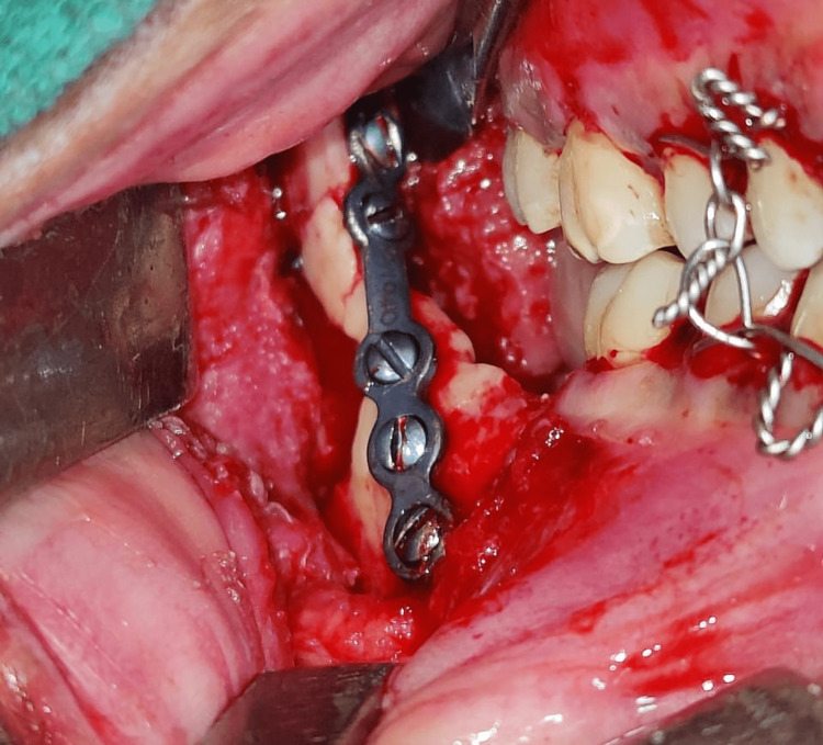
Third molar extractions are one of the most commonly performed dental procedures. It is associated with numerous complications, of which mandibular angle fracture is a rare but distressing complication. These can occur as intraoperative and postoperative (late) events. Iatrogenic fractures involving the angle of the mandible represent a unique challenge for management owing to their complex biomechanics and various anatomical factors. Intraoperative fractures occur due to various reasons, which include the position of the tooth, depth of impaction, extent of odontectomy performed, and injudicious use of dental elevators. This exhibited report describes a case of iatrogenic mandibular angle fracture (IFM) during excision of an impacted third molar in a 30-year-old female. Additionally, it discusses the various reasons and preventive strategies to avoid such complications.
Keywords: iatrogenic fracture; mandibular angle fracture; primum non nocere; third molar complications; third molar extraction.
Copyright © 2022, Agrawal et al.
Conflict of interest statement
The authors have declared that no competing interests exist.
Figures


Similar articles
-
Iatrogenic mandibular fracture associated with third molar removal after mandibular angle osteotectomy.J Craniofac Surg. 2014 May;25(3):e263-5. doi: 10.1097/SCS.0000000000000566. J Craniofac Surg. 2014. PMID: 24820729 Review.
-
Identifying the risk factors causing iatrogenic mandibular fractures associated with exodontia: a systemic meta-analysis of 200 cases from 1953 to 2015.Oral Maxillofac Surg. 2016 Dec;20(4):391-396. doi: 10.1007/s10006-016-0579-9. Epub 2016 Sep 23. Oral Maxillofac Surg. 2016. PMID: 27660249
-
Pattern of occurrence and treatment of impacted teeth at the Muhimbili National Hospital, Dar es Salaam, Tanzania.BMC Oral Health. 2013 Aug 6;13:37. doi: 10.1186/1472-6831-13-37. BMC Oral Health. 2013. PMID: 23914842 Free PMC article.
-
Prevalence of intraoperative and postoperative iatrogenic mandibular fractures after lower third molar extraction: A systematic review.J Clin Exp Dent. 2022 Jan 1;14(1):e85-e94. doi: 10.4317/jced.58390. eCollection 2022 Jan. J Clin Exp Dent. 2022. PMID: 35070129 Free PMC article. Review.
-
Management of iatrogenic dislodgment of a mandibular third molar into the pterygomandibular space.Gen Dent. 2019 May-Jun;67(3):e1-e4. Gen Dent. 2019. PMID: 31199751
Cited by
-
Accidental Intraoperative Mandibular Fracture in a Third Molar Surgery: When Surgical Skills Are Mandatory in the Face of Empiricism.Case Rep Dent. 2023 Jul 27;2023:2263554. doi: 10.1155/2023/2263554. eCollection 2023. Case Rep Dent. 2023. PMID: 37546575 Free PMC article.
References
-
- Pathological (late) fractures of the mandibular angle after lower third molar removal: a case series. Cutilli T, Bourelaki T, Scarsella S, Fabio DD, Pontecorvi E, Cargini P, Junquera L. https://jmedicalcasereports.biomedcentral.com/articles/10.1186/1752-1947.... J Med Case Rep. 2013;7:121. - PMC - PubMed
-
- Effectiveness of knotless suture as a wound closure agent for impacted third molar - a split mouth randomized controlled clinical trial. Ramkumar Ceyar KA, Thulasidoss GP, Raja Sethupathy Cheeman S, Sagadevan S, Panneerselvam E, Krishna Kumar Raja VB. J Craniomaxillofac Surg. 2020;48:1004–1008. - PubMed
-
- Characteristics of iatrogenic mandibular fractures associated with tooth removal: review and analysis of 189 cases. Bodner L, Brennan PA, McLeod NM. Br J Oral Maxillofac Surg. 2011;49:567–572. - PubMed
-
- A comparative study of surgical morbidity associated with mandibular third-molar surgery in young and aging populations. Adeyemo WL, Ogunlewe MO, Ladeinde AL, Hassan OO, Taiwo OA. https://pubmed.ncbi.nlm.nih.gov/20953558/ J Contemp Dent Pract. 2010;11:0–8. - PubMed
Publication types
LinkOut - more resources
Full Text Sources