. 2022 Aug 29:2022:7784148.
doi: 10.1155/2022/7784148. eCollection 2022.

The Renshen Chishao Decoction Could Ameliorate the Acute Lung Injury but Could Not Reduce the Neutrophil Extracellular Traps Formation

Affiliations

The Renshen Chishao Decoction Could Ameliorate the Acute Lung Injury but Could Not Reduce the Neutrophil Extracellular Traps Formation

Miao-En Yao et al. Evid Based Complement Alternat Med. .

Abstract

The acute lung injury (ALI) causes severe pulmonary diseases, leading to a high mortality rate. The Renshen and Chishao have protective and anti-inflammatory effects against the ALI. To explore the protective effects of the Renshen Chishao (RC) decoction against the ALI, we established the lipopolysaccharide-indued ALI model and randomly divided the mice into seven groups: control group, ALI group, high-dose RC group, middle-dose RC group, low-dose RC group, middle-dose RC group + CXCR2 antagonist group, and ALI + CXCR2 antagonist group. We estimated the lung injury by the hematoxylin and eosin staining, the neutrophil extracellular traps (NETs) formations by the immunofluorescence colocalization and enzyme-linked immunosorbent assay (ELISA), and the CXCR2/CXCL2 pathway by the flow cytometry, ELISA, and real-time polymerase chain reaction. We conducted the high-throughput sequencing and enrichment analyses to explore the potential mechanisms. The results showed that the RC decoction pathologically ameliorated the lipopolysaccharide-induced lung injury and inflammatory response but failed to reduce the circulating and lung tissue NETs formation and the blood neutrophil percent. The high-dose RC decoction increased the plasma CXCL2 level, but the RC decoction had no effects on the neutrophilic CXCR2 levels. Under the inhibition of the CXCR2, the middle-dose RC decoction still decreased the lung injury score but as yet had unobvious influence on the NETs formation. Other potential mechanisms of the RC decoction against the ALI involved the pathways of ribosome and coronavirus disease 2019 (COVID-19); the target genes of inflammatory factors, such as Ccl17, Cxcl17, Cd163, Cxcr5, and Il31ra, and lncRNAs; and the regulations of the respiratory cilia. In conclusion, the RC decoction pathologically ameliorated the lipopolysaccharide-induced lung inflammatory injury via upregulating the CXCL2/CXCR2 pathway but could not reduce the circulating or lung tissue NETs formation.


Conflict of interest statement

The authors declare that there are no conflicts of interest regarding the publication of this article.

Figures

Figure 1
Figure 1
Flowchart of the experiment. We built the LPS-induced ALI model, gave the drugs (two, 24 and 48 h after ALI), and conducted the sacrifice (60 h after ALI) for the mice with the same sequence in different groups in the same time periods. We randomly divided the mice into seven groups: (1) control group (n = 10, untreated); (2) ALI group (n = 10, ALI model + saline [0.1 ml/10 g/d]); (3) HRC group (n = 10, ALI model + high-dose RC decoction [2318 mg/kg/d]); (4) MRC group (n = 10, ALI model + middle-dose RC decoction [1159 mg/kg/d]); (5) LRC group (n = 10, ALI model + low-dose RC decoction [580 mg/kg/d]); (6) MRC + antagonist group (n = 10, ALI model + middle-dose RC decoction + CXCR2 antagonist); (7) ALI + antagonist group (n = 3, ALI model + CXCR2 antagonist).
Figure 2
Figure 2
The RC decoction effects against the ALI. (a) Lung wet weight/body weight ratio (LW/BW). (b) Lung HE staining (200x magnification, bar = 50 μm) and injury scores. Data is presented as mean ± SD; ∗∗P < 0.01, P < 0.05.
Figure 3
Figure 3
The RC decoction effects on the neutrophil extracellular traps (NETs). (a) Plasma NE-DNA relative concentration (ELISA). (b) Lung tissues NETs formations detected by the immunofluorescence (left, bar = 50 μm; red: cit-H3, green: myeloperoxidase, blue: DNA), with the colocalization quantitative analyses (right). Data is presented as mean ± SD or median (interquartile range); ∗∗P < 0.01, P < 0.05.
Figure 4
Figure 4
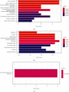
The differential expressed genes GO enrichment analyses after the MRC intervention. (a) Biological processes with top 10 significant P value. (b) Cellular components with top 10 significant P value. (c) Molecular functions.
Figure 5
Figure 5
The RC decoction target genes cluster heatmap against the ALI. The genes with the top 20 |log2FC| values (ALI group vs. MRC group) in the upregulated and downregulated RC decoction target genes against the ALI.
Figure 6
Figure 6
The inflammatory factors involved in the RC decoction effects against the ALI. The gene transcripts per million reads (TPM) comparisons among the control group, ALI group, and MRC group. Data is presented as mean ± SD; ∗∗P < 0.01, P < 0.05.
Figure 7
Figure 7
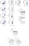
The RC decoction effects on the neutrophilic CXCL2/CXCR2 pathway. (a) The blood neutrophil (NEU) and CXCR2+ NEU (flow cytometry) percent. (b) Plasma CXCL2 concentration (ELISA). (c) The neutrophilic Cxcr2 gene relative mRNA expression (RT-PCR, normalized to β-actin). (d) The neutrophilic Cxcl2 gene relative mRNA expression (RT-PCR, normalized to β-actin). Data is presented as mean ± SD or median (interquartile range); ∗∗P < 0.01 and P < 0.05.
Figure 8
Figure 8
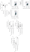
The CXCR2 antagonist SB 225002 suppressed the CXCR2 expression. (a) The blood CXCR2+ neutrophil (NEU) percent (flow cytometry). (b) The neutrophilic Cxcr2 gene relative mRNA expression (RT-PCR, normalized to β-actin). (c) Plasma CXCL2 concentration (ELISA). (d) The neutrophilic Cxcl2 gene relative mRNA expression (RT-PCR, normalized to β-actin). Data is presented as mean ± SD or median (interquartile range); ∗∗P < 0.01 and P < 0.05.
Figure 9
Figure 9
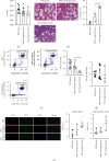
The RC decoction effects on the ALI and NETs under the CXCR2 expression suppression. (a) Lung wet weight/body weight ratio (LW/BW). (b) Lung HE staining (200x magnification, bar = 50 μm) and lung injury scores. (c) The blood neutrophil (NEU) percent (flow cytometry). (d) Plasma NE-DNA relative concentration (ELISA). (e) Lung tissues NETs formations detected by the immunofluorescence (left, bar = 50 μm; red: cit-H3, green: myeloperoxidase, blue: DNA), with the colocalization quantitative analyses (right). Data is presented as mean ± SD or median (interquartile range); P < 0.05.

Similar articles

References

    1. Bellani G., Laffey J. G., Pham T., et al. Epidemiology, patterns of care, and mortality for patients with acute respiratory distress syndrome in intensive care units in 50 countries. JAMA . 2016;315(8):788–800. doi: 10.1001/jama.2016.0291. - DOI - PubMed
    1. Meyer N. J., Gattinoni L., Calfee C. S. Acute respiratory distress syndrome. The Lancet . 2021;398(10300):622–637. doi: 10.1016/s0140-6736(21)00439-6. - DOI - PMC - PubMed
    1. Looney M. R., Su X., Van Ziffle J. A., Lowell C. A., Matthay M. A. Neutrophils and their Fc gamma receptors are essential in a mouse model of transfusion-related acute lung injury. Journal of Clinical Investigation . 2006;116(6):1615–1623. doi: 10.1172/jci27238. - DOI - PMC - PubMed
    1. Mowery N. T., Terzian W. H., Nelson A. C. Acute lung injury. Current Problems in Surgery . 2020;57(5) doi: 10.1016/j.cpsurg.2020.100777.100777 - DOI - PubMed
    1. Soy M., Keser G., Atagündüz P., Tabak F., Atagündüz I., Kayhan S. Cytokine storm in COVID-19: pathogenesis and overview of anti-inflammatory agents used in treatment. Clinical Rheumatology . 2020;39(7):2085–2094. doi: 10.1007/s10067-020-05190-5. - DOI - PMC - PubMed