. 2022 Aug 22:9:925058.
doi: 10.3389/fmolb.2022.925058. eCollection 2022.

Triblock copolymer micelle model of spherical paraspeckles

Affiliations

Triblock copolymer micelle model of spherical paraspeckles

Tetsuya Yamamoto et al. Front Mol Biosci. .

Abstract

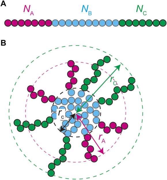
Paraspeckles are nuclear bodies scaffolded by RNP complexes of NEAT1_2 RNA transcripts and multiple RNA-binding proteins. The assembly of paraspeckles is coupled with the transcription of NEAT1_2. Paraspeckles form the core-shell structure, where the two terminal regions of NEAT1_2 RNP complexes compose the shell of the paraspeckle and the middle regions of these complexes compose the core. We here construct a theoretical model of paraspeckles by taking into account the transcription of NEAT1_2 in an extension of the theory of block copolymer micelles. This theory predicts that the core-shell structure of a paraspeckle is assembled by the association of the middle region of NEAT1_2 RNP complexes due to the multivalent interactions between RBPs bound to these regions and by the relative affinity of the terminal regions of the complexes to the nucleoplasm. The latter affinity results in the effective repulsive interactions between terminal regions of the RNA complexes and limits the number of complexes composing the paraspeckle. In the wild type, the repulsive interaction between the middle and terminal block dominates the thermal fluctuation. However, the thermal fluctuation can be significant in a mutant, where a part of the terminal regions of NEAT1_2 is deleted, and distributes the shortened terminal regions randomly between the shell and the core, consistent with our recent experiments. With the upregulated transcription, the shortened terminal regions of NEAT1_2 in a deletion mutant is localized to the core to decrease the repulsive interaction between the terminal regions, while the structure does not change with the upregulation in the wild type. The robustness of the structure of paraspeckles in the wild type results from the polymeric nature of NEAT1_2 complexes.

Keywords: NEAT1_2; architectural RNA; micellization; paraspeckle; transcription.


Conflict of interest statement

The authors declare that the research was conducted in the absence of any commercial or financial relationships that could be construed as a potential conflict of interest.

Figures

FIGURE 1
FIGURE 1
Super-resolution microscopic images of paraspeckles in HAP1 NEAT1 wild type (A) and D5′ mutant cells lacking their NEAT1 0–1.8 kb regions (B) detected by NEAT1_2k FISH probes against 5′ terminal region of NEAT1 (green) and NEAT1_3′ FISH probes (magenta) in the presence of MG132 (5 mM for 6 h). Scale bar, 500 nm. (C) The schematics of WT NEAT1_2 and mutants with deletions in the 5′ terminal regions. The positions of NEAT1 probes (NEAT1_2k and NEAT1_3′) are shown by the blue bars. See Yamazaki et al. 2021 for experimental details. Copolymer model is schematic and the borders of A, B, and C blocks remain to be experimentally characterized.
FIGURE 2
FIGURE 2
A spherical paraspeckle is modeled as a micelle of ABC block copolymers. The A, B, and C blocks are composed of NA , NB , and NC units, respectively (A). Each paraspeckle is composed of n copolymers. The B blocks of the copolymers are packed in the core of the paraspeckle and the C blocks are localized in the shell (B). A fraction α of the A blocks is localized at the shell and the other fraction is in the core. The A and C blocks are located in distinct domains in the shell. The size of the paraspeckle is characterized by the radius rc of the core, the distance rA between the top of an A domain and the center of the paraspeckle, and the distance rC between the top of a C domain and the center of the paraspeckle.
FIGURE 3
FIGURE 3
The free energy of a paraspeckle is composed of five terms: 1) the stretching free energy of blocks in the core Fstr , 2) the free energy due to the repulsive interactions between A and B units in the core FAB (χABNAn) , 3) the surface free energy Fsur (NB1/3n5/3) , 4) the free energy of the shell Fshl (NANB5/9n23/18) , and 5) the mixing free energy Fmix . The free energy contributions FAB and Fsur represent the (free) energetic penalty because B units at the vicinity of A units in the core and at the surface have a lesser number of partners of multivalent interactions.
FIGURE 4
FIGURE 4
The fraction α of A blocks in the shell of a paraspeckle is shown as a function of the (natural) logarithm of the transcription rate ktx for NA = 1.0 (cyan), 3.0 (light green), 5.2483 (black), 8.0 (orange), and 10.0 (magenta). The values of parameters used for the calculations are NB = 40.0, NC = 15.0, χB = 0.5, χAB = 1.0, vA/b3=vC/b3 = 1.0, and Cs = 1.5.
FIGURE 5
FIGURE 5
The phase diagram of paraspeckles is shown for the (natural) logarithm of production rate ktx of transcripts and the number NA of units in the A blocks. Paraspeckles are not stable in the region delineated by the red line. The values of parameters used for the calculations are NB = 40.0, NC = 15.0, χB = 0.5, χAB = 1.0. vA/b3=vC/b3 = 1.0, and Cs = 1.5. The vertical broken line indicates the critical number NAc of units in the A blocks.
FIGURE 6
FIGURE 6
The radius (A), the number n of copolymers (B), and the fraction α of A blocks in the shell (C) of paraspeckles are shown as functions of the number N A of units of the A blocks. In a, we showed the radius rc of the core of a paraspeckle (black), the distance rA between the top of the A blocks and the center of the paraspeckle (magenta), and the distance rC between the top of C blocks and the center of the paraspeckle (cyan). The (natural) logarithm log(ktx/k0) of the production rate of copolymers is fixed to -12.0. The values of parameters used for the calculations are NB = 40.0, NC = 15.0, χB = 0.5, χAB = 1.0, vA/b3=vC/b3 = 1.0, and Cs = 1.5. The vertical broken line indicates the critical number NAc of units in A blocks.
FIGURE 7
FIGURE 7
The radius rC/b (A), the number n of transcripts (B), and the fraction α of the A blocks in the shell (C) of a paraspeckle is shown as functions of the number NB of units in the B blocks for cases in which the number NA of units in the A blocks is 3.0 (light green), 5.33231 (black), and 8.0 (orange). The (natural) logarithm log(ktx/k0) of the transcription rate is fixed to −12.0. The values of parameters used for the calculations are NC = 15.0, χB = 0.5, χAB = 1.0, vA/b3=vC/b3 = 1.0, and Cs = 1.5.
FIGURE 8
FIGURE 8
The phase diagram of paraspeckles is shown for the number NB of units in the B blocks and the number NA of units in the A blocks. The values of parameters used for the calculations are log(ktx/k0) = −12.0, NC = 15.0, χB = 0.5, χAB = 1.0, vA/b3=vC/b3 = 1.0 and Cs = 1.5. The vertical broken line indicates the critical number NAc of units in the A blocks.
FIGURE 9
FIGURE 9
Summary of experimental results on paraspeckles in wild type and deletion mutants (Yamazaki et al., 2021). Copolymer model is schematic and the borders of A, B, and C blocks remain to be experimentally characterized.

References

    1. Alexander S. (1977). Adsorption of chain molecules with a polar head a scaling description. J. Phys. Fr. 28, 983–987. 10.1051/jphys:01977003808098300 10.1051/jphys:01977003808098300 | Google Scholar - DOI - DOI
    1. Annianson E. A. G., Wall S. N. (1974). On the kinetics of step-wise micelle association. J. Phys. Chem. 78, 1024–1030. 10.1021/j100603a016 10.1021/j100603a016 | Google Scholar - DOI - DOI
    1. Banani S. F., Lee H. O., Hyman A. A., Rosen M. K. (2017). Biomolecular condensates: Organizers of cellular biochemistry. Nat. Rev. Mol. Cell. Biol. 18, 285–298. 10.1038/nrm.2017.7 PubMed Abstract | 10.1038/nrm.2017.7 | Google Scholar - DOI - DOI - PMC - PubMed
    1. Berry J., Weber S. C., Vaidya N., Haataja M., Brangwynne C. P. (2015). RNA transcription modulates phase transition-driven nuclear body assembly. Proc. Natl. Acad. Sci. U. S. A. 112, E5237–E5245. 10.1073/pnas.1509317112 PubMed Abstract | 10.1073/pnas.1509317112 | Google Scholar - DOI - DOI - PMC - PubMed
    1. Bonetti A., Agostini F., Suzuki A. M., Hashimoto K., Pascarella G., Gimenez J., et al. (2020). RADICL-seq identifies general and cell type–specific principles of genome-wide RNA-chromatin interactions. Nat. Commun. 11, 1018. 10.1038/s41467-020-14337-6 PubMed Abstract | 10.1038/s41467-020-14337-6 | Google Scholar - DOI - DOI - PMC - PubMed

LinkOut - more resources