. 2022 Aug 22:14:963876.
doi: 10.3389/fnagi.2022.963876. eCollection 2022.

Trimethylamine N-oxide promotes demyelination in spontaneous hypertension rats through enhancing pyroptosis of oligodendrocytes

Affiliations

Trimethylamine N-oxide promotes demyelination in spontaneous hypertension rats through enhancing pyroptosis of oligodendrocytes

Xiaotan Ji et al. Front Aging Neurosci. .

Abstract

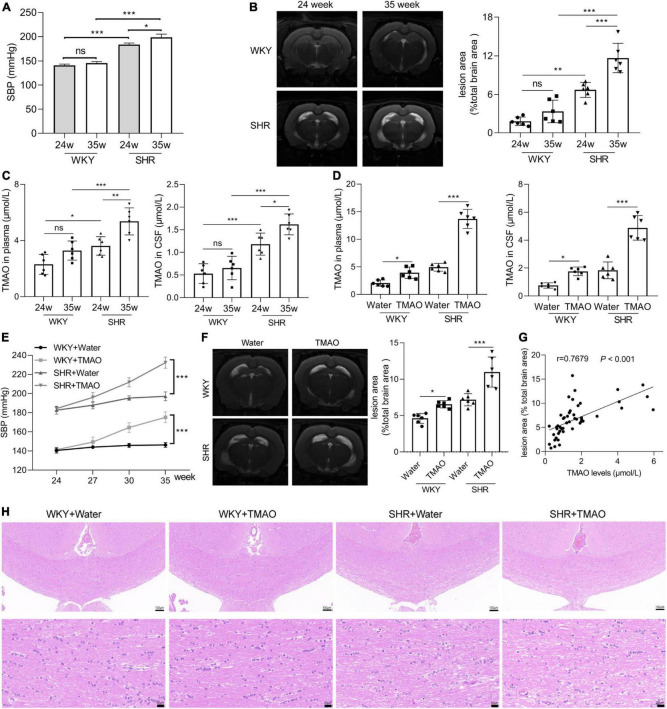
Background: Hypertension is a leading risk factor for cerebral small vessel disease (CSVD), a brain microvessels dysfunction accompanied by white matter lesions (WML). Trimethylamine N-oxide (TMAO), a metabolite of intestinal flora, is correlated with cardiovascular and aging diseases. Here, we explored the effect of TMAO on the demyelination of WML.

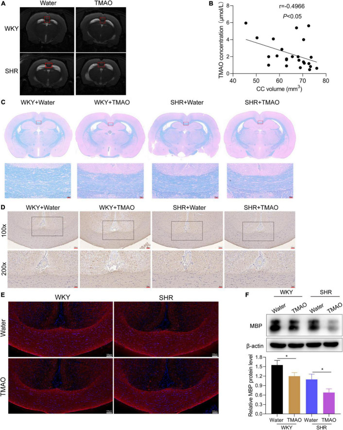
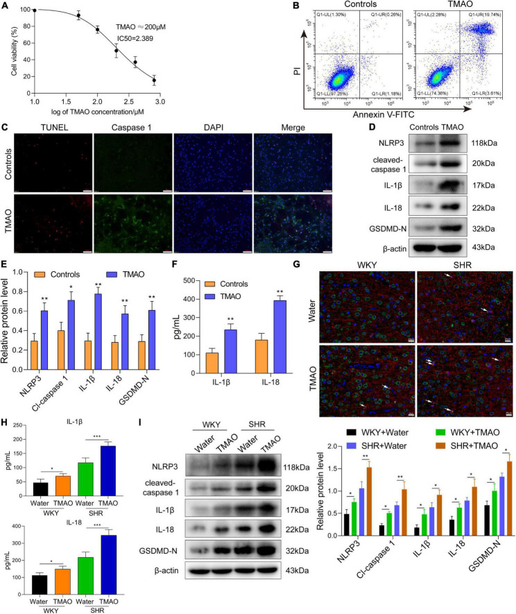
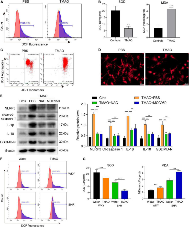
Methods: Spontaneous hypertension rats (SHRs) and primary oligodendrocytes were used to explore the effect of TMAO on demyelination in vivo and in vitro. T2-weighted magnetic resonance imaging (MRI) was applied to characterize the white matter hyperintensities (WMH) in rats. TMAO level was evaluated using LC-MS/MS assay. The histopathological changes of corpus callosum were measured by hematoxylin-eosin and luxol fast blue staining. And the related markers were detected by IHC, IF and western blot assay. Mito Tracker Red probe, DCFH-DA assay, flow cytometry based on JC-1 staining and Annexin V-FITC/PI double staining were conducted to evaluate the mitochondrial function, intracellular ROS levels and cell apoptosis.

Results: SHRs exhibited stronger WMH signals and a higher TMAO level than age-matched normotensive Wistar-kyoto rats (WKY). The corpus callosum region of SHR showed decreased volumes and enhanced demyelination when treated with TMAO. Furthermore, TMAO significantly elevated ROS production and induced NLRP3 inflammasome and impairment of mitochondrial function of oligodendrocytes. More importantly, TMAO enhanced the pyroptosis-related inflammatory death of oligodendrocytes.

Conclusion: TMAO could cross the blood-brain barrier (BBB) and promote oligodendrocytes pyroptosis via ROS/NLRP3 inflammasome signaling and mitochondrial dysfunction to promote demyelination, revealing a new diagnostic marker for WML under hypertension.

Keywords: demyelination; hypertension; pyroptosis; trimethylamine N-oxide; white matter lesions.


Conflict of interest statement

The authors declare that the research was conducted in the absence of any commercial or financial relationships that could be construed as a potential conflict of interest.

Figures

FIGURE 1
FIGURE 1
Increased TMAO promoted white matter lesions. (A) Systolic blood pressure (SBP) at 24 and 35 week of WKY and SHR (n = 6/group). (B) Images and quantation of T2-weighted MRI in 24- and 35-week SHR and age-matched normotension WKY rats. (C) TMAO level in plasma and CSF detected by LC-MS/MS (n = 6/group). 24-week-SHR and age-matched normotensive WKY rats were treated with TMAO for 11 weeks. (D) TMAO level in plasma and CSF detected by LC-MS/MS (n = 6/group). (E) SBP at different ages of WKY and SHR treated with TMAO (n = 6/group). (F) Images and quantation of T2-weighted MRI in SHR and WKY rats. (G) The relationship between TMAO levels and white matter lesion in brain was analyzed using Spearman correlation analysis. (H) Changes in white matter injury detected by H&E staining. Scale bar: upper, 100 μm; lower, 20 μm. The data were expressed as mean ± SD of three independent experiments. ns, no significance. *P < 0.05, **P < 0.01, ***P < 0.001.
FIGURE 2
FIGURE 2
TMAO promoted demyelination of white matter in hypertensive rats. (A) Images of T2-weighted MRI in SHR and WKY rats treated with TMAO or water. (B) The association between TMAO levels and corpus callosum volumes was analyzed using Spearman correlation analysis. (C) Luxol Fast Blue staining was used to evaluate myelin change. Scale bar: 50 μm. (D) MBP expressions were measured by IHC staining. Scale bar: upper, 100 μm; lower, 50 μm. (E) Immunofluorescence assay for MBP (red) staining in corpus callosum tissues. Scale bar: 100 μm (F) MBP expressions were measured by western blot. The data were expressed as mean ± SD of three independent experiments. *P < 0.05.
FIGURE 3
FIGURE 3
TMAO induced pyroptosis of oligodendrocytes to enhance demyelination in SHR. (A) CCK-8 kit was used to measure the cell viability of oligodendrocytes treated with TMAO. (B) Flow cytometry based on Annexin V-FITC/PI staining was used to detect the proportion of apoptotic cells. (C) Immunofluorescence assay for TUNEL (red) and caspase-1 (green) was carried out to measure the change of cell death. DAPI (blue) was used to stain neuclus. Scale bar = 50 μm. (D) The expressions and (E) quantitation of NLRP3, IL-1β, IL-18, cleaved-caspase 1 and GADMD-N terminal in cells were detected via western blot. (F) The levels of IL-1β and IL-18 in conditional medium were detected by ELISA assay. (G) GSDMD (green) and MBP (red) double staining immunofluorescence analysis in white matter region was performed. Arrows were for GSDMD+MBP+ oligodendrocytes in cerebrum. Scale bar = 20 μm. (H) The levels of IL-1β and IL-18 in CSF were detected by ELISA assay. (I) The expressions of NLRP3, IL-1β, IL-18, cleaved-caspase 1 and GADMD-N terminal were detected via western blot. The data were expressed as mean ± SD of three independent experiments. *P < 0.05, **P < 0.01, ***P < 0.001.
FIGURE 4
FIGURE 4
TMAO activated ROS/NLRP3 pathway to induce pyroptosis of oligodendrocytes. (A) The ROS level in oligodendrocyte was evaluated via DCFH-DA fluorescent probe. (B) The levels of SOD and MDA in oligodendrocyte were detected by ELISA assay. (C) Flow cytometry based on JC-1 staining was used to detect the change of mitochondrial membrane potential. (D) Mito-tracker red probe was used to evaluate the mitochondrial damage of oligodendrocytes in vitro. Scale bar = 20μm. (E) The expressions of NLRP3, IL-1β, IL-18, cleaved-caspase 1 and GADMD-N terminal in cells were detected via western blot. (F) The ROS level in cerebrum was evaluated via DCFH-DA fluorescent probe. (G) The levels of SOD and MDA were detected by ELISA assay. The data were expressed as mean ± SD of three independent experiments. **P < 0.01, ***P < 0.001.

Similar articles

Cited by

References

    1. Arrona Cardoza P., Spillane M. B., Morales Marroquin E. (2021). “Alzheimer’s disease and gut microbiota: does trimethylamine N-oxide (TMAO) play a role?”. Nutr. Rev. 2021:nuab022. 10.1093/nutrit/nuab022 - DOI - PubMed
    1. Bhattarai Y., Si J., Pu M., Ross O. A., McLean P. J., Till L., et al. (2021). “Role of gut microbiota in regulating gastrointestinal dysfunction and motor symptoms in a mouse model of Parkinson’s disease.”. Gut. Microbes 13:1866974. 10.1080/19490976.2020.1866974 - DOI - PMC - PubMed
    1. Brunt V. E., LaRocca T. J., Bazzoni A. E., Sapinsley Z. J., Miyamoto-Ditmon J., Gioscia-Ryan R. A., et al. (2021). “The gut microbiome-derived metabolite trimethylamine N-oxide modulates neuroinflammation and cognitive function with aging.”. Geroscience 43 377–94. 10.1007/s11357-020-00257-2 - DOI - PMC - PubMed
    1. Cannistraro R. J., Badi M., Eidelman B. H., Dickson D. W., Middlebrooks E. H., Meschia J. F. (2019). “CNS small vessel disease: a clinical review.”. Neurology 92 1146–56. 10.1212/WNL.0000000000007654 - DOI - PMC - PubMed
    1. Chen M. L., Zhu X. H., Ran L., Lang H. D., Yi L., Mi M. T. (2017). Trimethylamine-N-oxide induces vascular inflammation by activating the NLRP3 inflammasome through the SIRT3-SOD2-mtROS signaling pathway. J. Am. Heart Assoc. 6:6347. 10.1161/jaha.117.006347 - DOI - PMC - PubMed