Bilateral Optic Neuritis and Facial Palsy Following COVID-19 Infection
- PMID: 36072783
- PMCID: PMC9440666
- DOI: 10.7759/cureus.28735
Bilateral Optic Neuritis and Facial Palsy Following COVID-19 Infection
Abstract
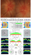
Cases of optic neuritis have been reported following the novel coronavirus disease 2019 (COVID-19), with most being unilateral and associated with demyelinating illness. We report a case of a 22-year-old woman who presented with sudden onset painless diminution of vision in both eyes six weeks following COVID-19 infection. She also had a history of left lower motor neuron (LMN) facial palsy immediately following COVID-19 disease that recovered fully on steroids. Ocular examination and ancillary and laboratory investigations pointed to bilateral atypical optic neuritis. The patient responded well to the standard optic neuritis treatment protocol. We diagnosed her as a case of left LMN facial palsy and parainfectious bilateral optic neuritis following COVID-19. Parainfectious bilateral optic neuritis and facial nerve palsy associated with COVID-19 can occur following COVID-19 disease. Ours is the first case to report the occurrence of both in a patient.
Keywords: bilateral optic neuritis; covid-19 disease; demyelinating disease; facial palsy; sars-cov-2.
Copyright © 2022, Behera et al.
Conflict of interest statement
The authors have declared that no competing interests exist.
Figures


References
-
- A case of COVID-19 with multiple cranial neuropathies. Gogia B, Gil Guevara A, Rai PK, Fang X. Int J Neurosci. 2020:1–3. - PubMed
Publication types
LinkOut - more resources
Full Text Sources
Research Materials
Miscellaneous
