. 2022 Oct;52(10):1662-1675.
doi: 10.1002/eji.202249972. Epub 2022 Sep 20.

Termination of CD40L co-stimulation promotes human B cell differentiation into antibody-secreting cells

Affiliations

Termination of CD40L co-stimulation promotes human B cell differentiation into antibody-secreting cells

Casper Marsman et al. Eur J Immunol. 2022 Oct.

Abstract

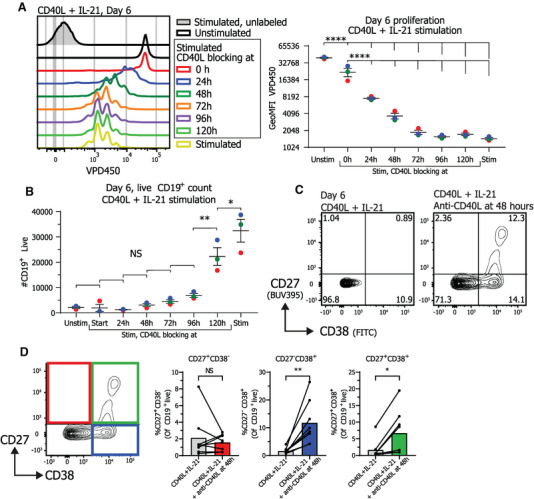
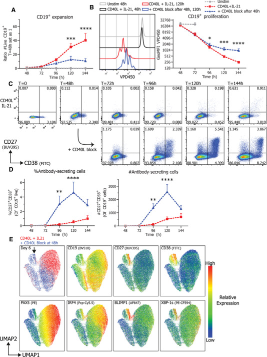
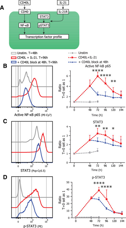
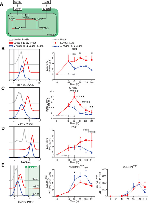
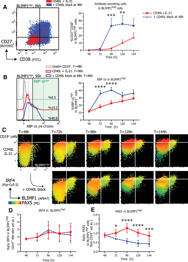
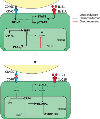
Human naïve B cells are notoriously difficult to differentiate into antibody-secreting cells (ASCs) in vitro while maintaining sufficient cell numbers to evaluate the differentiation process. B cells require T follicular helper (TFH ) cell-derived signals like CD40L and IL-21 during germinal center (GC) responses to undergo differentiation into ASCs. Cognate interactions between B and TFH cells are transient; after TFH contact, B cells cycle between GC light and dark zones where TFH contact is present and absent, respectively. Here, we elucidated that the efficacy of naïve B cells in ACS differentiation is dramatically enhanced by the release of CD40L stimulation. Multiparameter phospho-flow and transcription factor (TF)-flow cytometry revealed that termination of CD40L stimulation downmodulates NF-κB and STAT3 signaling. Furthermore, the termination of CD40 signaling downmodulates C-MYC, while promoting ASC TFs BLIMP1 and XBP-1s. Reduced levels of C-MYC in the differentiating B cells are later associated with crucial downmodulation of the B cell signature TF PAX5 specifically upon the termination of CD40 signaling, resulting in the differentiation of BLIMP1 high expressing cells into ASCs. The data presented here are the first steps to provide further insights how the transient nature of CD40 signaling is in fact needed for efficient human naïve B cell differentiation to ASCs.

Keywords: CD40L; IL-21; human B cells; phospho-signaling; transcriptional regulation.


Conflict of interest statement

The authors declare no commercial or financial conflict of interest.

Figures

Figure 1
Figure 1
Termination of CD40L stimulation after CD40L and IL‐21 stimulation inhibits proliferation and promotes differentiation into ASCs Naïve B cells were sorted and 25.000 cells were stimulated with a human‐CD40L expressing 3T3 feeder layer and recombinant IL‐21 (50 ng/ml). After the indicated culture duration CD40L blocking antibodies (13 μg/ml) were added and cells were analyzed at day 6 by flow cytometry. (A) Naïve B cells were labeled with proliferation dye VPD450 prior to culture. Representative histogram overlay (left) and quantification of VPD450 GeoMFI after 6 days of culture under indicated conditions. Data from a single independent experiment with the mean of three individual donors replicates marked as red, green, or blue dots with triplicate measurements (n = 3). (B) Number of live CD19+ cells was analyzed at day 6 after adding CD40L blocking antibodies at indicated time points (n = 3). Data from a single independent experiment with the mean of three individual donors marked as red, green or blue dots with triplicate measurements (n = 3), scatter plots show mean ± SD, p‐values were calculated using one‐way ANOVA with Tukey's multiple comparison test. (C) Representative contour plot of CD27 and CD38 expression profiles after 6 days of culture with indicated conditions. (D) Quantification of the relative percentages of CD27 and CD38 subpopulations after 6 days of culture with the indicated conditions (n = 8). Combined data from 7 independent experiments, each performed with 1–2 donors. Each data point represents the mean of an individual donor with duplicate or triplicate measurements. Mean values are represented as bars. P‐values were calculated using paired t‐test, *p ≤ 0.05, **p ≤ 0.01, ***p ≤ 0.001, ****p ≤ 0.0001.
Figure 2
Figure 2
Termination of CD40L stimulation inhibits CD19+ expansion and proliferation and results in efficient ASC differentiation Naïve B cells were sorted, labeled with VPD450, and 25.000 cells were stimulated with a human‐CD40L expressing 3T3 feeder layer and recombinant IL‐21 (50 ng/ml). After 48 h, CD40L blocking antibodies (13 μg/ml) were added or not. At indicated timepoints, cultures were harvested, stained for membrane markers, fixed, and stored until day 6 when all samples were analyzed by flow cytometry. (A) Quantification of live CD19+ cells per day (n = 8) (B) Representative histogram overlay (left) and quantification (right) of VPD450 GeoMFI per day (n = 8). Combined data from 4 independent experiments, each performed with two donors. Symbols represent the mean and error bars show ± SEM. (C) Representative pseudocolor dot plots of CD27 and CD38 expression profiles per day under indicated conditions. (D) Quantification of relative percentages (left) and absolute counts (right) of CD27+CD38+ ASC per day (n = 6). Combined data from three independent experiments, each performed with two donors. Symbols represent the mean and error bars show ± SEM. p‐Values were calculated by comparing the red CD40L + IL‐21 condition and the blue CD40L stimulation terminated after 48 h condition, using two‐way ANOVA with Sidak's multiple comparison test. *p ≤ 0.05, **p ≤ 0.01, ***p ≤ 0.001, ****p ≤ 0.0001. (E) UMAP clustering analysis on day 6 cultured B cells stained for CD19, CD27, CD38, PAX5, IRF4, BLIMP1, and XBP‐1s. The different conditions (red and blue) are shown overlaid at the top left, the ASC population is marked by a black arrow and relative protein expressions are represented as a heatmap. UMAP settings were as follows: distance function, Euclidean; number of neighbors, 30; minimal distance, 0.5; and number of components, 2. UMAP generated with data from a single representative donor measured in duplicate.
Figure 3
Figure 3
Termination of CD40L stimulation results in downregulation of NF‐kB and pSTAT3/STAT3 signaling Naïve B cells were sorted and 25.000 cells were stimulated with a human‐CD40L expressing 3T3 feeder layer and recombinant IL‐21 (50 ng/ml). After 48 hours CD40L blocking antibodies (13 ug/ml) were added or not. At indicated timepoints, cultures were harvested, stained for membrane markers, fixed, and stored until day 6 when all samples were stained intracellularly and analyzed by flow cytometry. (A) Schematic representation of CD40L and IL‐21 signaling pathways. (B) Representative histogram overlay (left) and quantification (right) of activated NF‐κB p65 GeoMFI per day under indicated conditions (n = 4). (C) Representative histogram overlay (left) and quantification (right) of STAT3 GeoMFI per day under indicated conditions (n = 4). (D) Representative histogram overlay (left) and quantification (right) of p‐STAT3 GeoMFI per day under indicated conditions (n = 4). p‐Values were calculated by comparing the red CD40L + IL‐21 condition and the blue CD40L stimulation terminated after 48 h condition, using two‐way ANOVA with Sidak's multiple comparison test Combined data from two independent experiments, each performed with two donors. Symbols represent the mean and error bars show ± SEM. *p ≤ 0.05, **p ≤ 0.01, ***p ≤ 0.001, ****p ≤ 0.0001.
Figure 4
Figure 4
Termination of CD40L stimulation inhibits expression of C‐MYC, IRF4 and PAX5 Naïve B cells were sorted and 25.000 cells were stimulated with a human‐CD40L expressing 3T3 feeder layer and recombinant IL‐21 (50 ng/ml). After 48 h, CD40L blocking antibodies (13 μg/ml) were added or not. At indicated timepoints, cultures were harvested, stained for membrane markers, fixed, and stored until day 6 when all samples were stained for TFs and analyzed by flow cytometry. (A) Schematic representation of CD40L and IL‐21 induced TF network. CD40L stimulation induces expression of IRF4, PAX5, and proliferation regulator C‐MYC while IL‐21 induces expression of BLIMP1 that is the crucial regulator of ASC differentiation. BLIMP1 expression inhibits and is inhibited by PAX5. Depending on the level of IRF4 expression either PAX5 or BLIMP1 expression is promoted. Additionally, TF XBP‐1s is repressed by PAX5 and promoted by BLIMP1. While B cells are known to express high levels of PAX5, ASCs express high levels of IRF4, BLIMP1, and XBP‐1s. (B) Representative histogram overlay (left) and quantification (right) of C‐MYC GeoMFI per day under indicated conditions (n = 4). (C) Representative histogram overlay (left) and quantification (right) of IRF4 GeoMFI per day under indicated conditions (n = 4). Combined data from 2 independent experiments, both performed with two donors. Symbols represent the mean and error bars show ±SEM. (D) Representative histogram overlay (left) and quantification (right) of PAX5 GeoMFI per day under indicated conditions (n = 6). (E) Representative histogram overlay (left), relative percentages (middle) and absolute counts (right) of BLIMP1High expressing cells under indicated conditions (n = 6). Combined data from three independent experiments, each performed with two donors. Symbols represent the mean and error bars show ±SEM. p‐Values were calculated by comparing the red CD40L + IL‐21 condition and the blue CD40L stimulation terminated after 48 h condition, using two‐way ANOVA with Sidak's multiple comparison test. *p ≤ 0.05, **p ≤ 0.01, ***p ≤ 0.001, ****p ≤ 0.0001
Figure 5
Figure 5
Termination of CD40L stimulation promotes differentiation of BLIMP1High B cells that express XBP‐1s while inhibiting PAX5 expression. Naïve B cells were sorted and 25.000 cells were stimulated with a human‐CD40L expressing 3T3 feeder layer and recombinant IL‐21 (50 ng/ml). After 48 h, CD40L blocking antibodies (13 ug/ml) were added or not. At indicated timepoints, cultures were harvested, stained for membrane markers, fixed, and stored until day 6 when all samples were stained for TFs and analyzed by flow cytometry. (A) Representative overlay of CD27 and CD38 expression profiles (left) of BLIMPHigh gated cells under indicated conditions after 96 h of culture and quantification (right) of the relative percentages of CD27+CD38+ ASCs within BLIMP1High gated cells per day (n = 6). (B) Representative histogram overlay (left) and quantification (right) of the relative percentages of XBP‐1sHigh expressing cells within BLIMP1High gated cells per day (n = 6). (C) Representative heatmap dot plots of IRF4, BLIMP1, and PAX5 expression profiles per day under indicated conditions gated on CD19+ cells. (D) Quantification of IRF4 GeoMFI per day under indicated conditions in BLIMP1High expressing cells (n = 4). (E) Quantification of PAX5 GeoMFI per day under indicated conditions in BLIMP1High expressing cells (n = 6). Combined data from two or three independent experiments, each performed with two donors. Symbols represent the mean and error bars show ±SEM. p‐Values were calculated by comparing the red CD40L + IL‐21 condition and the blue CD40L stimulation terminated after 48 h condition, using two‐way ANOVA with Sidak's multiple comparison test. *p ≤ 0.05, **p ≤ 0.01, ***p ≤ 0.001, ****p ≤ 0.0001
Figure 6
Figure 6
Schematic overview of CD40L stimulated and CD40L stimulation terminated B cells with phospho‐signaling and transcription factor profiles. Stimulation of CD40 by CD40L activates the NF‐κB pathway which in turn upregulates the expression of C‐MYC and IRF4. IL‐21 stimulation results in the phosphorylation of STAT3 which in turn can upregulate the expression of BLIMP1. Continuous CD40L stimulation results in an increased expression of NF‐κB, C‐MYC, IRF4, and PAX5 while preventing differentiation into ASCs. Upon termination of CD40L stimulation, NF‐κB, C‐MYC, and PAX5 expression is downregulated while expression of IRF4, BLIMP1, and XBP‐1s is increased, resulting in efficient ASC differentiation.

Similar articles

Cited by

References

    1. Ding, B. B. , Bi, E. , Chen, H. , Yu, J. J. and Ye, B. H. , IL‐21 and CD40L Synergistically Promote Plasma Cell Differentiation through Upregulation of Blimp‐1 in Human B Cells. The Journal of Immunology. 2013. 190: 1827–1836. - PMC - PubMed
    1. Moens, L. and Tangye, S. G. , Cytokine‐mediated regulation of plasma cell generation: IL‐21 takes center stage. Frontiers in Immunology. 2014. 5: 1–13. - PMC - PubMed
    1. Weinstein, J. S. , Herman, E. I. , Lainez, B. , Licona‐Limón, P., Esplugues, E. , Flavell, R. and Craft, J. , TFH cells progressively differentiate to regulate the germinal center response. Nature Immunology. 2016. 17: 1197–1205. - PMC - PubMed
    1. Weisel, F. J. , Zuccarino‐Catania, G.V. , Chikina, M. and Shlomchik, M. J. , A Temporal Switch in the Germinal Center Determines Differential Output of Memory B and Plasma Cells. Immunity. 2016. 44: 116–130. - PMC - PubMed
    1. McHeyzer‐Williams, L. J. , Pelletier, N. , Mark, L. , Fazilleau, N. and McHeyzer‐Williams, M. G. , Follicular helper T cells as cognate regulators of B cell immunity. Current Opinion in Immunology. 2009. 21: 266–273. - PMC - PubMed

Publication types