Review
. 2023 Feb;57(2):353-369.
doi: 10.1002/jmri.28426. Epub 2022 Sep 8.

Rectal MRI Interpretation After Neoadjuvant Therapy

Affiliations
Review

Rectal MRI Interpretation After Neoadjuvant Therapy

Natally Horvat et al. J Magn Reson Imaging. 2023 Feb.

Abstract

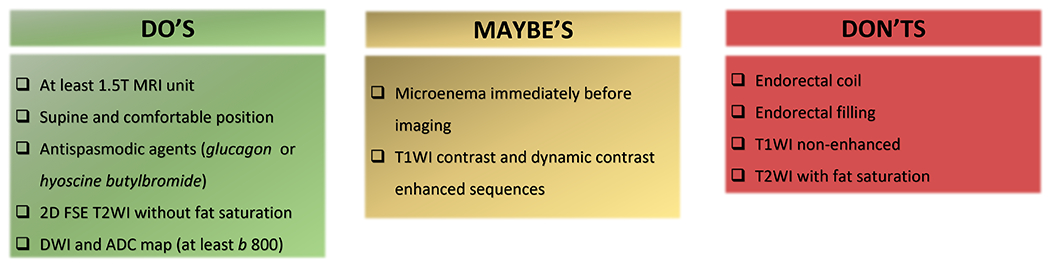
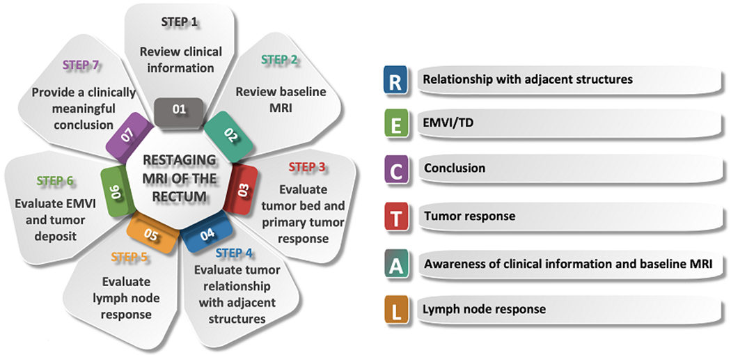
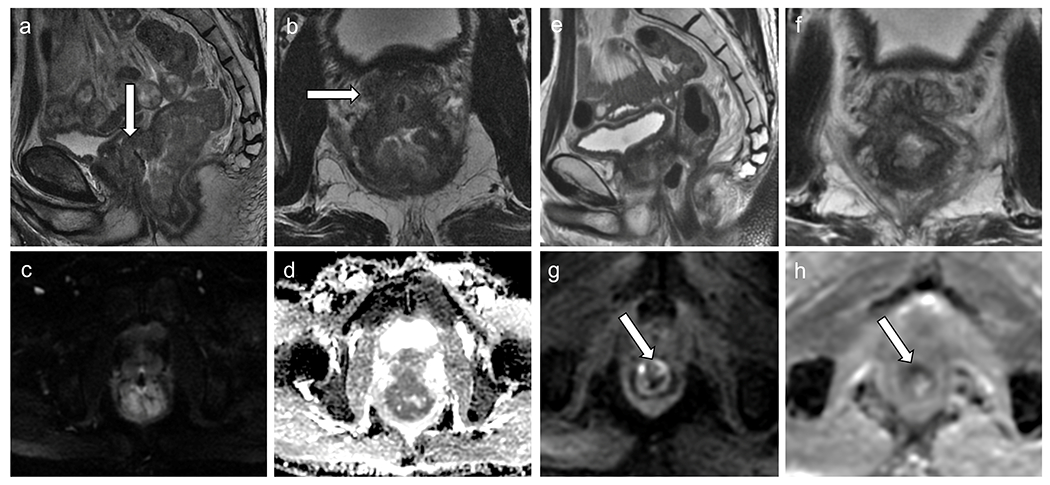
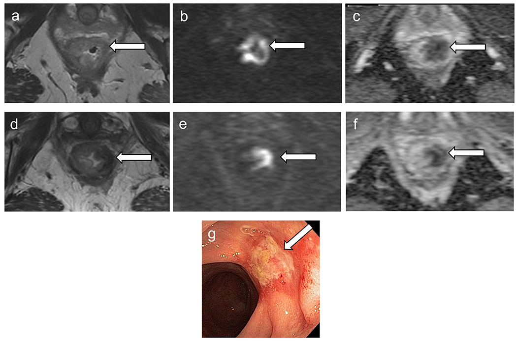
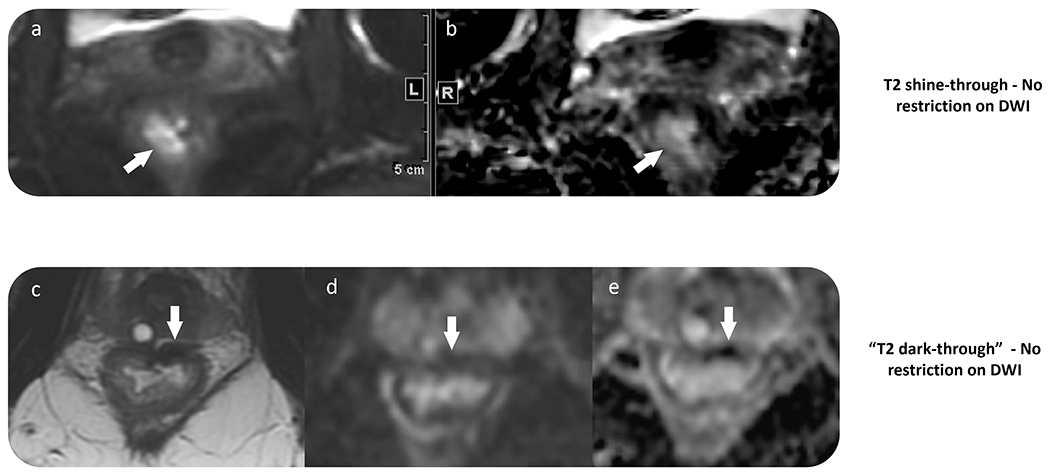
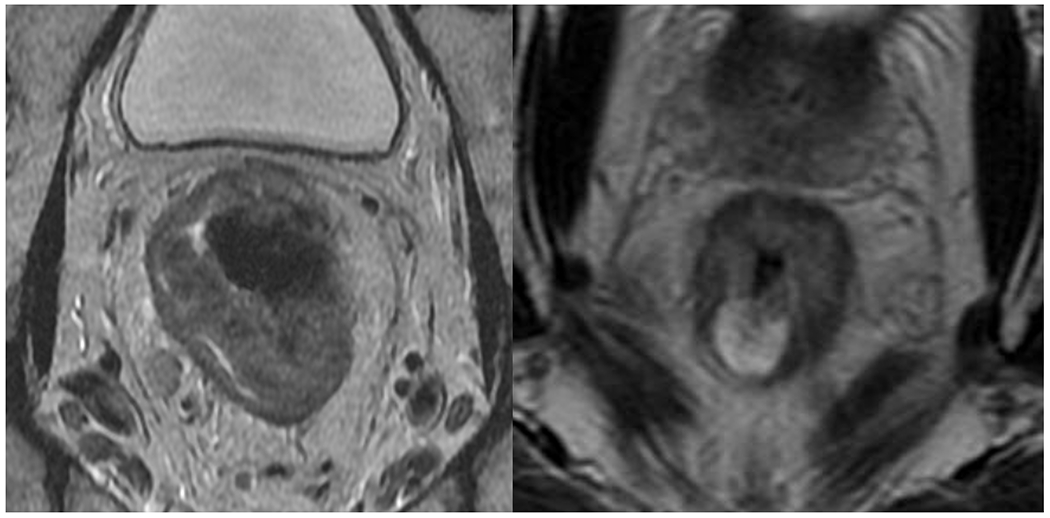
In recent years, several key advances in the management of locally advanced rectal cancer have been made, including the implementation of total mesorectal excision as the standard surgical approach; use of neoadjuvant chemoradiotherapy in selected patients with a high risk of local recurrence, and finally, adoption of organ preservation strategies, through either local excision or nonoperative management in selected patients with clinical complete response following neoadjuvant chemoradiotherapy. This review aims to shed light on the role of rectal MRI in the assessment of treatment response after neoadjuvant therapy, which is especially important given the growing feasibility of nonoperative management. First, an overview of current neoadjuvant therapies and response assessment based on digital rectal examination, endoscopy, and MRI will be provided. Second, the use of a high-quality restaging rectal MRI protocol will be presented. Third, a step-by-step approach to assessing treatment response on restaging rectal MRI following neoadjuvant treatment will be outlined, acknowledging challenges faced by radiologists during MRI interpretation. Finally, research related to response assessment will be discussed. LEVEL OF EVIDENCE: 4 TECHNICAL EFFICACY: Stage 3.

Keywords: magnetic resonance imaging; neoadjuvant therapy; rectal cancer.


Figures

Figure 1.
Figure 1.
Rectal MRI techniques that are recommended (“Do’s”), those that overall are not recommended (“Don’ts”), and some that are frequently used, with growing evidence of their added value in the restaging setting (“Maybe’s”).
Figure 2.
Figure 2.
Diagram showing a step-by-step approach to interpreting restaging MRI of the rectum. The mnemonic RECTAL summarizes the key findings to be addressed in the restaging report.
Figure 3.
Figure 3.
MRI of the rectum at baseline (a–c) as well as at restaging (d–f) after chemoradiation therapy. Baseline comparison allows for the delineation of the tumor bed (c and f, red shading). The opposite rectal wall shows mild submucosal edema (arrow).
Figure 4.
Figure 4.
MRI tumor regression grade (mrTRG) chart demonstrates the 5-point scoring system based on the proportion of residual tumor and fibrosis after neoadjuvant therapy on T2WI.
Figure 5.
Figure 5.
Baseline rectal MRI (a–d) shows, on T2WI, a low rectal tumor (a–b), T3N1, with diffusion restriction (c–d) infiltrating the prostate gland (white arrows). Restaging rectal MRI (e–h) demonstrates decreased rectal tumor, with few areas of fibrosis and mostly viable tumor with intermediate signal intensity on T2WI (mrTRG 4) (e–f), with areas of diffusion restriction (high signal intensity on DWI (g) and low signal intensity on ADC map (h)), inseparable from the prostate gland and seminal vesicles, consistent with incomplete response. . The patient underwent abdominoperineal resection, which demonstrated residual tumor infiltrating the prostate gland and seminal vesicles (ypT4b).
Figure 6.
Figure 6.
Baseline MRI (a–c) shows low rectal tumor with restriction on diffusion. Restaging MRI (d–f) demonstrates decreased size of primary tumor with thick scar and areas of intermediate signal intensity on T2 (mrTRG 3) (d) and diffusion restriction, high signal intensity on DWI (e) and low signal intensity on ADC map (f), consistent with incomplete response. (g) Restaging colonoscopy shows viable tumor. The patient underwent abdominoperineal resection and pT2N0 was confirmed.
Figure 7.
Figure 7.
Two examples of negative DWI obtained as part of restaging rectal MRI. (a–b) T2 shine-through effect, with high signal intensity within the rectal wall on DWI and the ADC map. (c–e) T2 dark-through shows the rectal wall scar with low signal intensity on T2 imaging, DWI, and ADC due to fibrosis. Viable tumor should be considered only in cases with high signal intensity on DWI and low signal intensity on ADC map within the tumor bed. Comparison with baseline scans may also be of value.
Figure 8.
Figure 8.
Baseline rectal MRI showing (a) a non-mucinous tumor. After neoadjuvant therapy, the patient developed (b) mucinous degeneration (black arrow). The patient underwent surgical resection and the pathology demonstrated viable tumor. Currently, MRI cannot differentiate cellular from acellular mucin.
Figure 9.
Figure 9.
A patient with T1-2N0 lower rectal tumor at baseline (a) underwent chemoradiotherapy with clinical complete response on nonoperative management. 12 months after chemoradiotherapy, there was no evidence of tumor regrowth on MRI (b), digital rectal examination (DRE), and endoscopy. 24 months after chemoradiotherapy (d), there was new isolated EMVI, without tumor regrowth within the tumor bed on MRI, DRE, or endoscopy, consistent with locoregional regrowth.
Figure 10.
Figure 10.
Baseline rectal MRI (a) shows on T2WI a c-shaped tumor (black arrow). Restaging MRI (b–c) shows decreased tumor with thin scar (mrTRG 1) without diffusion restriction. Endoscopy (d) shows telangiectasia and no residual tumor. The patient was classified as clinical complete response and is under nonoperative management without evidence of tumor regrowth.

References

    1. Bray F, Ferlay J, Soerjomataram I, Siegel RL, Torre LA, Jemal A. Global cancer statistics 2018: GLOBOCAN estimates of incidence and mortality worldwide for 36 cancers in 185 countries. CA Cancer J Clin 2018;68:394–424. - PubMed
    1. Kasi PM, Shahjehan F, Cochuyt JJ, Li Z, Colibaseanu DT, Merchea A. Rising Proportion of Young Individuals With Rectal and Colon Cancer. Clin Colorectal Cancer 2019;18:e87–e95. - PubMed
    1. Heald RJ, Ryall RD. Recurrence and survival after total mesorectal excision for rectal cancer. Lancet 1986;1:1479–1482. - PubMed
    1. Sauer R, Becker H, Hohenberger W, et al. Preoperative versus postoperative chemoradiotherapy for rectal cancer. N Engl J Med 2004;351:1731–1740. - PubMed
    1. Fokas E, Appelt A, Glynne-Jones R, et al. International consensus recommendations on key outcome measures for organ preservation after (chemo)radiotherapy in patients with rectal cancer. Nat Rev Clin Oncol 2021;18:805–816. - PubMed

Publication types

MeSH terms