. 2022 Nov 8;146(19):1444-1460.
doi: 10.1161/CIRCULATIONAHA.121.058901. Epub 2022 Sep 8.

ATIC-Associated De Novo Purine Synthesis Is Critically Involved in Proliferative Arterial Disease

Affiliations

ATIC-Associated De Novo Purine Synthesis Is Critically Involved in Proliferative Arterial Disease

Qian Ma et al. Circulation. .

Abstract

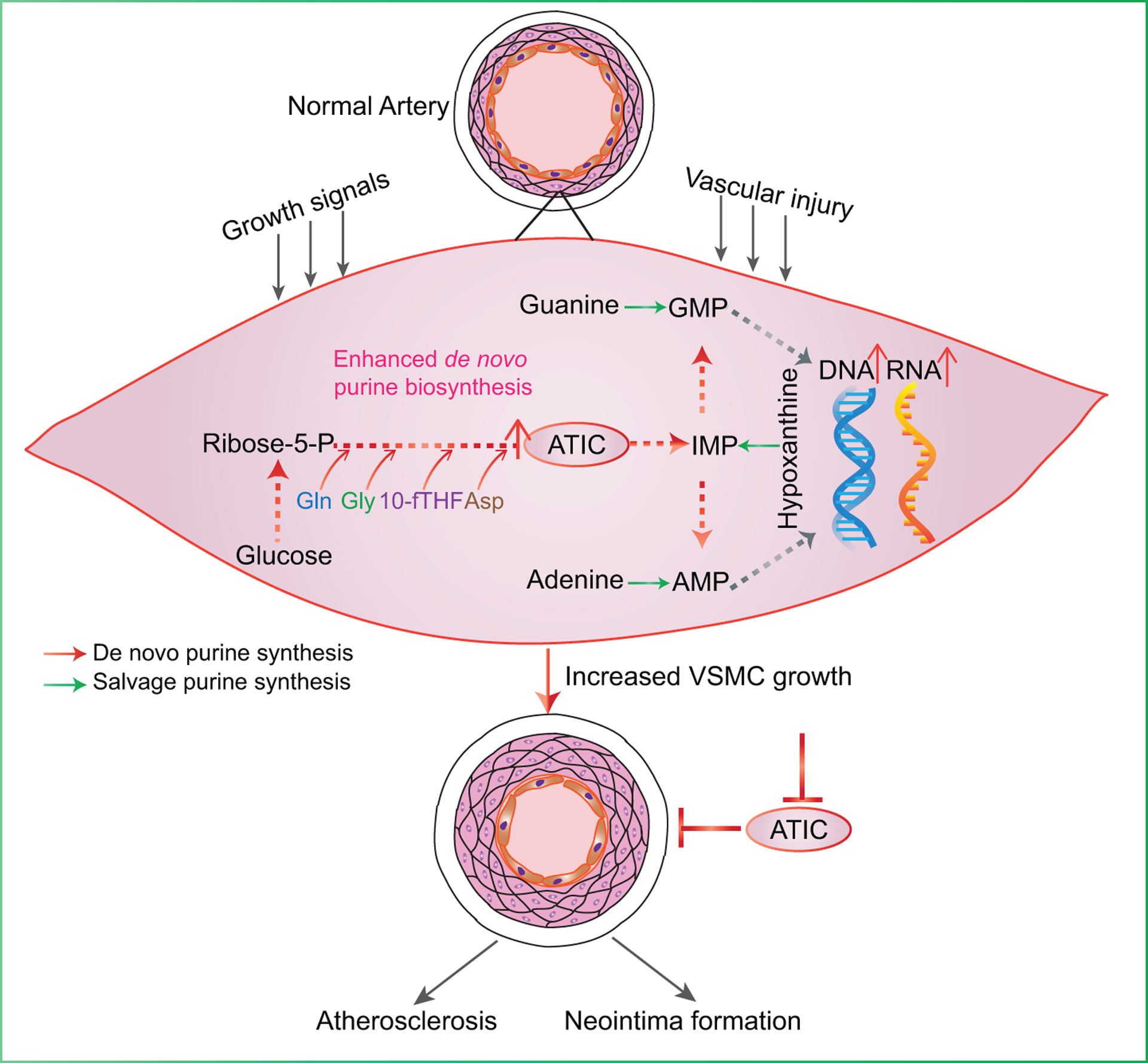
Background: Proliferation of vascular smooth muscle cells (VSMCs) is a hallmark of arterial diseases, especially in arterial restenosis after angioplasty or stent placement. VSMCs reprogram their metabolism to meet the increased requirements of lipids, proteins, and nucleotides for their proliferation. De novo purine synthesis is one of critical pathways for nucleotide synthesis. However, its role in proliferation of VSMCs in these arterial diseases has not been defined.

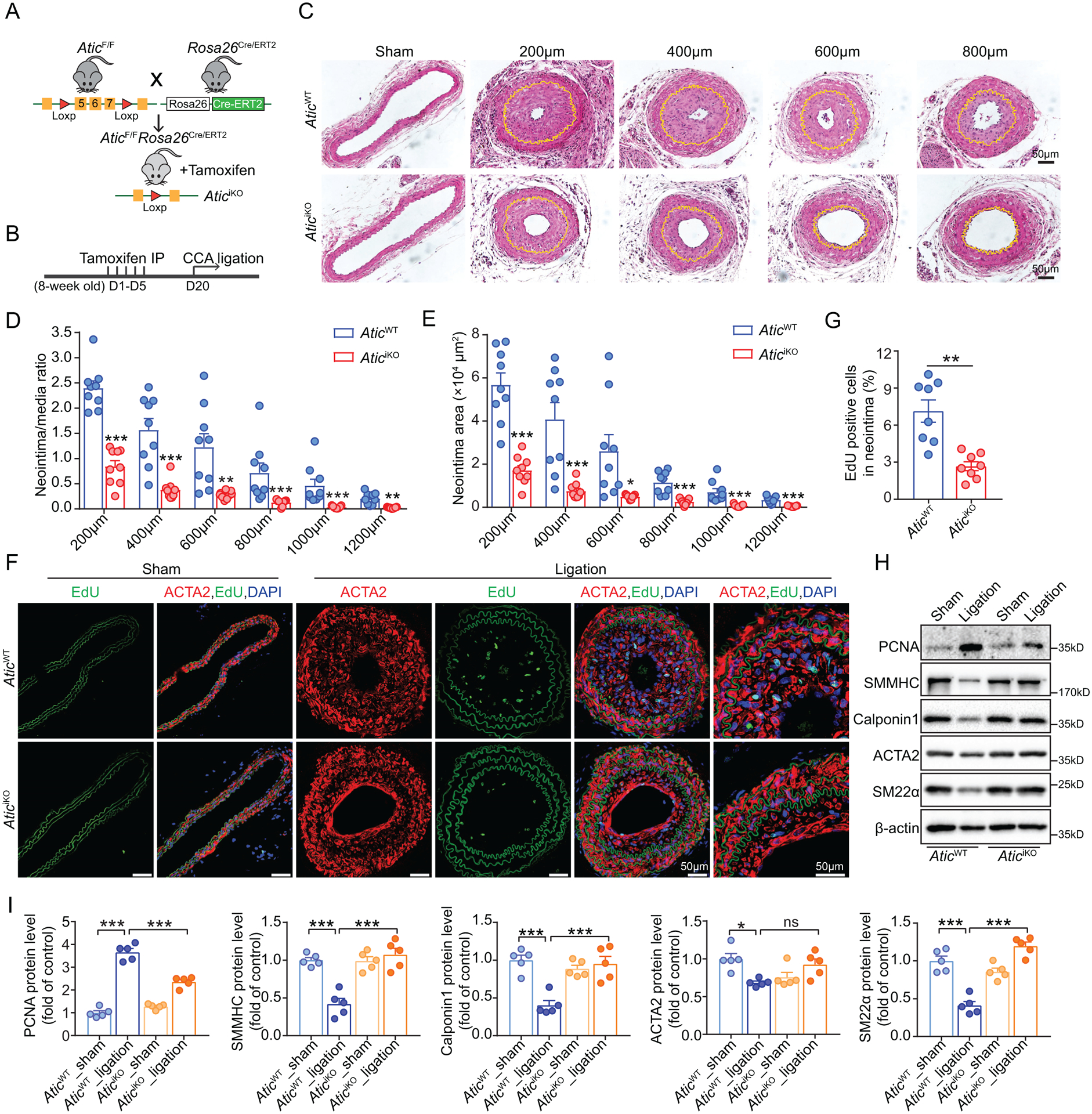
Methods: De novo purine synthesis in proliferative VSMCs was evaluated by liquid chromatography-tandem mass spectrometry. The expression of ATIC (5-aminoimidazole-4-carboxamide ribonucleotide formyltransferase/inosine monophosphate cyclohydrolase), the critical bifunctional enzyme in the last 2 steps of the de novo purine synthesis pathway, was assessed in VSMCs of proliferative arterial neointima. Global and VSMC-specific knockout of Atic mice were generated and used for examining the role of ATIC-associated purine metabolism in the formation of arterial neointima and atherosclerotic lesions.

Results: In this study, we found that de novo purine synthesis was increased in proliferative VSMCs. Upregulated purine synthesis genes, including ATIC, were observed in the neointima of the injured vessels and atherosclerotic lesions both in mice and humans. Global or specific knockout of Atic in VSMCs inhibited cell proliferation, attenuating the arterial neointima in models of mouse atherosclerosis and arterial restenosis.

Conclusions: These results reveal that de novo purine synthesis plays an important role in VSMC proliferation in arterial disease. These findings suggest that targeting ATIC is a promising therapeutic approach to combat arterial diseases.

Keywords: ATIC; arterial diseases; atherosclerosis; de novo purine synthesis; proliferation; vascular smooth muscle cells.


Conflict of interest statement

Conflict of Interest Disclosures

None.

Figures

Figure 1.
Figure 1.. De novo purine metabolism is increased in VSMCs of proliferative arterial diseases.
(A) Schematic showing the purine biosynthetic pathway and associated enzymes and metabolites. (B) Heat map showing the gene expression of venous bypass grafts from rabbit at indicated time points after transplantation by re-analysis of reported microarray dataset. (C) Heat map displaying the fold change of mRNA levels of indicated genes in the carotid arteries from mice with or without ligation injury for 5 days (n = 4–7). (D) Heat map showing fold change of mRNA levels of indicated genes in human saphenous vein grafts after ex vivo culture for 7 days (n = 3–6). (E) Representative Western blots and their quantification showing indicated protein expression in carotid arteries from mice after sham or ligation injury for 7 days (n = 6). (F) Representative Western blots and their quantification showing indicated protein expression in human saphenous vein grafts after ex vivo culture for 7 days (n = 6). (G) Schematic of the incorporation of nitrogen and carbon from glutamine, glycine and formate into the purine ring. (H) Normalized peak areas of 15N-labeled purine intermediates, as measured by targeted LC-MS/MS, in resting and proliferative HCSMCs labeled with 4 mM 15N-amide-glutamine for 4 h (n = 3). (I) Normalized peak areas of 13C-labeled purine intermediates, as measured by targeted LC-MS/MS, in resting and proliferative HCSMCs labeled with 1 mM 13C2-glycine for 4 h (n = 3). (J) Relative incorporation of 14C from glycine and formate and relative incorporation of 3H from hypoxanthine into DNA and RNA in resting and proliferative HCSMCs labeled with 2 μCi U-14C-glycine, 14C-formate and 3H-hypoxanthine for 12 hours, respectively (n = 3). The data are represented as mean ± SEM, *P < 0.05, **P < 0.01 and ***P < 0.001 for indicated comparisons.
Figure 2.
Figure 2.. ATIC expression is upregulated in proliferative VSMCs in arterial disease.
(A) Representative and quantification immunofluorescent staining of ATIC and EdU in the carotid arteries from mice after ligation injury for 4 weeks. The area highlighted with the white line is the arterial intima (I) and media (M) (n = 8–9). (B) Representative immunohistochemical (IHC) staining of ATIC in the carotid arteries from mice after ligation injury for 4 weeks. (C-D) Representative (C) and quantification (D) immunofluorescent staining of ATIC and EdU on the aortic sinus from Apoe−/− or wild-type (WT) mice fed with Western diet for 3 months (n = 6–9). The data are represented as mean ± SEM, *P < 0.05, **P < 0.01 and ***P < 0.001 for indicated comparisons.
Figure 3.
Figure 3.. ATIC knockdown decreases de novo purine metabolism-mediated nucleotide synthesis in proliferative VSMCs.
(A) Normalized peak areas of 15N-labeled purine intermediates, as measured by targeted LC-MS/MS, in proliferative HCSMCs transfected with control or ATIC siRNA and labeled with 4 mM 15N-amide-glutamine for 4 h (n = 3). (B) Relative incorporation of 14C from glycine into DNA and RNA in resting and proliferative HCSMCs transfected with control or ATIC siRNA and labeled with 2 μCi U-14C-glycine for 12 hours (n = 4). (C) Representative images and quantification of flow cytometry analysis showing EdU incorporation in HCSMCs transfected with control or ATIC siRNA for 48 hours and treated with PDGF (20 ng/ml) for 18 hours (n = 3). (D) Representative images and quantification of 5-EU staining showing newly synthesized RNA synthesis in HCSMCs transfected with control or ATIC siRNA for 48 hours and treated with vehicle or PDGF (20 ng/ml) for 12 hours (n = 5). (E) Representative images and quantification of flow cytometry analysis showing EdU incorporation in HCSMCs transfected with control or ATIC siRNA for 48 hours and treated with PDGF (20 ng/ml) and supplemented with adenine (20 μM) or hypoxanthine (100 μM) for 18 hours (n = 8). (F) Representative images and quantification of 5-EU staining showing new RNA synthesis in HCSMCs transfected with control or ATIC siRNA for 48 hours and treated with vehicle or PDGF (20 ng/ml) and supplemented with adenine (20 μM) or hypoxanthine (100 μM) for 12 hours (n = 5). The data are represented as mean ± SEM, *P < 0.05, **P < 0.01 and ***P < 0.001 for indicated comparisons.
Figure 4.
Figure 4.. ATIC is required for VSMC proliferation in vitro.
(A) Growth curves of HCSMCs transfected with control or ATIC siRNA for the indicated times (n = 6). (B) WST-1 cell proliferation assay of HCSMCs transfected with control or ATIC siRNA for 72 hours at indicated culture condition (n = 6–7). (C) Representative Western blot and quantification of the indicated proteins normalized to β-actin in HCSMCs transfected with control or ATIC siRNA for 48 hours and treated with PDGF (20 ng/ml) for 24 hours (n = 4). (D) Representative images and quantification of flow cytometry analysis of EdU staining in MASMCs isolated from AticWT or AticiKO mice (n = 7). (E) Representative EdU staining (green) and the percentage of EdU-positive MASMCs isolated from AticWT or AticiKO mice (n = 6–7). (F) Growth curves of MASMCs isolated from AticWT or AticiKO mice and cultured for the indicated times (n = 6). (G) WST-1 cell proliferation assay of MASMCs isolated from AticWT or AticiKO mice (n = 6–7). (H) Representative Western blot and quantification of the indicated proteins normalized to β-actin in MASMCs isolated from AticWT or AticiKO mice (n = 4–6). (I) Growth curves of HCSMCs transfected with control or ATIC siRNA and treated with adenine (20 μM) or hypoxanthine (100 μM) for the indicated times (n = 6). Black * for siATIC vs siCTRL, blue * for siATIC vs siATIC+Ade, green * for siATIC vs siATIC+Hyp. (J) Heat map showing the expression of cell cycle-associated genes in AticWT or AticiKO MASMCs as revealed by RNA sequencing. (K-L) Flow cytometry analysis of cell cycle for MASMCs isolated from AticWT or AticiKO mice (K) and for HCSMCs transfected with control or ATIC siRNA (L) (n = 6). The data are represented as mean ± SEM, *P < 0.05, **P < 0.01 and ***P < 0.001 for indicated comparisons.
Figure 5.
Figure 5.. VSMC-specific deletion of Atic attenuates neointima formation in a model of vascular injury.
(A) Strategy for generating AticiΔVSMC mice by crossing AticF/F mice with Myh11Cre/ERT2 mice and tamoxifen treatment of the consequent mice. (B) Representative Western blot and quantification showing ATIC expression in media of aortas from Myh11Cre/ERT2 and AticiΔVSMC mice. (C) Schematic timeline of tamoxifen treatment and carotid artery ligation model. (D) Representative HE-stained cross-sections of carotid arteries from mice with or without left common carotid artery ligation for 21 days. Sections are 200 to 600μm from the site of ligation. Yellow lines indicate the internal elastic lamina. (E) Quantification of the arterial neointima area and the ratio of neointima area to medial area of injured carotid arteries. (n = 5–6). (F) Representative EdU-positive stained cells in the neointima of carotid arteries from Myh11Cre/ERT2 and AticiΔVSMC mice and their quantification(n=7). (G) Representative Western blot and quantification of the indicated proteins normalized to β-actin in the sham and ligation-injured carotid arteries of Myh11Cre/ERT2 and AticiΔVSMC mice 7 days post ligation injury (n = 4). Data are represented as mean ± SEM, *P < 0.05, **P < 0.01 and ***P < 0.001 for indicated comparisons.
Figure 6.
Figure 6.. Global Atic deficiency attenuates neointima formation in a model of vascular injury.
(A) Strategy for generating AticiKO mice by crossing AticF/F mice with Rosa26Cre/ERT2 mice and tamoxifen treatment of the consequent mice. (B) Schematic timeline of tamoxifen treatment and carotid artery ligation model. (C) Representative HE-stained cross-sections of carotid arteries from the mice with or without left common carotid artery ligation for 28 days. Sections are 200 to 800 μm from the site of ligation. Yellow lines indicate the internal elastic lamina. (D-E) Quantification of the arterial neointima area and the ratio of neointima area to medial area of injured carotid arteries. (n = 9). (F-G) Representative EdU-positive stained cells in the neointima of carotid arteries from AticWT and AticiKO mice and their quantification (n = 8). (H-I) Representative Western blot (H) and quantification (I) of the indicated proteins normalized to β-actin in the sham and ligation-injured carotid arteries of AticWT and AticiKO mice 7 days post ligation injury (n = 5). Data are represented as mean ± SEM, *P < 0.05, **P < 0.01 and ***P < 0.001 for indicated comparisons.
Figure 7.
Figure 7.. VSMC-specific and global Atic deficiency alleviates atherosclerosis in Apoe−/− mice fed with Western diet for 12 weeks.
(A) Representative images and quantification of Oil Red O staining of en face aortas from Apoe−/−;Myh11Cre/ERT2 and Apoe−/−;AticiΔVSMC mice fed with Western diet for 12 weeks (n = 12–14). (B) Representative HE (upper) and Oil Red O (lower) staining of aortic sinuses from Apoe−/−;Myh11Cre/ERT2 and Apoe−/−;AticiΔVSMC mice fed with Western diet for 12 weeks (n = 9). (C) Representative ACTA2 staining of smooth muscle cells in aortic sinuses from Apoe−/−;Myh11Cre/ERT2 and Apoe−/−;AticiΔVSMC mice fed with Western diet for 12 weeks and their quantification (n = 8–9). (D) Representative images and quantification of Oil Red O staining of en face aortas from Apoe−/−;AticWT and Apoe−/−;AticiKO mice fed with Western diet for 12 weeks (n = 11). (E) Representative HE (upper) and Oil Red O (lower) staining of aortic sinuses from Apoe−/−;AticWT and Apoe−/−;AticiKO male mice fed with Western diet for 12 weeks (n = 7–11). (F) Representative ACTA2 staining of smooth muscle cells in aortic sinuses from Apoe−/−;AticWT and Apoe−/−;AticiKO male mice fed with Western diet for 12 weeks and their quantification (n = 8–9). Data are represented as mean ± SEM, **P < 0.01 and ***P < 0.001 for indicated comparisons.
Figure 8.
Figure 8.. Role of ATIC-mediated de novo purine synthesis in VSMC proliferation in arterial proliferative diseases.
ATIC-mediated de novo purine synthesis is enhanced when cells are exposed to the stimulation of injury or growth signals in VSMCs, which can supply purine nucleotides for incorporation into DNA/RNA synthesis to meet the demand for VSMC proliferation. Proliferative VSMCs ultimately contribute to the formation of arterial neointima and atherosclerotic lesions.

References

    1. Owens GK, Kumar MS and Wamhoff BR. Molecular regulation of vascular smooth muscle cell differentiation in development and disease. Physiol Rev. 2004;84:767–801. - PubMed
    1. Schwartz SM, deBlois D and O’Brien ER. The intima. Soil for atherosclerosis and restenosis. Circ Res. 1995;77:445–65. - PubMed
    1. Dzau VJ, Braun-Dullaeus RC and Sedding DG. Vascular proliferation and atherosclerosis: new perspectives and therapeutic strategies. Nat Med. 2002;8:1249–56. - PubMed
    1. Zhu J and Thompson CB. Metabolic regulation of cell growth and proliferation. Nature reviews Molecular cell biology. 2019;20:436–450. - PMC - PubMed
    1. Murray AW. The biological significance of purine salvage. Annu Rev Biochem. 1971;40:811–26. - PubMed

Publication types

Substances