. 2022 Sep 1;29(Pt 5):1273-1283.
doi: 10.1107/S1600577522006701. Epub 2022 Jul 21.

Pump-probe capabilities at the SPB/SFX instrument of the European XFEL

Affiliations

Pump-probe capabilities at the SPB/SFX instrument of the European XFEL

Jayanath C P Koliyadu et al. J Synchrotron Radiat. .

Abstract

Pump-probe experiments at X-ray free-electron laser (XFEL) facilities are a powerful tool for studying dynamics at ultrafast and longer timescales. Observing the dynamics in diverse scientific cases requires optical laser systems with a wide range of wavelength, flexible pulse sequences and different pulse durations, especially in the pump source. Here, the pump-probe instrumentation available for measurements at the Single Particles, Clusters, and Biomolecules and Serial Femtosecond Crystallography (SPB/SFX) instrument of the European XFEL is reported. The temporal and spatial stability of this instrumentation is also presented.

Keywords: European XFEL; megahertz pump and probe sources; pump–probe experiments; time-resolved experiments.


Figures

Figure 1
Figure 1
Top view of the general layout of the SPB/SFX instrument showing the central laser hutch, instrument laser hutch and experiment hutch. The two interactions regions are highlighted by orange boxes, the PP laser beam pipes and beam directions are marked in red, and the X-ray beam, marked in violet, goes from left to right.
Figure 2
Figure 2
Schematic of a pump–probe experimental setup at IRU of the SPB/SFX instrument.
Figure 3
Figure 3
Spectral (a) and temporal (b) profile of the PP laser at 15 fs FWHM and 50 fs FWHM pulse duration. The insets show the beam profile in the near-field (c) and far-field (d).
Figure 4
Figure 4
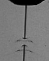
Visualizing the water jet explosion induced by an intense X-ray pulse with the PP laser. The image was captured by the side microscope with a 20× objective (20× Mitutoyo Plan Apo SL Infinity Corrected Objective, 378-810-3).
Figure 5
Figure 5
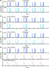
Several examples of pulse pattern setting for optical laser pulses. (a) Pump and probe source have the same pulse pattern. (b) Burst-mode operation where the optical laser pulses are down picked for so-called light and dark states for consecutive trains. (c) Burst-mode excitation with an arbitrary intra-train pulse pattern derived from a megahertz X-ray pulse pattern. (d) 10 Hz pump and sampling with a megahertz X-ray pulse pattern.
Figure 6
Figure 6
Output of TOPAS at different wavelengths. For this measurement, TOPAS was pumped by the PP laser with the following parameters: 800 nm, ∼54 fs, 215 µJ and 50 pulses per train at 1.1 MHz. (SH – second harmonic; SF – sum frequency; SIG – signal; IDL – idler).
Figure 7
Figure 7
Intensity stability of the TOPAS output measured at IRU over 12 h with the output beam set at a wavelength of 640 nm. The three lines show the 10 min moving average of intensity stability of the PP laser in the CLH, the ILH and the OPA output at IRU. Here the intensity is normalized to the mean intensity of each measurement.
Figure 8
Figure 8
2D histogram of the pointing stability of the TOPAS output beam in the horizontal and vertical directions.
Figure 9
Figure 9
Double exposure stroboscopic imaging of droplets with two nanosecond lasers with a delay of hundreds of nanoseconds between the two pulses. A zoomed view of one of the droplets imaged is shown in the inset. The image was collected using the side microscope with a 10× objective and an Andor Zyla 5.5 camera.
Figure 10
Figure 10
An aerosol particle beam with particles of size of a few tens of nanometres visualized by Rayleigh scattering imaging using the (a) inline microscope with 10× objective and (b) side microscope with 2× objective.
Figure 11
Figure 11
Schematic of (a) the PAM setup, and (b) the PAM sample holder.
Figure 12
Figure 12
Plot of temporal jitter measured over 15 min. The darker blue line shows the rolling mean over 5 s (Sato et al., 2020 ▸).
Figure 13
Figure 13
Time zero position determination using spatial encoding at SPB/SFX. (a) Laser transmission imaged with the side microscope with 10× magnification. (b) Line-out of the selected region highlighted by the white box in (a).

Similar articles

  • XFEL Beamline Optical Instrumentation for Ultrafast Science.
    Hutchison CDM, Perrett S, van Thor JJ. Hutchison CDM, et al. J Phys Chem B. 2024 Sep 19;128(37):8855-8868. doi: 10.1021/acs.jpcb.4c01492. Epub 2024 Aug 1. J Phys Chem B. 2024. PMID: 39087627 Free PMC article. Review.
  • The Single Particles, Clusters and Biomolecules and Serial Femtosecond Crystallography instrument of the European XFEL: initial installation.
    Mancuso AP, Aquila A, Batchelor L, Bean RJ, Bielecki J, Borchers G, Doerner K, Giewekemeyer K, Graceffa R, Kelsey OD, Kim Y, Kirkwood HJ, Legrand A, Letrun R, Manning B, Lopez Morillo L, Messerschmidt M, Mills G, Raabe S, Reimers N, Round A, Sato T, Schulz J, Signe Takem C, Sikorski M, Stern S, Thute P, Vagovič P, Weinhausen B, Tschentscher T. Mancuso AP, et al. J Synchrotron Radiat. 2019 May 1;26(Pt 3):660-676. doi: 10.1107/S1600577519003308. Epub 2019 Apr 12. J Synchrotron Radiat. 2019. PMID: 31074429 Free PMC article.
  • Megahertz pulse trains enable multi-hit serial femtosecond crystallography experiments at X-ray free electron lasers.
    Holmes S, Kirkwood HJ, Bean R, Giewekemeyer K, Martin AV, Hadian-Jazi M, Wiedorn MO, Oberthür D, Marman H, Adriano L, Al-Qudami N, Bajt S, Barák I, Bari S, Bielecki J, Brockhauser S, Coleman MA, Cruz-Mazo F, Danilevski C, Dörner K, Gañán-Calvo AM, Graceffa R, Fanghor H, Heymann M, Frank M, Kaukher A, Kim Y, Kobe B, Knoška J, Laurus T, Letrun R, Maia L, Messerschmidt M, Metz M, Michelat T, Mills G, Molodtsov S, Monteiro DCF, Morgan AJ, Münnich A, Peña Murillo GE, Previtali G, Round A, Sato T, Schubert R, Schulz J, Shelby M, Seuring C, Sellberg JA, Sikorski M, Silenzi A, Stern S, Sztuk-Dambietz J, Szuba J, Trebbin M, Vagovic P, Ve T, Weinhausen B, Wrona K, Xavier PL, Xu C, Yefanov O, Nugent KA, Chapman HN, Mancuso AP, Barty A, Abbey B, Darmanin C. Holmes S, et al. Nat Commun. 2022 Aug 11;13(1):4708. doi: 10.1038/s41467-022-32434-6. Nat Commun. 2022. PMID: 35953469 Free PMC article.
  • 3D printed devices and infrastructure for liquid sample delivery at the European XFEL.
    Vakili M, Bielecki J, Knoška J, Otte F, Han H, Kloos M, Schubert R, Delmas E, Mills G, de Wijn R, Letrun R, Dold S, Bean R, Round A, Kim Y, Lima FA, Dörner K, Valerio J, Heymann M, Mancuso AP, Schulz J. Vakili M, et al. J Synchrotron Radiat. 2022 Mar 1;29(Pt 2):331-346. doi: 10.1107/S1600577521013370. Epub 2022 Feb 15. J Synchrotron Radiat. 2022. PMID: 35254295 Free PMC article.
  • Dynamic Structural Biology Experiments at XFEL or Synchrotron Sources.
    Aller P, Orville AM. Aller P, et al. Methods Mol Biol. 2021;2305:203-228. doi: 10.1007/978-1-0716-1406-8_11. Methods Mol Biol. 2021. PMID: 33950392 Review.

Cited by

  • Microsecond time-resolved X-ray scattering by utilizing MHz repetition rate at second-generation XFELs.
    Konold PE, Monrroy L, Bellisario A, Filipe D, Adams P, Alvarez R, Bean R, Bielecki J, Bódizs S, Ducrocq G, Grubmueller H, Kirian RA, Kloos M, Koliyadu JCP, Koua FHM, Larkiala T, Letrun R, Lindsten F, Maihöfer M, Martin AV, Mészáros P, Mutisya J, Nimmrich A, Okamoto K, Round A, Sato T, Valerio J, Westphal D, Wollter A, Yenupuri TV, You T, Maia F, Westenhoff S. Konold PE, et al. Nat Methods. 2024 Sep;21(9):1608-1611. doi: 10.1038/s41592-024-02344-0. Epub 2024 Jul 5. Nat Methods. 2024. PMID: 38969722 Free PMC article.
  • Development of MHz X-ray phase contrast imaging at the European XFEL.
    Koliyadu JCP, Moško D, Asimakopoulou EM, Bellucci V, Birnšteinová Š, Bean R, Letrun R, Kim C, Kirkwood H, Giovanetti G, Jardon N, Szuba J, Guest T, Koch A, Grünert J, Szeles P, Villanueva-Perez P, Reuter F, Ohl CD, Noack MA, Garcia-Moreno F, Kuglerová-Valdová Z, Juha L, Nikl M, Yashiro W, Soyama H, Eakins D, Korsunsky AM, Uličný J, Meents A, Chapman HN, Mancuso AP, Sato T, Vagovič P. Koliyadu JCP, et al. J Synchrotron Radiat. 2025 Jan 1;32(Pt 1):17-28. doi: 10.1107/S160057752400986X. Epub 2025 Jan 1. J Synchrotron Radiat. 2025. PMID: 39565194 Free PMC article.
  • Kilohertz droplet-on-demand serial femtosecond crystallography at the European XFEL station FXE.
    Perrett S, Fadini A, Hutchison CDM, Bhattacharya S, Morrison C, Turkot O, Jakobsen MB, Größler M, Licón-Saláiz J, Griese F, Flewett S, Valerio J, Schulz J, Biednov M, Jiang Y, Han H, Yousef H, Khakhulin D, Milne C, Barty A, van Thor JJ. Perrett S, et al. Struct Dyn. 2024 Apr 17;11(2):024310. doi: 10.1063/4.0000248. eCollection 2024 Mar. Struct Dyn. 2024. PMID: 38638699 Free PMC article.
  • XFEL Beamline Optical Instrumentation for Ultrafast Science.
    Hutchison CDM, Perrett S, van Thor JJ. Hutchison CDM, et al. J Phys Chem B. 2024 Sep 19;128(37):8855-8868. doi: 10.1021/acs.jpcb.4c01492. Epub 2024 Aug 1. J Phys Chem B. 2024. PMID: 39087627 Free PMC article. Review.
  • Direct Observation of the Exciton-Polaron in Single CsPbBr3 Quantum Dots.
    Shen Z, Samoli M, Erdem O, Bielecki J, Samanta AK, E J, Estillore AD, Kim C, Kim Y, Koliyadu J, Letrun R, Locardi F, Lübke J, Mall A, Melo DVM, Mills G, Rafie-Zinedine S, Round A, Sato T, de Wijn R, Wollweber T, Worbs L, Zhuang Y, Mancuso AP, Bean R, Chapman HN, Küpper J, Infante I, Lange H, Hens Z, Ayyer K. Shen Z, et al. ACS Nano. 2025 Aug 12;19(31):28372-28382. doi: 10.1021/acsnano.5c06716. Epub 2025 Aug 2. ACS Nano. 2025. PMID: 40751687 Free PMC article.

References

    1. Allahgholi, A., Becker, J., Bianco, L., Delfs, A., Dinapoli, R., Goettlicher, P., Graafsma, H., Greiffenberg, D., Hirsemann, H., Jack, S., Klanner, R., Klyuev, A., Krueger, H., Lange, S., Marras, A., Mezza, D., Mozzanica, A., Rah, S., Xia, Q., Schmitt, B., Schwandt, J., Sheviakov, I., Shi, X., Smoljanin, S., Trunk, U., Zhang, J. & Zimmer, M. (2015). J. Instrum. 10, C01023.
    1. Ayyer, K., Xavier, P. L., Bielecki, J., Shen, Z., Daurer, B. J., Samanta, A. K., Awel, S., Bean, R., Barty, A., Bergemann, M., Ekeberg, T., Estillore, A. D., Fangohr, H., Giewekemeyer, K., Hunter, M. S., Karnevskiy, M., Kirian, R. A., Kirkwood, H., Kim, Y., Koliyadu, J., Lange, H., Letrun, R., Lübke, J., Michelat, T., Morgan, A. J., Roth, N., Sato, T., Sikorski, M., Schulz, F., Spence, J. C. H., Vagovic, P., Wollweber, T., Worbs, L., Yefanov, O., Zhuang, Y., Maia, F. R. N. C., Horke, D. A., Küpper, J., Loh, N. D., Mancuso, A. P. & Chapman, H. N. (2021). Optica, 8, 15–23.
    1. Bean, R. J., Aquila, A., Samoylova, L. & Mancuso, A. P. (2016). J. Opt. 18, 074011.
    1. Biasin, E., Fox, Z. W., Andersen, A., Ledbetter, K., Kjaer, K. S., Alonso-Mori, R., Carlstad, J. M., Chollet, M., Gaynor, J. D., Glownia, J. M., Hong, K., Kroll, T., Lee, J. H., Liekhus-Schmaltz, C., Reinhard, M., Sokaras, D., Zhang, Y., Doumy, G., March, A. M., Southworth, S. H., Mukamel, S., Gaffney, K. J., Schoenlein, R. W., Govind, N., Cordones, A. A. & Khalil, M. (2021). Nat. Chem. 13, 343–349.
    1. Bielecki, J., Hantke, M. F., Daurer, B. J., Reddy, H. K. N., Hasse, D., Larsson, D. S. D., Gunn, L. H., Svenda, M., Munke, A., Sellberg, J. A., Flueckiger, L., Pietrini, A., Nettelblad, C., Lundholm, I., Carlsson, G., Okamoto, K., Timneanu, N., Westphal, D., Kulyk, O., Higashiura, A., van der Schot, G., Loh, N. D., Wysong, T. E., Bostedt, C., Gorkhover, T., Iwan, B., Seibert, M. M., Osipov, T., Walter, P., Hart, P., Bucher, M., Ulmer, A., Ray, D., Carini, G., Ferguson, K. R., Andersson, I., Andreasson, J., Hajdu, J. & Maia, F. R. N. C. (2019). Sci. Adv. 5, eaav8801. - PMC - PubMed