. 2022 Oct 20;90(10):e0026322.
doi: 10.1128/iai.00263-22. Epub 2022 Sep 8.

Characterization of MroQ-Dependent Maturation and Export of the Staphylococcus aureus Accessory Gene Regulatory System Autoinducing Peptide

Affiliations

Characterization of MroQ-Dependent Maturation and Export of the Staphylococcus aureus Accessory Gene Regulatory System Autoinducing Peptide

Madison R Stock et al. Infect Immun. .

Abstract

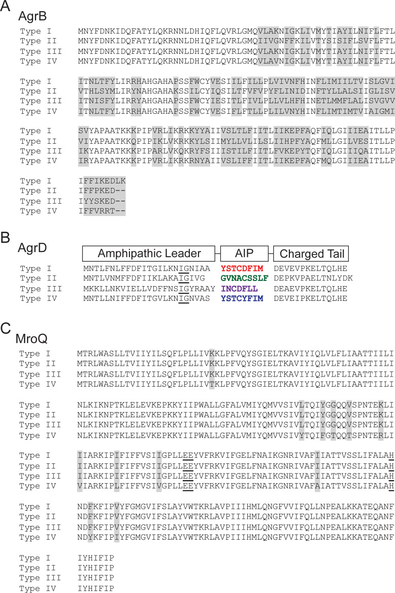
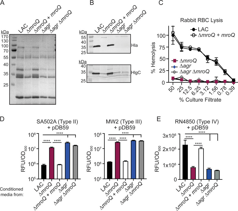
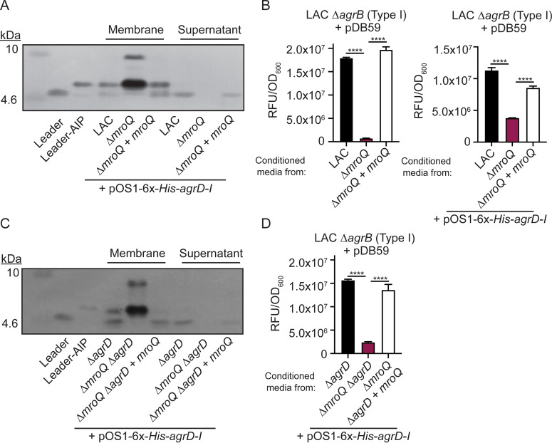
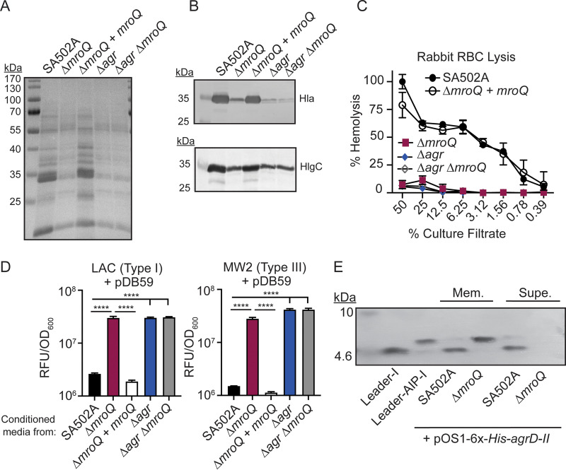
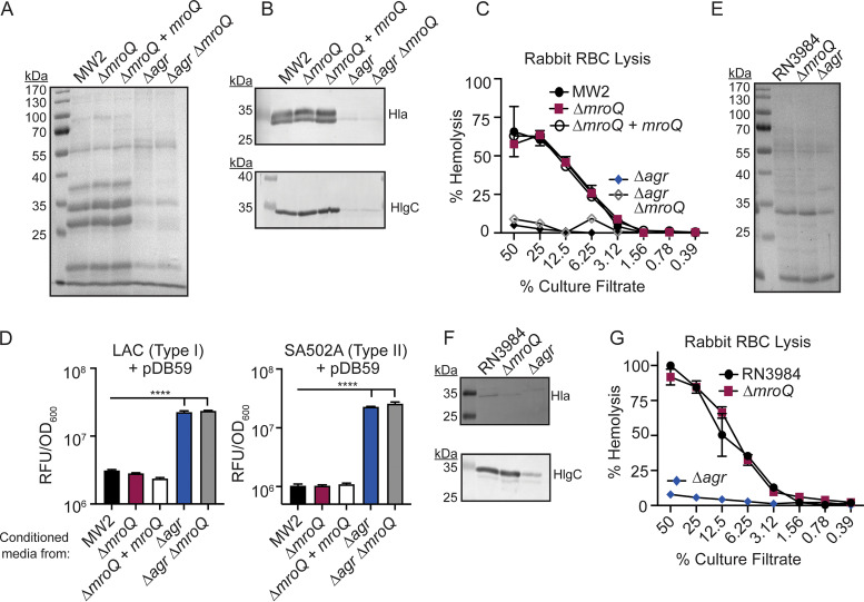
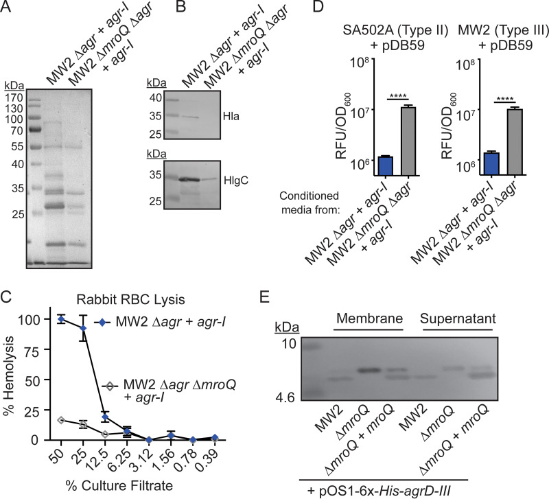
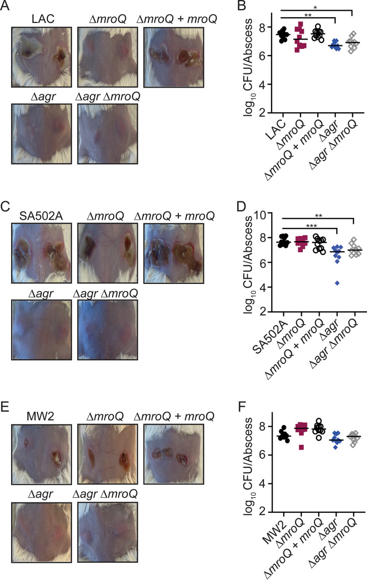
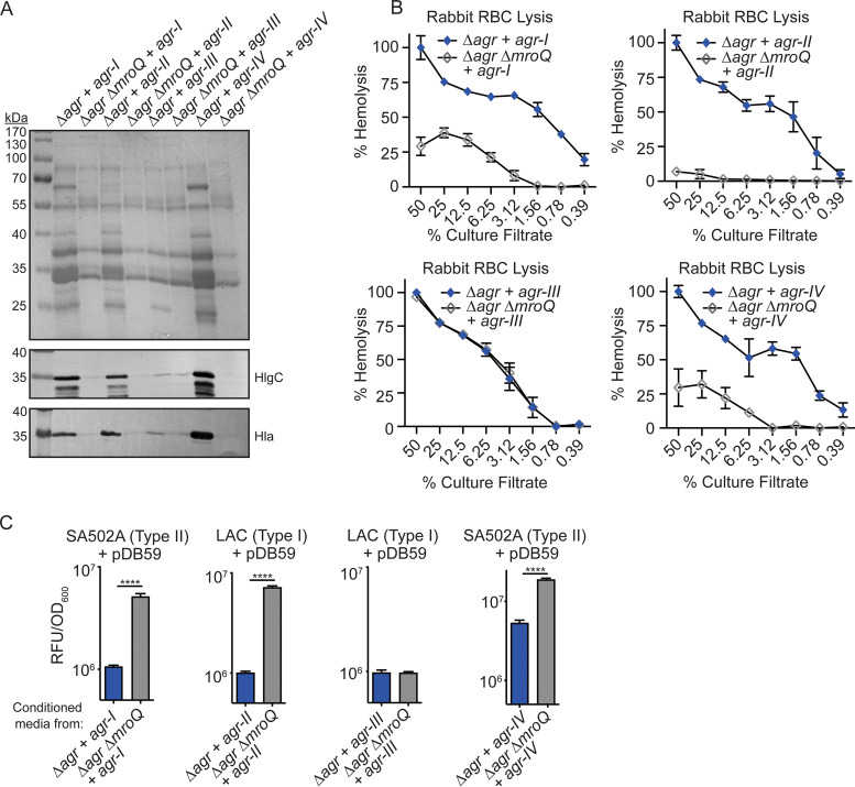
Gram-positive bacteria produce small autoinducing peptides (AIPs), which act to regulate expression of genes that promote adaptive traits, including virulence. The Gram-positive pathogen Staphylococcus aureus generates a cyclic AIP that controls expression of virulence factors via the accessory gene regulatory (Agr) system. S. aureus strains belong to one of four Agr groups (Agr-I, -II, -III, and -IV); each group harbors allelic variants of AgrD, the precursor of AIP. In a prior screen for S. aureus virulence factors, we identified MroQ, a putative peptidase. A ΔmroQ mutant closely resembled a Δagr mutant and had significant defects in AIP production in an Agr-I strain. Here, we show that expression of AgrD-I in a ΔmroQ mutant leads to accumulation of an AIP processing intermediate at the membrane that coincides with a loss of secreted mature AIP, indicating that MroQ promotes maturation of AgrD-I. MroQ is conserved in all Agr sequence variants, suggesting either identical function among all Agr types or activity specific to Agr-I strains. Our data indicate that MroQ is required for AIP maturation and activity in Agr-I, -II, and -IV strains irrespective of background. However, MroQ is not required for Agr-III activity despite an identifiable role in peptide maturation. Isogenic Δagr and Δagr ΔmroQ strains complemented with Agr-I to -IV validated the critical role of MroQ in the generation of active AIP-I, -II, and -IV but not AIP-III. These findings were reinforced by skin infection studies with mice. Our data substantiate the prevailing model that MroQ is a mediator of cyclic peptide maturation.

Keywords: Agr; MroQ; Staphylococcus aureus; peptidase; peptide; pheromone; quorum sensing; skin infection; virulence.


Conflict of interest statement

The authors declare no conflict of interest.

Figures

FIG 1
FIG 1
MroQ is strongly conserved among S. aureus strains harboring Agr allelic variants. Comparison of the amino acid sequences of AgrB (A) and AgrD (B) in LAC (type I), SA502A (type II), MW2 (type III), and RN4850 (type IV). An underlined “IG” shows the conserved “helix breaker”; colored regions correspond to AIPs I to IV. Type I and IV AIPs peptides differ by 1 amino acid. (C) Amino acid sequence alignments of MroQ from LAC (type I), SA502A (type II), MW2 (type III), and RN4850 (type IV). Gray shading shows regions of dissimilarity. Underlined amino acids correspond to conserved active-site residues in MroQ.
FIG 2
FIG 2
MroQ contributes to Agr type I and IV activation. (A and B) TCA-precipitated exoproteins (A) and Hla and HlgC immunoblots (B) from LAC, ΔmroQ, ΔmroQ + mroQ, Δagr::tet, and Δagr::tet ΔmroQ strains. (C) Rabbit red blood cell lysis of cell-free culture filtrates derived from LAC, ΔmroQ, ΔmroQ + mroQ, Δagr::tet, and Δagr::tet ΔmroQ strains. (D) pDB59 reporter activity (relative fluorescence units [RFU]/OD600) in SA502A (left) and MW2 (right) upon addition of conditioned medium from LAC, ΔmroQ, ΔmroQ + mroQ, Δagr::tet, and Δagr::tet ΔmroQ strains. (E) pDB59 reporter activity (RFU/OD600) in RN4850 (type IV) upon addition of conditioned medium from LAC, ΔmroQ, ΔmroQ + mroQ, Δagr::tet, and Δagr::tet ΔmroQ strains. Hemolysis and GFP reporter assay data are from one of at least three experiments conducted in triplicate. Immunoblots and GelCode blue-stained gels are a representative of at least four replicates. Means ± SD are shown (n = 3). ****, P < 0.0001 by one-way analysis of variance (ANOVA) with Tukey’s posttest.
FIG 3
FIG 3
A ΔmroQ mutant is compromised for AIP-I export and processing. (A) Immunoblots of supernatant and membrane fractions of LAC, ΔmroQ, and ΔmroQ + mroQ strains constitutively expressing 6×-His-AgrD-I (pOS1-PsarA-6×-His-agrD-I) using anti-His monoclonal antibody. 6×-His-leader-AIP-I (AgrB processing intermediate) and 6×-His-leader-I (AgrD-I leader peptide) were isolated from constitutively expressing S. aureus and are included as controls. (B) pDB59 reporter activity (RFU/OD600) in LAC ΔagrB upon addition of conditioned medium from LAC, ΔmroQ, and ΔmroQ + mroQ strains or LAC, ΔmroQ, and ΔmroQ + mroQ strains constitutively expressing 6×-His-AgrD-I. (C) Immunoblots of supernatant and membrane fractions of ΔagrD, ΔmroQ ΔagrD, and ΔmroQ ΔagrD + mroQ strains constitutively expressing 6×-His-AgrD-I (pOS1-PsarA-6×-His-agrD-I) using anti-His monoclonal antibody. (D) pDB59 reporter activity (RFU/OD600) in LAC ΔagrB upon addition of conditioned medium from ΔagrD, ΔmroQ ΔagrD, and ΔmroQ ΔagrD + mroQ strains constitutively expressing 6×-His-AgrD-I. Reporter assay data are from one of at least three experiments conducted in triplicate. Immunoblots are representative of at least three replicates. Means ± SD are shown (n = 3). ****, P < 0.0001 by one-way ANOVA with Tukey’s posttest.
FIG 4
FIG 4
A ΔmroQ mutant is defective for Agr type II activation and AIP-II maturation and export. (A) TCA-precipitated exoproteins from SA502A, ΔmroQ, ΔmroQ + mroQ, Δagr::tet, and Δagr::tet ΔmroQ strains. (B) Hla and HlgC immunoblots from SA502A, ΔmroQ, ΔmroQ + mroQ, Δagr::tet, and Δagr::tet ΔmroQ strains. (C) Rabbit red blood cell lysis of cell-free culture filtrates derived from SA502A, ΔmroQ, ΔmroQ + mroQ, Δagr::tet, and Δagr::tet ΔmroQ strains. (D) pDB59 reporter activity (RFU/OD600) of LAC (left) and MW2 (right) upon addition of conditioned medium from SA502A, ΔmroQ, ΔmroQ + mroQ, Δagr::tet, and Δagr::tet ΔmroQ strains. (E) Immunoblots of supernatant and membrane fractions from SA502A and ΔmroQ strains constitutively expressing 6×-His-AgrD-II (pOS1-PsarA-6×-His-agrD-II) using anti-His monoclonal antibody. 6×-His-leader-AIP-I (AgrB processing intermediate) and 6×-His-leader-I (AgrD-I leader peptide) were isolated from constitutively expressing S. aureus and were included as controls. Hemolysis and reporter assay data are from one of at least three experiments conducted in triplicate. Immunoblots and GelCode blue-stained gels are representative of at least four replicates. Means ± SD are shown (n = 3). ****, P < 0.0001 by one-way ANOVA with Tukey’s posttest.
FIG 5
FIG 5
MroQ is not required for Agr type III activation. (A) TCA-precipitated exoproteins and (B) Hla and HlgC immunoblots from MW2, ΔmroQ, ΔmroQ + mroQ, Δagr::tet, and Δagr::tet ΔmroQ strains. (C) Rabbit red blood cell lysis of cell-free culture filtrates derived from MW2, ΔmroQ, ΔmroQ + mroQ, Δagr::tet, and Δagr::tet ΔmroQ strains. (D) pDB59 reporter activity (RFU/OD600) of LAC (left) and SA502A (right) upon addition of conditioned medium from MW2, ΔmroQ, ΔmroQ + mroQ, Δagr::tet, and Δagr::tet ΔmroQ. (E) TCA-precipitated exoproteins from RN3984, ΔmroQ, and Δagr::tet strains. (F) Hla and HlgC immunoblots of from RN3984, ΔmroQ, and Δagr::tet strains. (G) Rabbit red blood cell lysis of cell-free culture filtrates derived from RN3984, ΔmroQ, and Δagr::tet strains. Hemolysis and reporter assay data are from one of at least three experiments conducted in triplicate. Immunoblots and GelCode blue-stained gels are representative of at least four replicates. Means ± SD are shown (n = 3). ****, P < 0.0001 by one-way ANOVA with Tukey’s posttest.
FIG 6
FIG 6
MroQ contributes to AIP-I processing in an Agr type III strain. (A) TCA-precipitated exoproteins from MW2 Δagr::tet + agr-I and MW2 Δagr::tet ΔmroQ + agr-I strains. (B) Hla and HlgC immunoblots from MW2 Δagr::tet + agr-I and MW2 Δagr::tet ΔmroQ + agr-I strains. (C) Rabbit red blood cell lysis of cell-free culture filtrates derived from MW2 Δagr::tet + agr-I and MW2 Δagr::tet ΔmroQ + agr-I strains. (D) pDB59 reporter activity (RFU/OD600) of SA502A (left) and MW2 (right) upon addition of conditioned medium from MW2 Δagr::tet + agr-I and MW2 Δagr::tet ΔmroQ + agr-I strains. (E) Immunoblots of supernatant and membrane fractions from MW2, ΔmroQ, and ΔmroQ + mroQ strains constitutively expressing 6×-His-AgrD-III (pOS1-PsarA-6×-His-agrD-III) using anti-His monoclonal antibody. Hemolysis and reporter assay data are from one of at least three experiments conducted in triplicate. Immunoblots and GelCode blue-stained gels are representative of at least four replicates. Means ± SD are shown (n = 3). ****, P < 0.0001 by a two-tailed t test.
FIG 7
FIG 7
MroQ is important for S. aureus skin and soft tissue infection in Agr type I strains. (A) Representative images of skin abscesses at 96 h after infection with LAC (WT), ΔmroQ, ΔmroQ + mroQ, Δagr::tet, and Δagr::tet ΔmroQ strains. (B) Bacterial burden in skin abscesses of mice at 96 h after infection with LAC (WT) (n = 10), ΔmroQ (n = 10), ΔmroQ + mroQ (n = 10), Δagr::tet (n = 10), and Δagr::tet ΔmroQ (n = 10) strains. (C) Representative images of skin abscesses at 96 h after infection with SA502A (WT), ΔmroQ, ΔmroQ + mroQ, Δagr::tet, and Δagr::tet ΔmroQ strains. (D) Bacterial burden in skin abscesses of mice at 96 h after infection with SA502A (WT) (n = 10), ΔmroQ (n = 10), ΔmroQ + mroQ (n = 10), Δagr::tet (n = 10), and Δagr::tet ΔmroQ (n = 10) strains. (E) Representative images of skin abscesses at 96 h after infection with MW2 (WT), ΔmroQ, ΔmroQ + mroQ, Δagr::tet, and Δagr::tet ΔmroQ. (F) Bacterial burden in skin abscesses of mice at 96 h after infection with MW2 (WT) (n = 10), ΔmroQ (n = 10), ΔmroQ + mroQ (n = 10), Δagr::tet (n = 10), and Δagr::tet ΔmroQ (n = 10) strains. P values were determined by a nonparametric one-way ANOVA (Kruskal-Wallis test) with Dunn’s posttest. *, P < 0.05; **, P < 0.01; ***, P < 0.001.
FIG 8
FIG 8
MroQ is required for Agr activity of isogenic strains containing Agr-I, -II, and -IV but not Agr-III. (A) TCA-precipitated exoproteins and Hla and HlgC immunoblots from Δagr::tet and Δagr::tet ΔmroQ strains in LAC (type I) complemented with the entire Agr locus from each Agr variant (+ agr-I, agr-II, agr-III, or agr-IV). (B) Rabbit red blood cell lysis of cell-free culture filtrates derived from Δagr::tet and Δagr::tet ΔmroQ strains from LAC (type I) reconstituted with the entire Agr locus from each Agr variant (+ agr-I, agr-II, agr-III, or agr-IV). (C) pDB59 reporter activity (RFU/OD600) of SA502A (type II) or LAC (type I) upon addition of conditioned medium from Δagr::tet and Δagr::tet ΔmroQ strains in LAC (type I) reconstituted with the entire Agr locus from each Agr variant (+ agr-I, agr-II, agr-III, or agr-IV). Hemolysis and reporter assay data are from one of at least three experiments conducted in triplicate. Immunoblots and GelCode blue-stained gels are representative of at least four replicates. Means ± SD are shown (n = 3). ****, P < 0.0001 by a two-tailed t test.

Similar articles

Cited by

References

    1. Monnet V, Juillard V, Gardan R. 2016. Peptide conversations in Gram-positive bacteria. Crit Rev Microbiol 42:339–351. 10.3109/1040841X.2014.948804. - DOI - PubMed
    1. Waters CM, Bassler BL. 2005. Quorum sensing: cell-to-cell communication in bacteria. Annu Rev Cell Dev Biol 21:319–346. 10.1146/annurev.cellbio.21.012704.131001. - DOI - PubMed
    1. Cook LC, Federle MJ. 2014. Peptide pheromone signaling in Streptococcus and Enterococcus. FEMS Microbiol Rev 38:473–492. 10.1111/1574-6976.12046. - DOI - PMC - PubMed
    1. Thoendel M, Kavanaugh JS, Flack CE, Horswill AR. 2011. Peptide signaling in the staphylococci. Chem Rev 111:117–151. 10.1021/cr100370n. - DOI - PMC - PubMed
    1. Lyon GJ, Novick RP. 2004. Peptide signaling in Staphylococcus aureus and other Gram-positive bacteria. Peptides (NY) 25:1389–1403. 10.1016/j.peptides.2003.11.026. - DOI - PubMed

Publication types

LinkOut - more resources