Review
. 2022 Dec 21;35(4):e0023321.
doi: 10.1128/cmr.00233-21. Epub 2022 Sep 8.

Paracoccidioidomycosis: Current Status and Future Trends

Affiliations
Review

Paracoccidioidomycosis: Current Status and Future Trends

Rosane Christine Hahn et al. Clin Microbiol Rev. .

Abstract

Paracoccidioidomycosis (PCM), initially reported in 1908 in the city of São Paulo, Brazil, by Adolpho Lutz, is primarily a systemic and neglected tropical mycosis that may affect individuals with certain risk factors around Latin America, especially Brazil. Paracoccidioides brasiliensis sensu stricto, a classical thermodimorphic fungus associated with PCM, was long considered to represent a monotypic taxon. However, advances in molecular taxonomy revealed several cryptic species, including Paracoccidioides americana, P. restrepiensis, P. venezuelensis, and P. lutzii, that show a preference for skin and mucous membranes, lymph nodes, and respiratory organs but can also affect many other organs. The classical diagnosis of PCM benefits from direct microscopy culture-based, biochemical, and immunological assays in a general microbiology laboratory practice providing a generic identification of the agents. However, molecular assays should be employed to identify Paracoccidioides isolates to the species level, data that would be complemented by epidemiological investigations. From a clinical perspective, all probable and confirmed cases should be treated. The choice of treatment and its duration must be considered, along with the affected organs, process severity, history of previous treatment failure, possibility of administering oral medication, associated diseases, pregnancy, and patient compliance with the proposed treatment regimen. Nevertheless, even after appropriate treatment, there may be relapses, which generally occur 5 years after the apparent cure following treatment, and also, the mycosis may be confused with other diseases. This review provides a comprehensive and critical overview of the immunopathology, laboratory diagnosis, clinical aspects, and current treatment of PCM, highlighting current issues in the identification, treatment, and patient follow-up in light of recent Paracoccidioides species taxonomic developments.

Keywords: Paracoccidioides; Paracoccidioides brasiliensis; Paracoccidioides lutzii; diagnostics; dimorphic fungi; endemic mycosis; epidemiology; mycology.


Conflict of interest statement

The authors declare no conflict of interest.

Figures

FIG 1
FIG 1
Patient with the chronic form of paracoccidioidomycosis. (A) Chest radiograph showing bilateral symmetrical infiltrates of the mixed type, with a predominance of alveolar lesions. (B) The same patient after 56 days of itraconazole therapy.
FIG 2
FIG 2
Young adult patient with an acute/subacute form of paracoccidioidomycosis (juvenile type). Shown is intense lymph node enlargement in the cervical and submandibular chains, of the suppurative type.
FIG 3
FIG 3
Patient with the acute/subacute form (juvenile form) of paracoccidioidomycosis. (A) Very thin patient with intense adenopathy of the suppurative type and multiple nodular and papular skin lesions. (B) The same patient after 3 months of itraconazole therapy. Note the weight gain and the disappearance of the skin and lymph node lesions.
FIG 4
FIG 4
Patient with the chronic form of paracoccidioidomycosis. The oral mucous membrane lesions are characterized by an ulcerative-infiltrative process, with red points covered with a whitish secretion, called ulcerative mulberry-like stomatitis. (Latin morus, for mulberry fruit, leading to the term moriform).
FIG 5
FIG 5
Patient with the acute/subacute form of paracoccidioidomycosis. An endoscopic view of the ascending colon shows luminal narrowing.
FIG 6
FIG 6
Magnetic resonance imaging with four views. Intense encephalic lesions in a patient with the chronic form of paracoccidioidomycosis are shown.
FIG 7
FIG 7
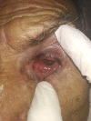
Patient with the chronic form of paracoccidioidomycosis. Shown is unilateral eye involvement, reminiscent of the moriform aspect of Aguiar Pupo’s stomatitis.
FIG 8
FIG 8
Patients with the chronic form of paracoccidioidomycosis after efficacious treatment. Computed tomography scans of the thorax show pulmonary sequelae, including axial and peripheral interstitial sequelae, with septal, centrilobular, and subpleural thickening; fibrosis; and distortion of the parenchymal architecture. Dilatation and thickening of the bronchial wall can be seen. Bullae of emphysema are located at the periphery and interior of the parenchyma.
FIG 9
FIG 9
Patients with the chronic form of paracoccidioidomycosis after efficacious treatment. Computed tomography scans of the thorax show pulmonary sequelae, including axial and peripheral interstitial sequelae, with septal, centrilobular, and subpleural thickening; fibrosis; and distortion of the parenchymal architecture. Bronchiectasis is present.
FIG 10
FIG 10
Flowchart for the laboratory diagnosis of paracoccidioidomycosis. BAL, bronchoalveolar lavage; CSF, cerebrospinal fluid; HE, hematoxylin and eosin; PAS, periodic acid-Schiff; GMS, Gomori methenamine silver; ELISA, enzyme-linked immunosorbent assay; DID, double immunodiffusion; WB, Western blotting.
FIG 11
FIG 11
Morphological aspects of Paracoccidioides yeast cells. (A) Paracoccidioides species showing multiple buds with a “helm-like” aspect (lymph node aspirate [KOH]). (B) Positive direct mycological examination of pus showing large yeast cells (10 to 30 μm) that have a thick, birefringent cell wall with single or multiple buds. (C) Paracoccidioides species (arrow) in tissue stained with hematoxylin and eosin (HE). (D) Paracoccidioides species in tissue stained with Gomori methenamine silver (GMS) showing a Mickey Mouse aspect. Bars = 20 μm.
FIG 12
FIG 12
SDS-PAGE of Paracoccidioides brasiliensis exoantigen showing the major antigen, a glycoprotein of 43,000 Da (gp43).
FIG 13
FIG 13
Immunodiffusion test. Paracoccidioides brasiliensis exoantigen (Ag) is in the center well; different serum samples from patients with paracoccidioidomycosis are in the peripherical wells (wells 1 to 6).
FIG 14
FIG 14
Scheme of the ELISA for antibody (Ab) detection. Ag, antigen; OPD, o-phenylenediamine dihydrochloride; E, enzyme; S, substrate; P, product of substrate degradation.
FIG 15
FIG 15
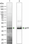
Western blotting of P. brasiliensis exoantigen. Lane 1, SDS-PAGE of P. brasiliensis exoantigen; lane 2, gp43 recognized by the serum of a patient with paracoccidioidomycosis.
FIG 16
FIG 16
Treatment regimen and follow-up of paracoccidioidomycosis patients according to data described previously by Mendes et al. (180, 323). DID, double-agar gel immunodiffusion (levels are the inverse of the dilution); ESR, erythrocyte sedimentation rate.

References

    1. Lutz A. 2004. Uma micose pseudococcídica localizada na boca e observada no Brasil. Contribuição ao conhecimento das hifoblastomicoses americanas, p 483–494. In Benchimol JL, Sá MR (ed), Adolpho Lutz obra completa, vol 1. Dermatalogia e micologia. FIOCRUZ, Rio de Janeiro, Brazil.
    1. Splendore A. 1912. Zymonematosi con localizzazione nella cavita della bocca, osservata in Brasile. Bull Soc Pathol Exot 5:313–319.
    1. de Almeida F. 1930. Estudos comparativos do granuloma coccidióidico nos Estados Unidos e no Brasil. Novo gênero para o parasito brasileiro. An Fac Med Sao Paulo 5:125–141.
    1. Moore M. 1935. A new species of the Paracoccidioides Almeida. Rev Biol Hig 6:148–162.
    1. Gezuele E. 1989. Aislamiento de Paracoccidioides sp. de heces de pinguino de la Antartida. In Proceedings of the IV International Symposium on Paracoccidioidomycosis. Instituto Venezolano de Investigaciones Cientificas, Caracas, Venezuela.

Publication types

LinkOut - more resources