Review
. 2022 Dec;20(12):2722-2732.
doi: 10.1111/jth.15873. Epub 2022 Sep 22.

Mechanisms of ADAMTS13 regulation

Affiliations
Review

Mechanisms of ADAMTS13 regulation

Veronica DeYoung et al. J Thromb Haemost. 2022 Dec.

Abstract

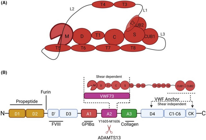
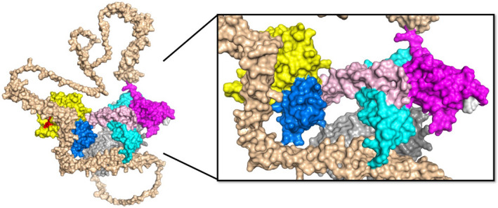
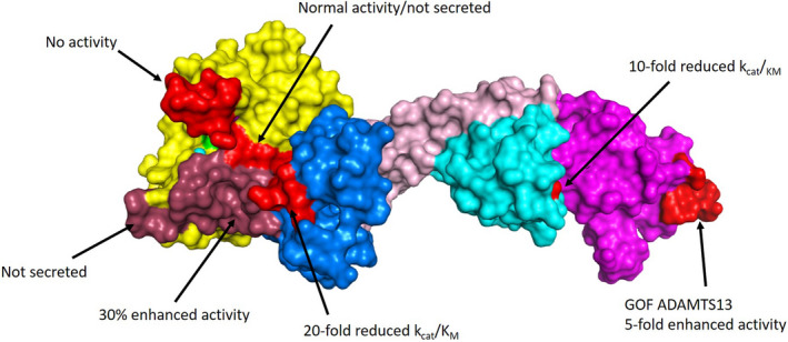
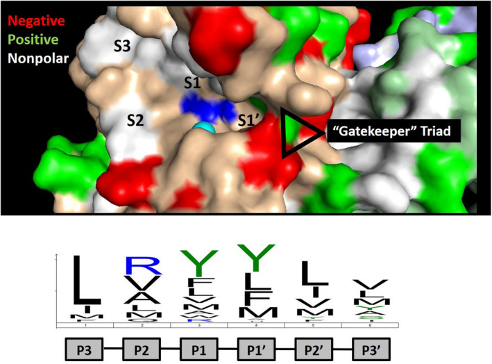
Recombinant ADAMTS13 is currently undergoing clinical trials as a treatment for hereditary thrombotic thrombocytopenic purpura, a lethal microvascular condition resulting from ADAMTS13 deficiency. Preclinical studies have also demonstrated its efficacy in treating arterial thrombosis and inflammation without causing bleeding, suggesting that recombinant ADAMTS13 may have broad applicability as an antithrombotic agent. Despite this progress, we currently do not understand the mechanisms that regulate ADAMTS13 activity in vivo. ADAMTS13 evades canonical means of protease regulation because it is secreted as an active enzyme and has a long half-life in circulation, suggesting that it is not inhibited by natural protease inhibitors. Although shear can spatially and temporally activate von Willebrand factor to capture circulating platelets, it is also required for cleavage by ADAMTS13. Therefore, spatial and temporal regulation of ADAMTS13 activity may be required to stabilize von Willebrand factor-platelet strings at sites of vascular injury. This review outlines potential mechanisms that regulate ADAMTS13 in vivo including shear-dependency, local inactivation, and biochemical and structural regulation of substrate binding. Recently published structural data of ADAMTS13 is discussed, which may help to generate novel hypotheses for future research.

Keywords: ADAMTS13; VWF; antithrombotic; protease regulation; thrombosis.


Conflict of interest statement

All of the authors declare that there are no conflicts of interest.

Figures

FIGURE 1
FIGURE 1
ADAMTS13 binding to VWF and domain organization. A, ADAMTS13 proximal domains include the metalloprotease domains (M), disintegrin‐like domain (D), type‐1 thrombospondin domain (T), cysteine‐rich domain (C), and spacer domain (S). The distal CUB domains are connected to the proximal domains through seven additional type‐1 thrombospondin domains and three linker regions, which provide ADAMTS13 with conformational flexibility. ADAMTS13 adopts a closed conformation in the absence of VWF binding in which the C‐terminal CUB domains bind to the VWF‐binding exosite on the spacer domain. The closed conformation is facilitated by inherent flexibility provided by the linker regions connecting T2‐T3, T4‐T5, and T8‐CUB1. B, ADAMTS13 binds to VWF in both a shear‐independent and a shear‐dependent mechanism. The CUB domains bind to the D4‐CK interval of VWF in a shear‐independent mechanism, which positions MDTCS near the A2 domain. This binding interaction facilitates localization of ADAMTS13 to VWF strings under flow. Upon shear‐activation of the substrate, the spacer, cysteine‐rich, and disintegrin‐like domains bind to the unfolded VWF A2 domain, stabilizing its denatured state and facilitating proteolysis of the Tyr1605‐Met1605 scissile bond by the metalloprotease domain. VWF73, comprising residues Asp1596‐Arg1668 of VWF, is a biochemical tool used to study ADAMTS13 activity in the absence of shear, and only engages the proximal MDTCS domains of ADAMTS13.
FIGURE 2
FIGURE 2
AlphaFOLD2 prediction of full length ADAMTS13 structure. The predicted three‐dimensional structure for full‐length human ADAMTS13 was obtained from AlphaFOLD2 (https://alphafold.ebi.ac.uk/). The domains are indicated as follows: metalloprotease (yellow), disintegrin‐like (blue), TSP1‐1 (rose), cysteine‐rich (cyan), spacer (magenta), TSP1‐2 to TSP1‐8 (wheat), CUB1 (light gray), CUB2 (dark gray). The RRY motif within the spacer domains (white) is primarily associated with autoantibodies in patients with immune TTP and partially occupies the suspected binding site for the CUB domains, but this intramolecular interaction is not represented in this model. Long linker regions extend outward following TSP1‐4 and TSP1‐8, providing flexibility to ADAMTS13 that may facilitate interdomain contacts that promote a closed conformation and confer global latency. This model predicts multiple long‐range interactions between distal TSP1 domains and proximal metalloprotease, disintegrin‐like, and cysteine‐rich domains that may lead to novel hypotheses for the study of ADAMTS13 regulation. (PDB: AF‐Q76LX8‐F1)
FIGURE 3
FIGURE 3
ADAMTS13 exosites. The crystal structure of MDTCS is shown, with domains indicated: metalloprotease (yellow), disintegrin‐like (blue), TSP1 (rose), cysteine‐rich (cyan), and spacer (magenta). The active site zinc (cyan) and catalytic glutamic acid (mutated to glutamine; green) are also indicated within the metalloprotease domain. The structure was obtained using a stabilizing antibody fragment to the metalloprotease domain (not shown). Compared with the AlphaFOLD2 model, this structure is lacking two loops in the cysteine‐rich domain that did not resolve (Asp453‐Tyr468 and I490‐K497). A summary of important mutagenesis work is indicated, with modified residues indicated in light red and dark red for clarity. These studies highlight important contact points for the VWF A2 domain to exosites on ADAMTS13. (PDB: 6qig)
FIGURE 4
FIGURE 4
ADAMTS13 active site specificity. The active site of ADAMTS13 is shown with the catalytic zinc (cyan) and glutamic acid (mutated to glutamine; blue) indicated. The active site specificity motif determined by substrate phage display is indicated below, which is dominated by leucine at position P3 and bulky aliphatic amino acids at positions P1 and P1′. These residues are expected to bind to subsites and pockets within the active site, which are indicated. The crystal structure provides evidence of a “gatekeeper” triad at the entrance to the S1′ subsite comprised of Arg193, Asp217, and Asp252 that is predicted to limit access to the active site of ADAMTS13. This ionic interaction is likely overcome by VWF following processive docking to distal exosites, permitting access to the catalytic motif within the active site.

References

    1. Robertson J, Lillicrap D, Von James PD. Willebrand disease. Pediatr Clin North Am. 2008;55:377‐392, viii‐ix. doi:10.1016/j.pcl.2008.01.008 - DOI - PubMed
    1. Chen X, Cheng X, Zhang S, Wu D. ADAMTS13: an emerging target in stroke therapy. Front Neurol. 2019;10:772. doi:10.3389/fneur.2019.00772 - DOI - PMC - PubMed
    1. Favaloro EJ, Henry BM, Lippi G. Increased VWF and decreased ADAMTS‐13 in COVID‐19: creating a milieu for (micro)thrombosis. Semin Thromb Hemost. 2021;47:400‐418. doi:10.1055/s-0041-1727282 - DOI - PubMed
    1. Groeneveld DJ, Poole LG, Luyendyk JP. Targeting von Willebrand factor in liver diseases: a novel therapeutic strategy? J Thromb Haemost. 2021;19:1390‐1408. doi:10.1111/jth.15312 - DOI - PMC - PubMed
    1. Katneni UK, Ibla JC, Hunt R, Schiller T, Kimchi‐Sarfaty C. von Willebrand factor/ADAMTS‐13 interactions at birth: implications for thrombosis in the neonatal period. J Thromb Haemost. 2019;17:429‐440. doi:10.1111/jth.14374 - DOI - PubMed

Publication types

MeSH terms

Grants and funding