Editorial
. 2023 Jan 4;29(1):174-182.
doi: 10.1158/1078-0432.CCR-22-1138.

Preclinical and Clinical Efficacy of Trastuzumab Deruxtecan in Breast Cancer Brain Metastases

Affiliations
Editorial

Preclinical and Clinical Efficacy of Trastuzumab Deruxtecan in Breast Cancer Brain Metastases

Sheheryar Kabraji et al. Clin Cancer Res. .

Abstract

Purpose: Brain metastases can occur in up to 50% of patients with metastatic HER2-positive breast cancer. Because patients with active brain metastases were excluded from previous pivotal clinical trials, the central nervous system (CNS) activity of the antibody-drug conjugate trastuzumab deruxtecan (T-DXd) is not well characterized.

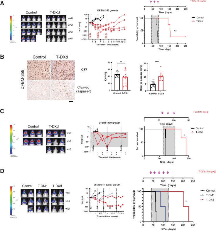
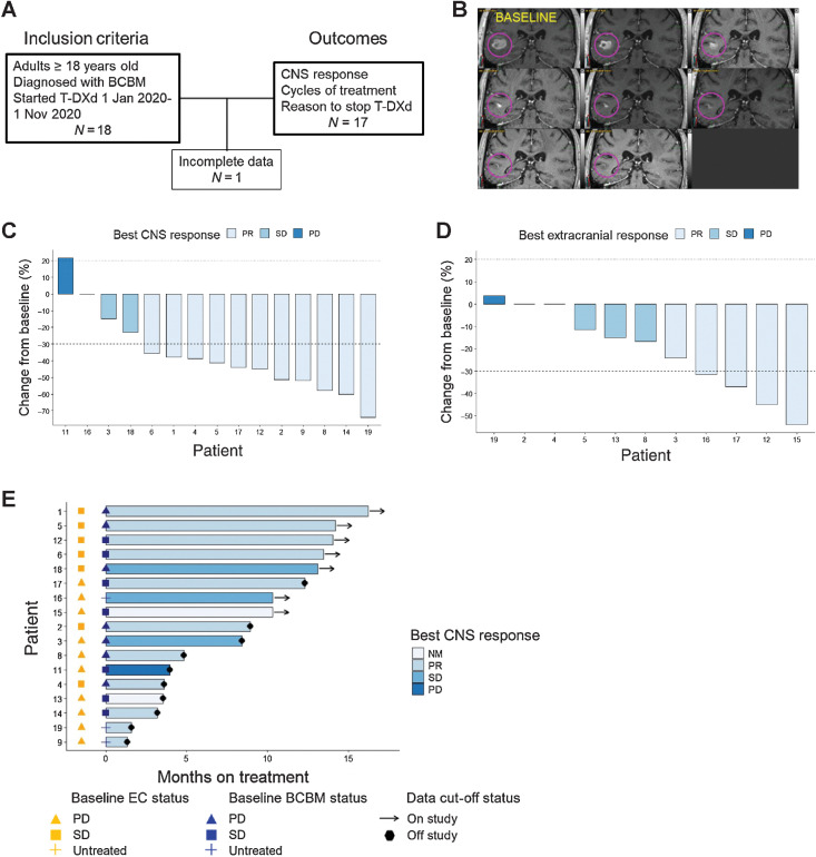
Experimental design: We studied how T-DXd affects growth and overall survival in orthotopic patient-derived xenografts (PDX) of HER2-positive and HER2-low breast cancer brain metastases (BCBM). Separately, we evaluated the effects of T-DXd in a retrospective cohort study of 17 patients with stable or active brain metastases.

Results: T-DXd inhibited tumor growth and prolonged survival in orthotopic PDX models of HER2-positive (IHC 3+) and HER2-low (IHC 2+/FISH ratio < 2) BCBMs. T-DXd reduced tumor size and prolonged survival in a T-DM1-resistant HER2-positive BCBM PDX model. In a retrospective multi-institutional cohort study of 17 patients with predominantly HER2-positive BCBMs, the CNS objective response rate (ORR) was 73% (11/15) while extracranial response rate was 45% (5/11). In the subset of patients with untreated or progressive BCBM at baseline, the CNS ORR was 70% (7/10). The median time on treatment with T-DXd was 8.9 (1.3-16.2) months, with 42% (7/17) remaining on treatment at data cutoff.

Conclusions: T-DXd demonstrates evidence of CNS activity in HER2-positive and HER2-low PDX models of BCBM and preliminary evidence of clinical efficacy in a multi-institution case series of patients with BCBM. Prospective clinical trials to further evaluate CNS activity of T-DXd in patients with active brain metastases are warranted. See related commentary by Soffietti and Pellerino, p. 8.


Figures

Figure 1. In vivo effects of T-DXd on HER2-positive BCBM PDXs. A, Representative bioluminescence images (left), spider plots tumor response (middle), and Kaplan–Meier survival analysis of mice bearing intracranial DFBM-355 cells treated with vehicle control (black line) or T-DXd (red line) as indicated (right). n = 4/group. The gray area indicates the treatment period. The treatment schedule and doses were indicated on the graph. **, P < 0.01, log-rank (Mantel–Cox) test; B, IHC analyses of Ki67 and cleaved caspase-3 of DFBM-355 tumor samples harvested from tumor-bearing mice treated 4 days after one dose of T-DXd (10 mg/kg, i.v.) (Scale bar = 50 μmol/L). Data show mean ± SD of quantification of Ki67 and cleaved caspase-3 in tumors. Mann–Whitney U test; ***, P < 0.001. C, T-DXd effect on intracranial DFBM-1409 (HER2-low) BCBM PDX. Bioluminescence images (left), spider plots of tumor response (middle), and Kaplan–Meier survival analysis of mice bearing DFBM-1409 (right) treated with vehicle control (black line) or T-DXd (red line) as indicated. n = 3/group. The treatment schedule and doses were indicated on the graph. *, P < 0.05, log-rank (Mantel–Cox) test. D, Representative bioluminescence images (left) in each group mice at indicated imaging timepoints with spider plots of tumor response (middle). Kaplan–Meier survival analysis of mice bearing T-DM1–resistant model DFBM-355TDM1R (right) intracranially, treated with vehicle control (black line), T-DM1 (blue line), or T-DXd (red line) as indicated. n = 2–3/group.
Figure 1.
In vivo effects of T-DXd on HER2-positive BCBM PDXs. A, Representative bioluminescence images (left), spider plots of tumor response (middle), and Kaplan–Meier survival analysis of mice bearing intracranial DFBM-355 cells treated with vehicle control (black line) or T-DXd (red line) as indicated (right). n = 4/group. The gray area indicates the treatment period. The treatment schedule and doses are indicated on the graph. **, P < 0.01, log-rank (Mantel–Cox) test. B, IHC analyses of Ki67 and cleaved caspase-3 of DFBM-355 tumor samples harvested from tumor-bearing mice treated 4 days after one dose of T-DXd (10 mg/kg, i.v.) (Scale bar = 50 μmol/L). Data show mean ± SD of quantification of Ki67 and cleaved caspase-3 in tumors. Mann–Whitney U test; ***, P < 0.001. C, T-DXd effect on intracranial DFBM-1409 (HER2-low) BCBM PDX. Bioluminescence images (left), spider plots of tumor response (middle), and Kaplan–Meier survival analysis of mice bearing DFBM-1409 (right) treated with vehicle control (black line) or T-DXd (red line) as indicated. n = 3/group. The treatment schedule and doses were indicated on the graph. *, P < 0.05, log-rank (Mantel–Cox) test. D, Representative bioluminescence images (left) in each group of mice at indicated imaging timepoints with spider plots of tumor response (middle). Kaplan–Meier survival analysis of mice bearing T-DM1–resistant model DFBM-355TDM1R (right) intracranially, treated with vehicle control (black line), T-DM1 (blue line), or T-DXd (red line) as indicated. n = 2–3/group.
Figure 2. Response to T-DXd in patients with BCBMs. A, Study scheme showing study entry criteria and reported outcomes. B, Contrast-enhanced brain MRIs of patient response to T-DXd. Baseline image shown at top left and images arranged in serpentine succession at restaging intervals show evidence of durable response. The lesion shown had not received prior stereotactic radiosurgery and the patient had progressed through prior treatment with T-DM1, neratinib, and tucatinib. C, Best CNS response to T-DXd. Waterfall plot of best CNS response in patients with measurable disease (n = 15). D, Best extracranial response to T-DXd. Waterfall plot of best extracranial response in patients with measurable disease (n = 11). E, Months on treatment with T-DXd. Swimmer plot showing patient (n = 17) cycles on treatment with T-DXd by baseline extracranial (EC) disease (gold shapes) and BCBM status (navy blue shapes). A total of 10 of 17 (59%) had progressive or untreated CNS disease at baseline. NM = nonmeasurable, PR = partial response, SD = stable disease, PD = progressive disease.
Figure 2.
Response to T-DXd in patients with BCBMs. A, Study scheme showing study entry criteria and reported outcomes. B, Contrast-enhanced brain MRIs of patient response to T-DXd. Baseline image shown at top left and images arranged in serpentine succession at restaging intervals show evidence of durable response. The lesion shown had not received prior stereotactic radiosurgery, and the patient had progressed through prior treatment with T-DM1, neratinib, and tucatinib. C, Best CNS response to T-DXd. Waterfall plot of best CNS response in patients with measurable disease (n = 15). D, Best extracranial response to T-DXd. Waterfall plot of best extracranial response in patients with measurable disease (n = 11). E, Months on treatment with T-DXd. Swimmer plot showing patient (n = 17) cycles on treatment with T-DXd by baseline extracranial (EC) disease (gold shapes) and BCBM status (navy blue shapes). A total of 10 of 17 (59%) had progressive or untreated CNS disease at baseline. NM, nonmeasurable; PR, partial response; SD, stable disease; PD, progressive disease.

Comment in

Comment on

References

    1. Pestalozzi BC, Holmes E, de Azambuja E, Metzger-Filho O, Hogge L, Scullion M, et al. CNS relapses in patients with HER2-positive early breast cancer who have and have not received adjuvant trastuzumab: a retrospective substudy of the HERA trial (BIG 1-01). Lancet Oncol 2013;14:244–8. - PubMed
    1. Wolff AC, Hammond MEH, Allison KH, Harvey BE, Mangu PB, Bartlett JMS, et al. Human epidermal growth factor receptor 2 testing in breast cancer: American Society of Clinical Oncology/College of American Pathologists Clinical Practice Guideline Focused Update. J Clin Oncol 2018;36:2105–22. - PubMed
    1. Nabors LB, Portnow J, Ammirati M, Brem H, Brown P, Butowski N, et al. Central nervous system cancers, version 2.2014. Featured updates to the NCCN Guidelines. J Natl Compr Canc Netw 2014;12:1517–23. - PMC - PubMed
    1. Kennecke H, Yerushalmi R, Woods R, Cheang MC, Voduc D, Speers CH, et al. Metastatic behavior of breast cancer subtypes. J Clin Oncol 2010;28:3271–7. - PubMed
    1. Drago JZ, Modi S, Chandarlapaty S. Unlocking the potential of antibody–drug conjugates for cancer therapy. Nat Rev Clin Oncol 2021;18:327–44. - PMC - PubMed