Review
. 2022 Aug;82(12):1251-1276.
doi: 10.1007/s40265-022-01766-4. Epub 2022 Sep 8.

Acute Pancreatitis: Diagnosis and Treatment

Affiliations
Review

Acute Pancreatitis: Diagnosis and Treatment

Peter Szatmary et al. Drugs. 2022 Aug.

Abstract

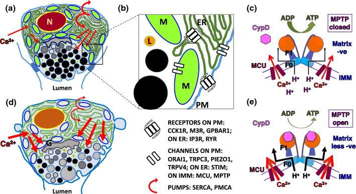
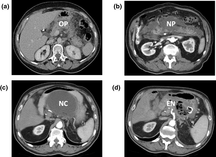
Acute pancreatitis is a common indication for hospital admission, increasing in incidence, including in children, pregnancy and the elderly. Moderately severe acute pancreatitis with fluid and/or necrotic collections causes substantial morbidity, and severe disease with persistent organ failure causes significant mortality. The diagnosis requires two of upper abdominal pain, amylase/lipase ≥ 3 ×upper limit of normal, and/or cross-sectional imaging findings. Gallstones and ethanol predominate while hypertriglyceridaemia and drugs are notable among many causes. Serum triglycerides, full blood count, renal and liver function tests, glucose, calcium, transabdominal ultrasound, and chest imaging are indicated, with abdominal cross-sectional imaging if there is diagnostic uncertainty. Subsequent imaging is undertaken to detect complications, for example, if C-reactive protein exceeds 150 mg/L, or rarer aetiologies. Pancreatic intracellular calcium overload, mitochondrial impairment, and inflammatory responses are critical in pathogenesis, targeted in current treatment trials, which are crucially important as there is no internationally licenced drug to treat acute pancreatitis and prevent complications. Initial priorities are intravenous fluid resuscitation, analgesia, and enteral nutrition, and when necessary, critical care and organ support, parenteral nutrition, antibiotics, pancreatic exocrine and endocrine replacement therapy; all may have adverse effects. Patients with local complications should be referred to specialist tertiary centres to guide further management, which may include drainage and/or necrosectomy. The impact of acute pancreatitis can be devastating, so prevention or reduction of the risk of recurrence and progression to chronic pancreatitis with an increased risk of pancreas cancer requires proactive management that should be long term for some patients.


Conflict of interest statement

TG has consulted for Albireo (ongoing). WH has received research funding from Cypralis, Farsight, and Corvidia, with all funds paid to West China Hospital of Sichuan University. RS has consulted for AbbVie CalciMedica, GlaxoSmithKline (GSK), Novartis and Takeda, and has received research funding from CalciMedica, EA Pharma, GSK, Lilly, Merck/MSD, Pfizer as well as multiple public sources in the last three years. RS is collaborating in the IMI2 TransBioLine Consortium with Janssen, Lilly, Merck/MSD, Novartis, Pfizer, Roche, and Sanofi-Aventis. All funds received have been paid to the University of Liverpool and/or Liverpool University Hospitals NHS Foundation Trust.

Figures

Fig. 1
Fig. 1
Pathophysiology of acute pancreatitis illustrating effects of calcium overload within acinar cells and the consequent failure of adenosine triphosphate (ATP) generation from adenosine diphosphate (ADP); Ca2+ calcium, CypD cyclophilin D, ER endoplasmic reticulum, G Golgi, IMM inner mitochondrial membrane, L lysosome, MCU mitochondrial calcium uniporter, MPTP mitochondrial permeability transition pore, M mitochondrion, N nucleus, PM plasma membrane, Z zymogen granule. a Cartoon of normal pancreatic acinar cell showing typical apical granular pole facing lumen, with peri-granular, peri-nuclear and sub-plasmalemmal mitochondria; red arrows indicate calcium flux into the cell, which triggers apical stimulus-secretion and stimulus-metabolism coupling; the semi-circular arrows represent sarco-endoplasmic reticulum calcium ATP-ase (SERCA) and plasmalemmal ATP-ase (PMCA), pumps that restore low resting calcium levels within the cell. b Higher power cartoon of small part of an acinar cell representing PM receptors for the two secretagogues cholecystokin (cholecystokin 1 receptor, CCK1R) and acetylcholine (muscarinic type 3 receptor, M3R), and for bile acid (G-protein bile acid receptor 1, GPBAR1). ER receptors are those for second messengers, released after CCK1R and M3R ligation, which are the inositol trisphosphate receptor (IP3R) and ryanodine receptor (RYR); after their ligation, IP3R and RYR release calcium from the ER. Also represented are the PM calcium channels Orai1, transient receptor potential cation channel 3 (TRPC3), Piezo1, and transient receptor potential vanilloid subtype 4 (TRPV4); the former two coordinate normal calcium entry through stromal interaction molecules 1 and 2 (STIM), calcium channels on the ER. Calcium uptake into the mitochondria is mediated by the MCU on the IMM for stimulus-metabolism coupling, and while the sodium calcium exchanger removes this calcium, the MPTP in low conductance mode may assist mitochondrial calcium removal. c Normal mitochondrial generation of ATP is undertaken by the F0F1ATP synthase using the H+ electrochemical gradient produced by the electron transport chain in response to calcium entry through the MCU into the electronegative mitochondrial matrix; cyclophilin D in the matrix is not bound to the F0F1ATP synthase and the MPTP is closed. d Calcium overload within acinar cells is caused by excessive intracellular release of calcium from the ER induced by pancreatitis toxins, for example bile acids, fatty acid ethyl esters or hyperstimution, provoking excessive calcium entry in response to low ER concentrations, or is caused by excessive calcium entry induced by pressure activation of Piezo1 and TRPV4. Thick red arrows indicate excessive calcium flux disrupting organellar and regulatory functions, resulting in impaired, basolateral secretion; SERCA and PMCA function are diminished, exacerbating calcium overload. e Impaired mitochondrial generation of ATP is a result of mitochondrial calcium overload, which induces binding of the MPTP regulator cyclophilin D and disruption of the F0F1ATP synthase forming the MPTP, which in high conductance mode allows particles up to 1,500 daltons into the matrix, with collapse of the H+ electrochemical gradient that normally drives ATP production. Derived from [, –75, 79, 81, 96]
Fig. 2
Fig. 2
Contrasts of severity in uncomplicated acute pancreatitis (‘mild’ in the Revised Atlanta Classification [13]) versus severe acute pancreatitis that features persistent failure of respiratory, cardiac or renal function and, typically, pancreatic necrosis; SIRS = systemic inflammatory response syndrome; MODS = multi-organ dysfunction syndrome. Upper panel shows a cartoon of acute pancreatitis with inflammatory swelling and circulating inflammation represented by neutrophils, but no local complications and no significant impact on respiratory, cardiac or renal function. Lower panel shows cartoon of acute pancreatitis with marked glandular change and necrosis in the pancreatic body, also found in some patients with moderately severe acute pancreatitis (‘moderate’ in the Revised Atlanta Classfication); in severe acute pancreatitis extensive circulatory inflammation with profuse neutrophilia extends the disease impact systemically, with exacerbation through vicious circles of damage [7], inducing dysfunction and failure of lungs, heart and/or kidneys that persists > 48 h (‘severe’ in the Revised Atlanta Classification)
Fig. 3
Fig. 3
Rationale and plan of investigation for patients with acute pancreatitis to guide treatment. a Determining aetiology is a priority in initial investigations [e.g., serum triglyceride measurement, planning early laparoscopic cholecystectomy, contrast-enhanced CT to differentiate perforation from acute pancreatitis after endoscopic retrograde cholangiopancreatography (ERCP)]. There are many tools to assess if acute pancreatitis is severe, more likely in the very young, very old or those with co-morbidities; identification of complications is central to planning management and assessing when and how to prevent recurrence. b Early investigations should ensure differentiation of acute pancreatitis from other emergencies, confirmed by two of characteristic abdominal pain, amylase or lipase ≥ 3 × upper normal limit, and characteristic 3D imaging that may be done if there is diagnostic uncertainty. The listed assessments are important in establishing aetiology and severity. c Further tests for aetiology, severity and the identification and/or assessment of progression of complications. The extent of testing is dependent on whether an aetiology is evident from b, and disease severity, as more severe disease requires more extensive investigation. Specific investigations may include those to identify or exclude further complications, for example, pulmonary or mesenteric angiography, further contrast-enhanced CT or MRI or other tests after discharge from hospital during early or long-term follow-up
Fig. 4
Fig. 4
Generalised treatment strategy for acute pancreatitis at the admitting hospital, after referral to specialist, tertiary services for complex pancreatic disease, and subsequently to prevent recurrence as well as address the aftermath of the disease. a Initial treatment at the admitting hospital, scaled to the severity of disease. A low index of suspicion is recommended for 3D abdominal imaging, otherwise complications are likely to be missed and patients readmitted in a more compromised state, with delay in referral for specialist opinion and/or transfer. Specific treatments include insulin and/or plasmapheresis for hypertriglyceridaemia and antivenom for scorpion or snake bites in endemic areas throughout the world. b There are many options required for the specialist management of complex acute pancreatitis, many of which are itemised. Necrosectomy is better delayed for some 4 weeks but may have to be brought forward if there is uncontrollable sepsis or other organ injury. Embolisation is required for pseudoaneurysms; contrarily, anticoagulation is indicated for recent thrombosis at or near the portal venous confluence. Pancreatic ductal stenting together with glyceryl trinitrate (GTN, to relax the smooth muscle of the sphincter of Oddi) and octreotide (to reduce secretion) may assist healing of pancreatic ductal rupture. c Measures to prevent recurrent acute pancreatitis are displayed as these save lives, reduce morbidity, reduce healthcare costs, and halt or slow progression of disease. Local complications of complex acute pancreatitis include recurrent pseudocyst formation, recollection of abscesses, ductal strictures, progression to chronic pancreatitis—all of which can be causes of recurrent pain and/or sepsis, and for which patients should be kept under review. Appropriately thorough investigation of acute pancreatitis may have identified benign or malignant neoplasms, for which surgical resection +/– chemotherapy may be most appropriate
Fig. 5
Fig. 5
Contrast-enhanced computerised tomography scans of patients with acute pancreatitis, with application of specialist interventions for pancreatic necrosis. a Uncomplicated acute oedematous pancreatitis (OP) is seen with perfusion of the pancreatic parenchyma, surrounded by inflammation. b Acute necrotising pancreatitis (NP) with loss of most of the parenchyma, excepting a small portion in the head and separate small portion of the tail of the gland. This scan was taken 2 weeks into the attack and the necrosis with associated inflammation is diffuse and poorly localised. c More than 4 weeks after the onset of acute pancreatitis in the same patient as in b, the necrotic collection (NC) has become localised and walled off close to the posterior wall of the stomach, making it suitable for endoscopic drainage. (d) In the same patient as in b, infection has supervened, identified by the presence of radiolucent black gas bubbles within the collection, which was treated by endoscopic necrosectomy (EN). The endoscopically inserted self-expanding metal stent between the stomach and necrotic cavity is visible as a radiopaque white ring; flushing the cavity endoscopically every 7–10 days was necessary to empty and allow collapse of the cavity, following which the stent was removed
Fig. 6
Fig. 6
Prevalence of exocrine pancreatic insufficiency (EPI) during inpatient stay and over 5 or more years after mild or severe acute pancreatitis. Data are derived from a systematic review of multiple studies during follow-up after an attack of acute pancreatitis [214]. Separate data are given on mild and severe acute pancreatitis as two categories of Original Atlanta Classification [104], and combining as severe acute pancreatitis data on the moderate and severe categories in studies that used the Revised Atlanta Classification [13]. Studies were either prospective observational or interventional, the latter assessing the effectiveness of pancreatic enzyme replacement therapy. The number of patients with either mild or severe acute pancreatitis for whom there were data are given underneath the histogram

References

    1. Petrov MS, Yadav D. Global epidemiology and holistic prevention of pancreatitis. Nat Rev Gastroenterol Hepatol. 2019;16(3):175–184. doi: 10.1038/s41575-018-0087-5. - DOI - PMC - PubMed
    1. Iannuzzi JP, King JA, Leong JH, Quan J, Windsor JW, Tanyingoh D, et al. Global incidence of acute pancreatitis is increasing over time: a systematic review and meta-analysis. Gastroenterology. 2022;162(1):122–134. doi: 10.1053/j.gastro.2021.09.043. - DOI - PubMed
    1. Andersson B, Appelgren B, Sjodin V, Ansari D, Nilsson J, Persson U, et al. Acute pancreatitis–costs for healthcare and loss of production. Scand J Gastroenterol. 2013;48(12):1459–1465. doi: 10.3109/00365521.2013.843201. - DOI - PubMed
    1. Della Corte C, Faraci S, Majo F, Lucidi V, Fishman DS, Nobili V. Pancreatic disorders in children: new clues on the horizon. Dig Liver Dis. 2018;50(9):886–893. doi: 10.1016/j.dld.2018.06.016. - DOI - PubMed
    1. Roberts SE, Morrison-Rees S, John A, Williams JG, Brown TH, Samuel DG. The incidence and aetiology of acute pancreatitis across Europe. Pancreatology. 2017;17(2):155–165. doi: 10.1016/j.pan.2017.01.005. - DOI - PubMed