Review
. 2022 Sep;44 Suppl 1(Suppl 1):101-113.
doi: 10.1111/ijlh.13954.

Diagnosis and treatment of thrombotic microangiopathy

Affiliations
Review

Diagnosis and treatment of thrombotic microangiopathy

Gemma L Thompson et al. Int J Lab Hematol. 2022 Sep.

Abstract

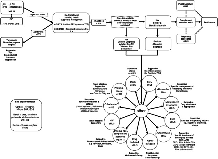
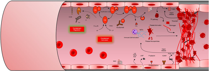
Thrombotic microangiopathy (TMA) is characterized by thrombocytopenia, microangiopathic haemolytic anaemia and end organ damage. TMAs have varying underlying pathophysiology and can therefore present with an array of clinical presentations. Renal involvement is common as the kidney is particularly susceptible to the endothelial damage and microvascular occlusion. TMAs require rapid assessment, diagnosis, and commencement of appropriate treatment due to the high morbidity and mortality associated with them. Ground-breaking research into the pathogenesis of TMAs over the past 20 years has driven the successful development of targeted therapeutics revolutionizing patient outcomes. This review outlines the clinical presentations, pathogenesis, diagnostic tests and treatments for TMAs.

Keywords: STEC-HUS; haemolytic uraemic syndrome; thrombotic microangiopathy; thrombotic thrombocytopenic purpura.


Conflict of interest statement

David Kavanagh has received consultancy income from Gyroscope Therapeutics, Silence Therapeutics, Alexion Pharmaceuticals, Novartis, Apellis, and Sarepta.

Figures

FIGURE 1
FIGURE 1
Diagnostic and treatment algorithm for thrombotic microangiopathy (TMA). In a patient presenting with MAHA and thrombocytopenia suggestive of a TMA a thorough diagnostic evaluation will usually reveal the underlying aetiology and guide treatment. As many of the investigations will not be available at initial presentation the initial focus should be on the consideration of TTP given the high mortality if untreated. In adults PE should be instituted on the presumption that it is TTP unless other evidence is available that strongly suggests an alternative aetiology. In children, in whom TTP is rarer, first‐line treatment with eculizumab should be considered if complement‐mediated aHUS is suspected and should not be delayed whilst ADAMTS13 activity is determined. In the absence of a defined aetiology, complement‐mediated aHUS is presumed and treatment with eculizumab is recommended pending the complete evaluation. +ve, positive; Ab, antibody; ACA, anti‐centromere antibody; ACEI, angiotensin converting enzyme inhibitor; ADAMTS13, a disintegrin and metalloproteinase with a thrombospondin type 1 motif, member 13; Ag, antigen; aHUS, atypical haemolytic uraemic syndrome; AKI, acute kidney injury; ANA antinuclear antibody; Anti‐ds DNA, anti‐double stranded DNA; anti‐Scl‐70, anti‐topoisomerase I antibody; BMT; bone marrow transplant; CAPS, catastrophic antiphospholipid syndrome; CMV, cytomegalovirus; DIC, disseminated intravascular coagulation; FACS, flow cytometry; FH, factor H; FI, factor I; Hb, haemoglobin; HIV, human immunodeficiency virus; HUS, haemolytic uraemic syndrome; LDH, lactate dehydrogenase; MAHA, microangiopathic haemolytic anaemia; MLPA, multiplex ligation‐dependent probe amplification; MMA, methylmalonic acid; Nt‐proBNP; brain natriuretic peptide PCR, polymerase chain reaction; PE, plasma exchange; SLE, systemic lupus erythematosus; SRC, scleroderma renal crisis; STEC, Shiga toxin producing Escherichia coli; Stx, Shiga toxin; T‐Ag, Thomsen‐Friedenreich antigen; TMA, thrombotic microangiopathy; TTP, thrombotic thrombocytopenic purpura
FIGURE 2
FIGURE 2
Pathogenesis of complement‐mediated atypical haemolytic uraemic syndrome (aHUS). Complement is activated by the classical (CP) lectin (LP) and alternative (AP) pathways. The AP is a positive amplification loop. C3b interacts with factor B, which is then cleaved by factor D to form the C3 convertase C3bBb. Unchecked this leads to activation of the terminal complement pathway with generation of the membrane attack complex (MAC, C5b‐9) and the anaphylatoxin C5a. Complement regulators including factor H (FH), factor I (FI) and CD46 protect the glomerular endothelium from collateral damage from the AP. In complement‐mediated aHUS, activating mutations in C3 and CFB, loss‐of‐function mutations in CFH, CFI and CD46, and autoantibodies to FH, result in over‐activation of the AP. C5a induces tissue factor (TF) expression endothelial cells and increases tissue plasminogen activator inhibitor −1 in mast cells and basophils. C5a also promotes endothelial cell retraction, exposing underlying basement membrane. Sublytic membrane attack complex (MAC) also induces TF expression and increases adhesion molecule expression on the endothelium (VWF). Downstream effects of sublytic MAC on endothelium additionally include secretion of multimers of endothelial VWF and release of heparan sulphate proteoglycans from endothelial glycocalyx. Complement activation on platelets increases activation markers (CD40L; CD62P) with resulting in activation of the platelets with subsequent release of pro‐thrombogenic TF‐expressing platelet micro‐particles. Haem release from intravascular haemolysis stimulates TF upregulation and Neutrophil extra cellular traps (NETs) formation. NETs released by damaged or activated neutrophils and red cell degradation products have been shown to contribute to thrombus formation. Together these mechanisms lead to immune cell and platelet activation and endothelial cell damage and swelling, with consequent thrombus formation, platelet consumption, vascular occlusion mechanical haemolysis. Eculizumab & ravulizumab binds to C5 to prevent activation of the terminal pathway inhibiting the generation of the effector molecules that cause TMA. Modified from the National Renal Complement Therapeutics Centre 2020/2021 annual report (http://www.atypicalhus.co.uk/)

Similar articles

Cited by

References

    1. Brocklebank V, Wood KM, Kavanagh D. Thrombotic microangiopathy and the kidney. Clin J Am Soc Nephrol. 2018;13(2):300‐317. - PMC - PubMed
    1. Moake JL. Thrombotic microangiopathies. N Engl J Med. 2002;347(8):589‐600. - PubMed
    1. Scully M, Yarranton H, Liesner R, et al. Regional UK TTP registry: correlation with laboratory ADAMTS 13 analysis and clinical features. Br J Haematol. 2008;142(5):819‐826. - PubMed
    1. Alwan F, Vendramin C, Vanhoorelbeke K, et al. Presenting ADAMTS13 antibody and antigen levels predict prognosis in immune‐mediated thrombotic thrombocytopenic purpura. Blood. 2017;130(4):466‐471. - PubMed
    1. Joly BS, Coppo P, Veyradier A. Thrombotic thrombocytopenic purpura. Blood. 2017;129(21):2836‐2846. - PubMed

MeSH terms