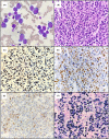
Plasmablastic lymphoma: An update
- PMID: 36074710
- PMCID: PMC9545967
- DOI: 10.1111/ijlh.13863
Plasmablastic lymphoma: An update
Erratum in
-
Plasmablastic lymphoma: An update.Int J Lab Hematol. 2022 Dec;44(6):1121. doi: 10.1111/ijlh.13981. Int J Lab Hematol. 2022. PMID: 36380470 Free PMC article. No abstract available.
Abstract
Plasmablastic lymphoma (PBL) is a highly aggressive B cell non-Hodgkin lymphoma frequently associated with immunosuppression, particularly human immunodeficiency virus (HIV) infection. Although PBL is rare globally, South Africa has a high burden of HIV infection leading to a higher incidence of PBL in the region. Laboratory features in PBL may overlap with plasmablastic myeloma and other large B cell lymphomas with plasmablastic or immunoblastic morphology leading to diagnostic dilemmas. There are, however, pertinent distinguishing laboratory features in PBL such as a plasma cell immunophenotype with MYC overexpression, expression of Epstein-Barr virus-encoded small RNAs and lack of anaplastic lymphoma kinase (ALK) expression. This review aims to provide a summary of current knowledge in PBL, focusing on the epidemiology, pathophysiology, laboratory diagnosis and clinical management.
Keywords: EBV; HIV; MYC; aggressive lymphoma; plasmablastic lymphoma; plasmablastic myeloma.
© 2022 The Authors. International Journal of Laboratory Hematology published by John Wiley & Sons Ltd.
Conflict of interest statement
The authors declare no conflict of interest.
Figures


References
-
- Castillo JJ, Bibas M, Miranda RN. The biology and treatment of plasmablastic lymphoma. Blood. 2015;125(15):2323‐2330. - PubMed
-
- Campo ESH, Harris NL. Plasmablastic lymphoma. WHO Classification of Tumour of Haematopoietic and Lymphoid Tissues. 4th ed. IARC; 2017:321‐322.
-
- Delecluse HJ, Anagnostopoulos I, Dallenbach F, et al. Plasmablastic lymphomas of the oral cavity: a new entity associated with the human immunodeficiency virus infection. Blood. 1997;89(4):1413‐1420. - PubMed
-
- Pather S, Mashele T, Willem P, et al. MYC status in HIV‐associated plasmablastic lymphoma: dual‐colour CISH, FISH and Immunohistochemistry. Histopathology. 2021;79(1):86‐95. - PubMed
Publication types
MeSH terms
LinkOut - more resources
Full Text Sources
Medical

