. 2022 Sep 22;65(18):12055-12067.
doi: 10.1021/acs.jmedchem.2c00772. Epub 2022 Sep 8.

Balancing Affinity, Selectivity, and Cytotoxicity of Hydrazone-Based G-Quadruplex Ligands for Activation of Interferon β Genes in Cancer Cells

Affiliations

Balancing Affinity, Selectivity, and Cytotoxicity of Hydrazone-Based G-Quadruplex Ligands for Activation of Interferon β Genes in Cancer Cells

Simona Marzano et al. J Med Chem. .

Abstract

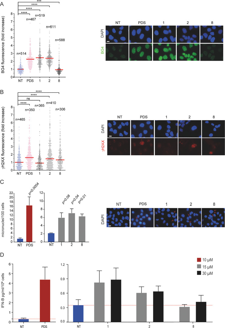
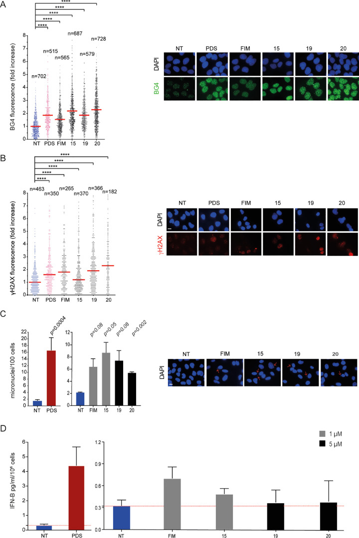
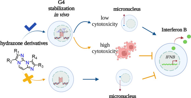
G-quadruplex (G4) ligands are investigated to discover new anticancer drugs with increased cell-killing potency. These ligands can induce genome instability and activate innate immune genes at non-cytotoxic doses, opening the discovery of cytostatic immune-stimulating ligands. However, the interplay of G4 affinity/selectivity with cytotoxicity and immune gene activation is not well-understood. We investigated a series of closely related hydrazone derivatives to define the molecular bases of immune-stimulation activity. Although they are closely related to each other, such derivatives differ in G4 affinity, cytotoxicity, genome instability, and immune gene activation. Our findings show that G4 affinity of ligands is a critical feature for immune gene activation, whereas a high cytotoxic potency interferes with it. The balance of G4 stabilization versus cytotoxicity can determine the level of immune gene activation in cancer cells. Thus, we propose a new rationale based on low cell-killing potency and high immune stimulation to discover effective anticancer G4 ligands.


Conflict of interest statement

The authors declare no competing financial interest.

Figures

Chart 1
Chart 1. Chemical Structures of the Lead Compounds FG and FIM (1 and 3 by Amato et al., respectively)
Chart 2
Chart 2. Chemical Structures of New FG and FIM Derivatives Synthesized in This Study
Figure 1
Figure 1
G4 stabilization, DNA damage, and IFN-B stimulation induced by FG derivatives. (A) Quantification of fluorescence signals of BG4 foci in U2OS cells being treated for 10 min with PDS or FG derivatives (compounds 1, 2, and 8) at 10 μM concentration. The graph shows the fold increase reported as the mean ± SEM of three biological replicates, and the IF representative images are reported (left). (B) Quantification of fluorescence signals of γH2AX in U2OS cells being treated with PDS (10 μM) and FG derivatives at IC50 concentrations (46, 100, and 20 μM for 1, 2, and 8, respectively) for 24 h of treatment. The graph shows the fold increase reported as the median ± SEM of two biological replicates and the IF representative images are reported (left). (C) Micronuclei quantification by DAPI staining in MNMCA1 cells treated (15 μM) after 24 h of treatment followed by 24 h of drug-free recovery. PDS (10 μM)-treated cells are also shown. The graph shows the mean ± SEM of two biological replicates, and the IF representative images are reported (left). Above the bar chart, the p-value are reported. The scale bar is 10 μm. (D) Quantification of IFN-B produced by MNMCA1 cells treated with FG derivatives at different concentrations (15 and 30 μM). PDS (10 μM)-treated cells are also shown. The IFN-B detection was performed with ELISA assay after 24 h of compounds treatment followed by 48 h of recovery. The bar chart reports the mean ± SEM of three biological replicates. Significance in all the graph was calculated by Mann–Witney test (*p < 0.05, **p > 0.01, ***p > 0.001, and ****p < 0.0001).
Figure 2
Figure 2
G4 stabilization, DNA damage, and IFN-B stimulation induced by FIM derivatives. (A) Quantification of fluorescence signals of BG4 foci in U2OS cells treated for 10 min with PDS or FIM derivatives at 10 μM concentration. Graphs show the fold increase reported as the mean ± SEM of three biological replicates. The images are representative of IF assays performed at reported concentrations (left). (B) Quantification of fluorescence signals of γH2AX in U2OS cells treated with PDS (10 μM) and FIM derivatives at IC50 concentrations (4, 2.5, 24, and 14 μM for FIM, 15, 19, and 20, respectively). The graph shows the fold increase reported as the median ± SEM of two biological replicates, and the IF representative images are reported (left). (C) Micronuclei quantification by DAPI staining in MNMCA1 cells treated with 1 μM of compounds FIM and 15 and 5 μM for the analogues 19 and 20. PDS (10 μM)-treated cells are also shown. Left, the graph shows the mean ± SEM of two biological replicates; right, representative cell images. The scale bar is 10 μm. Above the bar chart, the p-value are reported. (D) Quantification of IFN-B stimulated at the reported concentration has been detected after 24 h of treatment followed by 48 h of recovery. PDS (10 μM)-treated cells are also shown. The IFN-B protein levels were detected with ELISA assay. The bar chart reports the mean ± SEM of two biological replicates. Significance in all the graphs was calculated by Mann–Whitney test (*p < 0.05, **p > 0.01, ***p > 0.001, and ****p < 0.0001).
Figure 3
Figure 3
Schematic representation of cellular effects of hydrazone derivatives.

References

    1. Balasubramanian S.; Hurley L. H.; Neidle S. Targeting G-Quadruplexes in Gene Promoters: A Novel Anticancer Strategy?. Nat. Rev. Drug Discovery 2011, 10, 261–275. 10.1038/nrd3428. - DOI - PMC - PubMed
    1. Neidle S. Quadruplex Nucleic Acids as Novel Therapeutic Targets. J. Med. Chem. 2016, 59, 5987–6011. 10.1021/acs.jmedchem.5b01835. - DOI - PubMed
    1. Carvalho J.; Mergny J.-L.; Salgado G. F.; Queiroz J. A.; Cruz C. G-Quadruplex, Friend or Foe: The Role of the G-Quartet in Anticancer Strategies. Trends Mol. Med. 2020, 26, 848–861. 10.1016/j.molmed.2020.05.002. - DOI - PubMed
    1. Miglietta G.; Russo M.; Capranico G. G-Quadruplex–R-Loop Interactions and the Mechanism of Anticancer G-Quadruplex Binders. Nucleic Acids Res. 2020, 48, 11942–11957. 10.1093/nar/gkaa944. - DOI - PMC - PubMed
    1. Miglietta G.; Marinello J.; Russo M.; Capranico G.. Ligands Stimulating Antitumour Immunity as the next G-Quadruplex Challenge. Mol. Cancer 2022, in press. - PMC - PubMed

Publication types