. 2022 Sep 8;18(9):e1010329.
doi: 10.1371/journal.ppat.1010329. eCollection 2022 Sep.

Arbovirus-vector protein interactomics identifies Loquacious as a co-factor for dengue virus replication in Aedes mosquitoes

Affiliations

Arbovirus-vector protein interactomics identifies Loquacious as a co-factor for dengue virus replication in Aedes mosquitoes

Benoit Besson et al. PLoS Pathog. .

Abstract

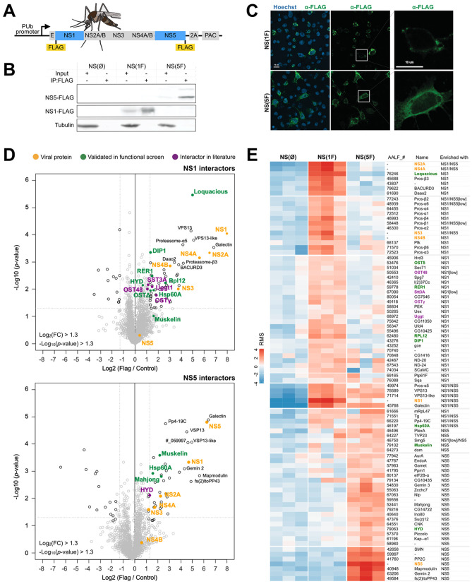
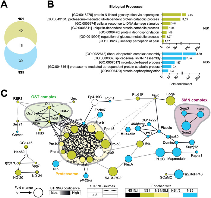
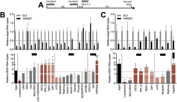
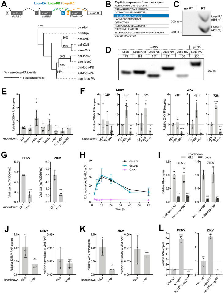
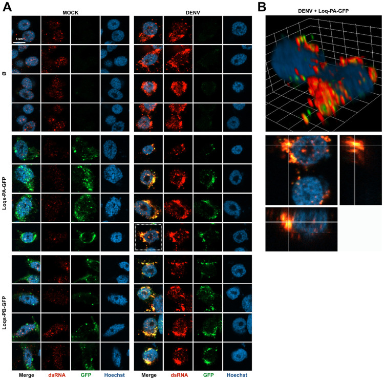
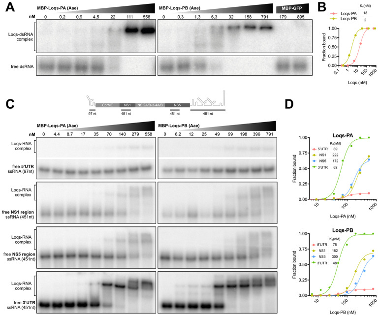
Efficient virus replication in Aedes vector mosquitoes is essential for the transmission of arboviral diseases such as dengue virus (DENV) in human populations. Like in vertebrates, virus-host protein-protein interactions are essential for viral replication and immune evasion in the mosquito vector. Here, 79 mosquito host proteins interacting with DENV non-structural proteins NS1 and NS5 were identified by label-free mass spectrometry, followed by a functional screening. We confirmed interactions with host factors previously observed in mammals, such as the oligosaccharyltransferase complex, and we identified protein-protein interactions that seem to be specific for mosquitoes. Among the interactors, the double-stranded RNA (dsRNA) binding protein Loquacious (Loqs), an RNA interference (RNAi) cofactor, was found to be essential for efficient replication of DENV and Zika virus (ZIKV) in mosquito cells. Loqs did not affect viral RNA stability or translation of a DENV replicon and its proviral activity was independent of its RNAi regulatory activity. Interestingly, Loqs colocalized with DENV dsRNA replication intermediates in infected cells and directly interacted with high affinity with DENV RNA in the 3' untranslated region in vitro (KD = 48-62 nM). Our study provides an interactome for DENV NS1 and NS5 and identifies Loqs as a key proviral host factor in mosquitoes. We propose that DENV hijacks a factor of the RNAi mechanism for replication of its own RNA.


Conflict of interest statement

The authors have declared that no competing interests exist.

Figures

Fig 1
Fig 1. Interactome of NS1 and NS5 in Aedes mosquito cells.
A. Schematic representation of constructs used to express DENV non-structural proteins in mosquito cells. Constructs were generated with a 3xFLAG tag at the N-terminus of NS1 (NS(1F)) or the C-terminus of NS5 (NS(5F)). A construct without a tag (NS(ØF)) was included as a control. 2A, self-cleaving peptide from foot-and-mouth disease virus; PAC, Puromycin N-acetyltransferase. Mosquito image by Mariana Ruiz Villarreal, licensed under CC0. Fig: https://bioicons.com/?query=mosquito. B. Western blot of input and FLAG immunoprecipitation samples of C6/36 cells expressing NS(ØF), NS(1F) and NS(5F), stained with FLAG and α-tubulin (Tub) antibodies. See uncropped gel in S5 Fig. C. Confocal microscopy image of FLAG-tagged NS1 and NS5 in C6/36 cells at 24 h after transgene transfection. Cells were stained with anti-FLAG M2 antibody (green) and Hoechst to stain nuclei (blue). D. Volcano plot of proteins interacting with 3xFLAG-tagged NS1 (top) or NS5 (bottom) in C6/36 cell lysates as determined by label-free quantitative mass spectrometry. The X-axis shows the log2 fold change (FC) over untagged NS(ØF) (control), and the Y-axis shows -log10(p-value). Proteins in the top right are identified as significantly enriched proteins. Colored dots indicate proteins of interest. Each condition was performed in triplicate. Proteins are named according to the fly ortholog, as defined in S1 Table. E. Heatmap of the relative enrichment (red) and depletion (blue) of proteins in each sample, based on row-mean subtraction and K-means clustering. Statistically significant enrichment in the volcano plot analysis is indicated, p < 0.05. Ost48 and Stt3A were included, although not significantly enriched. ‘Low’ indicates an enrichment between 2 and 2.5-fold, below the threshold of the volcano plot.
Fig 2
Fig 2. Characterization of DENV NS1 and NS5 interactomes.
A. Venn diagram of 85 interactors of NS1 and/or NS5 identified by mass spectrometry (Fig 1E). B. GO term analyses of interactors of NS1 (top panel) and NS5 (lower panel). Enrichment of biological processes was based on D. melanogaster ortholog annotation. Numbers indicate -log10 p values. See complete list of GO terms in S2 Table. C. Functional STRING networks based on D. melanogaster ortholog annotation, using the following four sources: text mining, experiments, databases or co-expression. Hits were classified and colored according to their enrichment in ≥ 2 out of 3 NS(1F) or NS(5F) samples in the heatmap of Fig 1E. Node sizes represent the fold enrichment in NS(1F) or NS(5F) immunoprecipitation, keeping the highest value if the interactor was present in both. Edges are representative of the number of sources (solid or dashed) and the confidence (color) supporting the interaction as defined by STRING. Font indicates hits confirmed (bold) or not (italic) as modulators of DENV in the functional screening (Fig 3).
Fig 3
Fig 3. Functional screen identifies Loqs as a DENV proviral host factor in Aedes mosquitoes.
A. Schematic outline of the functional RNAi screen. B-C. Relative quantification of target gene expression (top panel) and DENV RNA levels (lower panel) in U4.4 cells upon silencing of the indicated genes. Selected genes from the initial screen (B) were tested in an independent validation screen (C) using dsRNA targeting a different region of the gene. Expression was quantified by RT-qPCR, normalized to the house-keeping gene ribosomal protein L5, and expressed relative to expression in cells treated with dsRNA targeting firefly luciferase (GL3). Ago2 was used as a positive control. Data represent means and standard deviation of three replicates. Color coding represents classification of hits based on gene knockdown efficiency, phenotype, and consistency between screens. Light grey, inefficient knockdown (< 0.5-fold); dark grey, no phenotype despite efficient knockdown (> 0.5-fold); dark red, strong hit with efficient knockdown and DENV RNA levels < 0.66-fold or >1.5-fold in both dsRNA sets; light red, weak hits for which one of the criteria was not met. One-way ANOVA were used to determine statistically significant differences with the GL3 control, with: * p < 0.05; ** p < 0.01; *** p < 0.001; **** p < 0.0001.
Fig 4
Fig 4. Loquacious is an essential co-factor for flavivirus replication.
A. Structure of Loquacious splice variants in Ae. aegypti and maximum likelihood phylogenic tree based on the protein sequence of Loqs-PA and its orthologs and paralogs. ce, Caenorhabditis elegans; dm, Drosophila melanogaster; h, Homo sapiens; aae, Ae. aegypti; aal, Ae. albopictus. Percentages indicates the identity between protein sequences compared to Ae. aegypti Loqs-PA. Branch lengths are proportional to the number of substitutions per site. Loqs transcript annotation is according to the reference genome AaegL5, which differs from the annotation used in [24]. dsRBD, dsRNA-binding domain. B. Loqs peptides identified by mass spectrometry in NS1 immunoprecipitations. The peptide unique to Loqs-PB is indicated in blue; other peptides are shared between Loqs isoforms. C. Loqs-RA and Loqs-RB specific amplicons from PCR using primers spanning exon 5 on cDNA from Aag2 cells. D. PCR amplification of Loqs splice variants with various set of primers on cDNA or genomic DNA isolated from Aag2 cells. Numbers indicate expected sizes. E. Relative quantification of DENV RNA at 72 h infection of Aag2 cells in which siRNA and miRNA pathway genes were silenced. F. Relative quantification of DENV RNA or ZIKV RNA at the indicated time after infection in Ago2 or Loqs depleted Aag2 cells. G. Titration of DENV (left) and ZIKV (right) from the supernatant of Loqs depleted Aag2 cells after 48h of infection at an MOI of 0.01. H. Renilla luciferase activity in Aag2 cells transfected with DENV2 replicon RNA and dsRNA targeting Loqs (dsLoqs) or firefly luciferase (dsGL3) as a control. Luciferase activity was assessed at the indicated time points and normalized to dsGL3-treated cells at 3 h after transfection. Cells treated with cycloheximide (CHX) were included as control. I. Relative quantification of DENV (left) or ZIKV (right) total viral RNA or antisense viral RNA after infection of Loqs depleted Aag2 cells of infection at an MOI of 0.01. J-K. Relative quantification of DENV (H) and ZIKV (I) RNA (left) and vsiRNAs (right) at 48 h after infection of Loqs depleted or control (GL3 dsRNA treated) Aag2 cells. vsiRNAs were normalized to the cellular piRNA tapiR1 and viral RNA to correct for library size and viral RNA levels. L. Relative quantification of DENV (left) and ZIKV (right) RNA levels at 48 h after infection in wild type (wt), U4.4-Ago2KO, or U4.4-Ago2KO; LoqsKO cells at an MOI of 0.01. ZIKV was undetectable (n.d.) in U4.4-Ago2KO; LoqsKO cells and relative viral RNA copies were calculated using imputed Ct values of 40. Viral RNA levels were quantified by RT-qPCR, normalized to the housekeeping gene lysosomal aspartic protease (E-F, I-L) and expressed relative to cells treated with dsRNA targeting luciferase (dsGL3) as negative control. See relative knockdown efficiencies in S2 Fig. Bars represent mean and standard deviation from at least three biological replicates. Non-parametric one-way ANOVA (E) and two-tailed student’s t-tests (F, I-L) were used to determine statistically significant differences with the control: * p < 0.05; ** p < 0.01; *** p < 0.001; **** p < 0.0001.
Fig 5
Fig 5. Loquacious colocalizes with DENV replication organelles.
A. Confocal microscopy images of mock or DENV infected Aag2 cells. Cells were transfected at 72 h post infection with plasmids encoding the indicated transgenes or mock transfected (Ø) and processed for microscopy 24 h later. Cells were stained with anti-dsRNA J2 antibody (red) and Hoechst to stain nuclei (blue). Scale bar corresponds to 5 μm and is the same for all panels. In mock infected cells, a nuclear background signal is detectable that is distinct from the cytoplasmic viral dsRNA signal in DENV infected cells. B. Three-dimensional visualizations of two DENV infected Aag2 cells transfected with Loqs-PA-GFP indicated with a dashed square in panel A. The dsRNA and GFP signals were optimized for visualization in the three-dimensional projection. The grid is scaled with 2 μm xyz units.
Fig 6
Fig 6. Loquacious interacts directly with DENV 3’UTR.
A. Electrophoretic mobility assay of Ae. aegypti Loqs-PA (left panel), Loqs-PB (right panel) and GFP control with a 117 bp 32P labeled dsRNA corresponding to the firefly luciferase sequence. dsRNA was incubated with 5-fold dilutions of recombinant maltose-binding protein (MBP)-Loqs or MBP-GFP and complexes were resolved on native polyacrylamide gels. B. Quantification of (A) with dissociation constants for the indicated protein. C. Electrophoretic mobility assay of Ae. aegypti Loqs-PA (left panels) and Loqs-PB (right panels) with the indicated ssRNAs corresponding to DENV2 5’ UTR, NS1, NS5 and 3’ UTR sequences. Top panel indicates a schematic representation of the position of the probes on the DENV genome (not to scale). ssRNA was incubated with 2-fold dilutions of recombinant MBP-Loqs and complexes were resolved on native polyacrylamide gels. The images of the free ssRNA and the Loqs-RNA complex for the 5’ UTR are cropped from the same gel. Full images are provided in S5 Fig. D. Quantification of (C) with dissociation constants for the indicated RNAs.

References

    1. Bhatt S, Gething PW, Brady OJ, Messina JP, Farlow AW, Moyes CL, et al.. The global distribution and burden of dengue. Nature. 2013;496: 504–507. doi: 10.1038/nature12060 - DOI - PMC - PubMed
    1. Fischer D, Thomas SM, Neteler M, Tjaden NB, Beierkuhnlein C. Climatic suitability of Aedes albopictus in Europe referring to climate change projections: comparison of mechanistic and correlative niche modelling approaches. Eurosurveillance. 2014;19: 20696. doi: 10.2807/1560-7917.es2014.19.6.20696 - DOI - PubMed
    1. Aranda C, Martínez MJ, Montalvo T, Eritja R, Navero-Castillejos J, Herreros E, et al.. Arbovirus surveillance: first dengue virus detection in local aedes albopictus mosquitoes in Europe, Catalonia, Spain, 2015. Eurosurveillance. 2018;23: e08347. doi: 10.2807/1560-7917.ES.2018.23.47.1700837 - DOI - PMC - PubMed
    1. Kraemer MUG, Reiner RC, Brady OJ, Messina JP, Gilbert M, Pigott DM, et al.. Past and future spread of the arbovirus vectors Aedes aegypti and Aedes albopictus. Nat Microbiol. 2019;4: 854–863. doi: 10.1038/s41564-019-0376-y - DOI - PMC - PubMed
    1. Kraemer MUG, Sinka ME, Duda KA, Mylne AQN, Shearer FM, Barker CM, et al.. The global distribution of the arbovirus vectors Aedes aegypti and Ae. albopictus. Elife. 2015;4: e08347. doi: 10.7554/eLife.08347 - DOI - PMC - PubMed

Publication types

Substances