Review
. 2022 Oct 11;22(20):3801-3816.
doi: 10.1039/d2lc00262k.

Integrated biosensors for monitoring microphysiological systems

Affiliations
Review

Integrated biosensors for monitoring microphysiological systems

Lei Mou et al. Lab Chip. .

Erratum in

Abstract

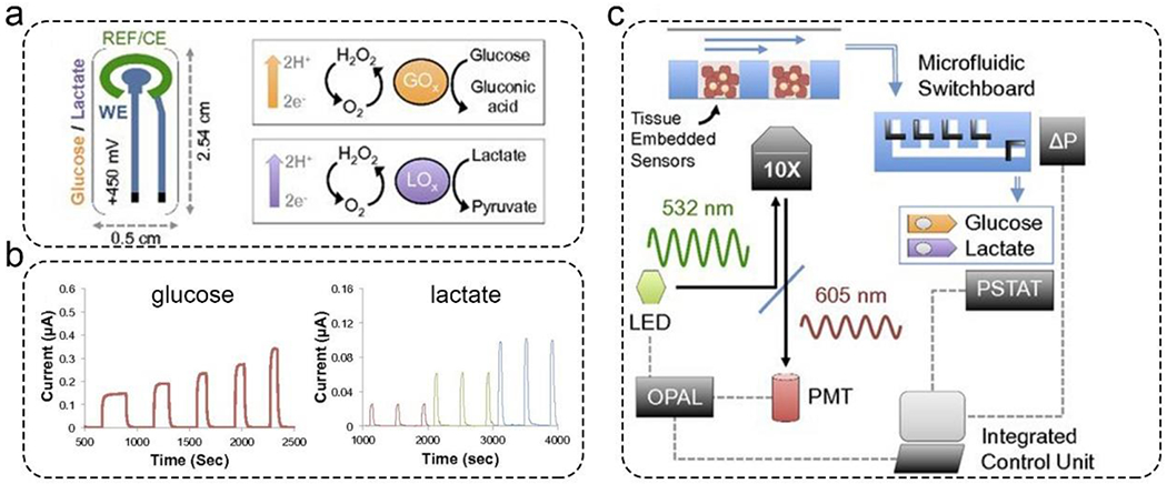
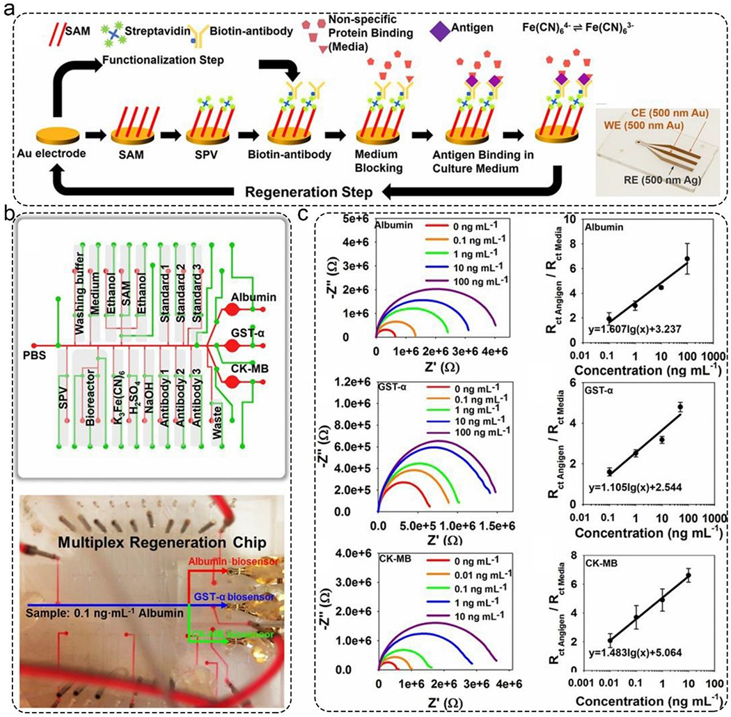
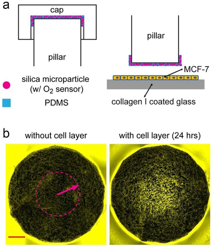
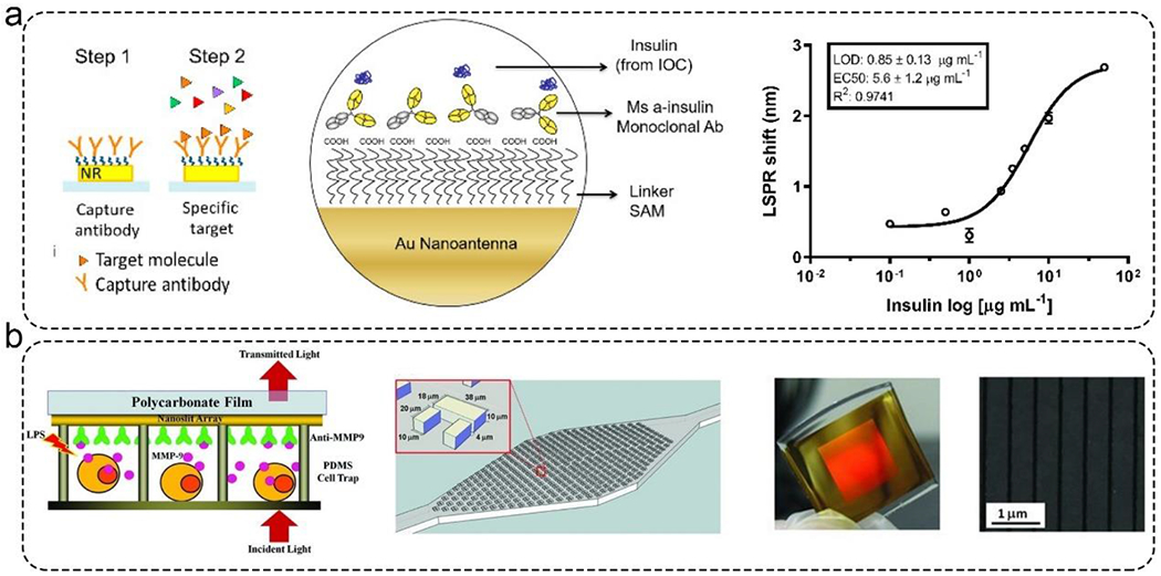
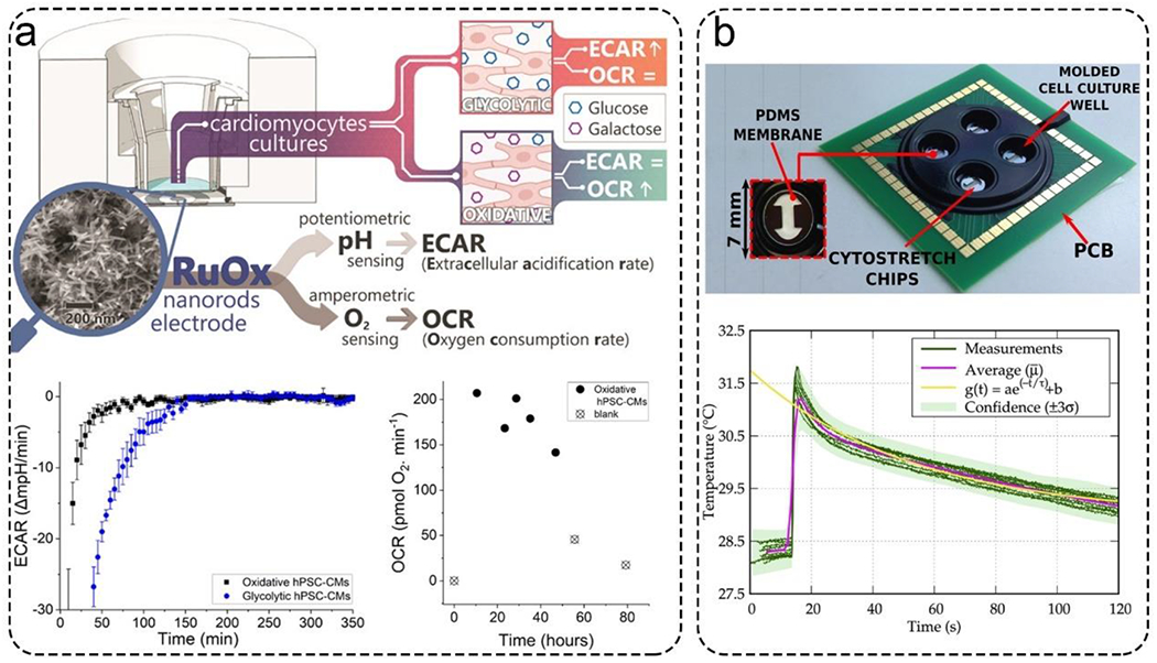
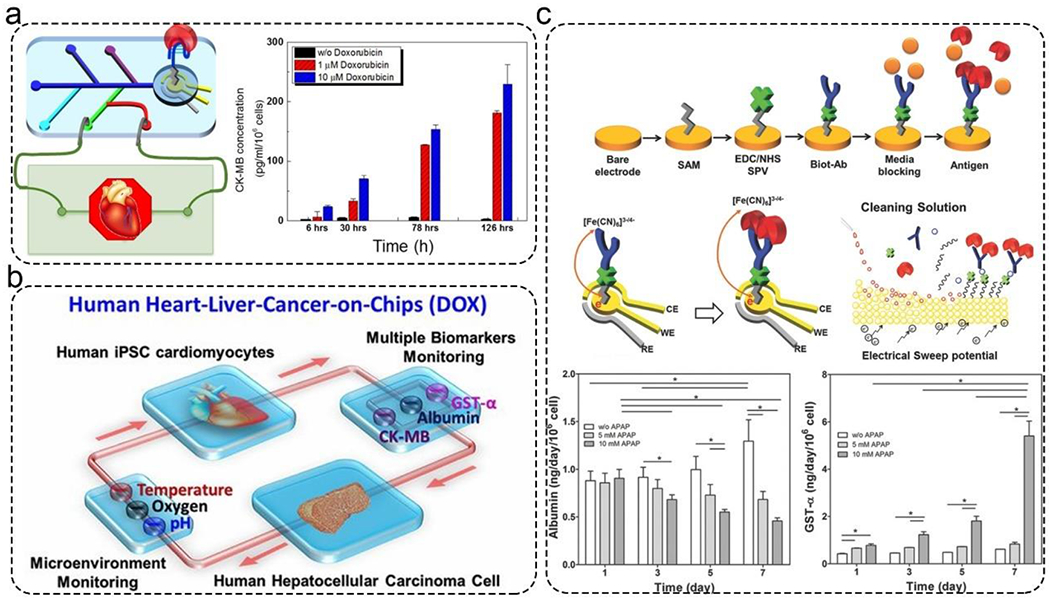
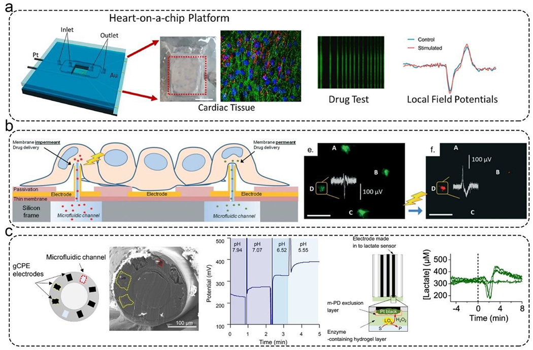
Microphysiological systems (MPSs), also known as organ-on-a-chip models, aim to recapitulate the functional components of human tissues or organs in vitro. Over the last decade, with the advances in biomaterials, 3D bioprinting, and microfluidics, numerous MPSs have emerged with applications to study diseased and healthy tissue models. Various organs have been modeled using MPS technology, such as the heart, liver, lung, and blood-brain barrier. An important aspect of in vitro modeling is the accurate phenotypical and functional characterization of the modeled organ. However, most conventional characterization methods are invasive and destructive and do not allow continuous monitoring of the cells in culture. On the other hand, microfluidic biosensors enable in-line, real-time sensing of target molecules with an excellent limit of detection and in a non-invasive manner, thereby effectively overcoming the limitation of the traditional techniques. Consequently, microfluidic biosensors have been increasingly integrated into MPSs and used for in-line target detection. This review discusses the state-of-the-art microfluidic biosensors by providing specific examples, detailing their main advantages in monitoring MPSs, and highlighting current developments in this field. Finally, we describe the remaining challenges and potential future developments to advance the current state-of-the-art in integrated microfluidic biosensors.


Conflict of interest statement

Conflicts of Interest:

The authors declare no conflict of interest.

Figures

Figure 1.
Figure 1.
Amperometric glucose and lactate sensors for the real-time analysis of mitochondrial stress in a liver-on-a-chip model. a). The principle of the amperometric glucose and lactate sensors. Platinum electrodes were immobilized with an enzyme (i.e., the bioreceptor), which can catalyze glucose and lactate to produce hydrogen peroxide. b). The standard curve of the amperometric glucose and lactate sensors. c). Automated microfluidic switchboard. This switchboard can operate automatically with an integrated control unit. Reproduced with permission from ref (42). Copyright 2016 National Academy of Sciences.
Figure 2.
Figure 2.
Electrochemical impedance biosensors for automated and in-line detection of albumin, GST-α, and CK-MB secreted by MPSs. a). The functionalization, detection, and regeneration steps of the impedance biosensors. The inset image is the photo of the fabricated gold electrode. b). Schematic illustration of the microfluidic breadboard design. This allowed multiple sensing cycles, including biofunctionalization, washing, sensing, and regeneration. c). The detection performance of these three impedance biosensors. All of them can detect targets from 0 to 100 ng/mL. Reproduced with permission from ref (51). Copyright 2016 National Academy of Sciences.
Figure 3.
Figure 3.
Oxygen levels in the microchamber were measured by silica microparticles encapsulated with an oxygen-sensitive dye. a). Schematic illustration of the silica microparticles inside the microchamber. b). Fluorescent image captured by a camera. These images demonstrated the distribution of oxygen concentration. Reproduced with permission from ref (78). Copyright 2017 Springer.
Figure 4.
Figure 4.
Representative SPR sensor for monitoring MPSs. a). Left: the detection principle of gold nanorods-based SPR sensor. Right: the detection performance of this sensor. This sensor showed a LOD of 0.85± 0.13 μg/ mL when detecting insulin. Reproduced with permission from ref (93). Copyright 2021 MDPI. b). Fano Resonance optofluidic sensor was used to monitor live cell secretomes. Left: the detection principle of gold nanoslits-based SPR sensor. Middle: microchamber contains an array of cell traps for capturing cells. Right: The photo and SEM image of gold nanoslits. Reproduced with permission from ref (94). Copyright 2013 Wiley.
Figure 5.
Figure 5.
Representative biosensors for monitoring the microenvironment. a). A ruthenium oxide (RuOx) based electrode measured extracellular acidification rate (ECARs) and oxygen consumption rates (OCRs). This nanorods electrode continuously measured ECARs and OCRs in cardiomyocytes cultures over 48 hours. Reproduced with permission from ref (114). Copyright 2021 American Chemical Society. b). A silicon-based sensor integrated into an organ-on-a-chip for temperature monitoring. The sensor could respond in about 15 seconds. Reproduced with permission from ref (115). Copyright 2021 Elsevier.
Figure 6.
Figure 6.
Applications of microfluidic biosensors for monitoring MPSs. a). CK-MB was detected by an aptamer-based electrochemical impedance spectroscopy biosensor. Reproduced with permission from ref (135) Copyright 2016 American Chemical Society. b). Automated multiple biosensors for monitoring acetaminophen-induced toxicity in normal human heart-liver-on-chips and doxorubicin-induced toxicity from heart-liver-cancer-on-chip MPSs. Reproduced with permission from ref (51) Copyright 2016 National Academy of Sciences. c). Sensors for monitoring the metabolic activity of the liver organoids in response to drugs for 7 d. Top: The principle and surface chemistry of electrochemical impedance spectroscopy-based biosensor. Middle: Schematic illustration of the electrode and regeneration process. Bottom: Real-time monitoring of GST-α and albumin for 7 d. Reproduced with permission from ref (136) Copyright 2017 Wiley.
Figure 7.
Figure 7.
Applications of microfluidic biosensors for monitoring MPSs. a). An integrated cardiac MPS with a platinum wire electrode for applying electrical stimulation, a gold MEAs for acquiring electrophysiological signals, and a microfluidic chamber for long-term culturing cells. This platform can test drug responses with local field potentials. Reproduced with permission from ref (138) Copyright 2021 Elsevier. b). An integrated cardiac MPS. The MEAs were decorated with 3D hollow nanostructures that could deliver calcein-AM and propidium iodide into cardiac cells. The MEAs could monitor this process. Reproduced with permission from ref (139) Copyright 2018 Royal Society of Chemistry. c). Potentiometric fiber electrodes were used to monitor pH and transient neurometabolic lactate in neural tissue. Left: Schematic illustration of the fiber-based biosensor. Middle: Real-time monitoring pH. Right: The detection principle of lactate sensor and the real-time monitoring lactate. Reproduced with permission from ref (142) Copyright 2021 American Chemical Society.

References

    1. Wang K, Man K, Liu J, et al., ACS Biomater. Sci. Eng, 2020, 6, 3231–3257. - PMC - PubMed
    1. Roth A and Berlin M-W, Science, 2021, 373, 1304–1306. - PubMed
    1. Ashammakhi N, Darabi MA, Çelebi-Saltik B, et al., Small Methods, 2020, 4, 1900589. - PMC - PubMed
    1. Huh D, Matthews BD, Mammoto A, et al., Science, 2010, 328, 1662–1668. - PMC - PubMed
    1. Grosberg A, Alford PW, McCain ML, et al., Lab Chip, 2011, 11, 4165–4173. - PMC - PubMed

Publication types

Substances