. 2022 Oct 7;378(6615):eabn5648.
doi: 10.1126/science.abn5648. Epub 2022 Oct 7.

The human disease gene LYSET is essential for lysosomal enzyme transport and viral infection

Affiliations

The human disease gene LYSET is essential for lysosomal enzyme transport and viral infection

Christopher M Richards et al. Science. .

Abstract

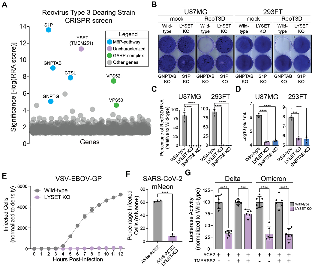
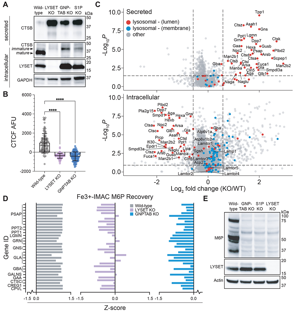
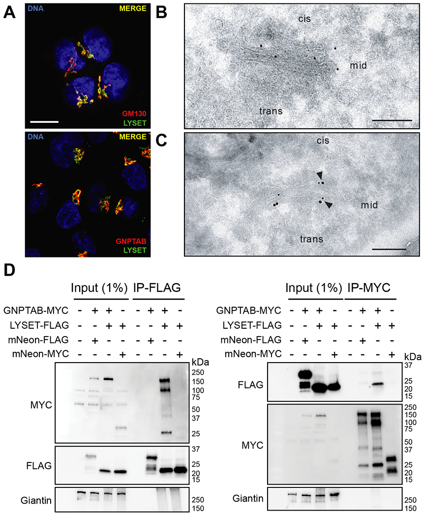
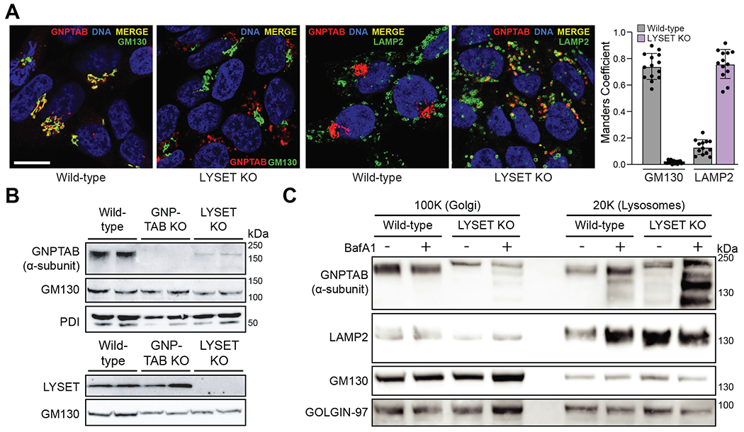
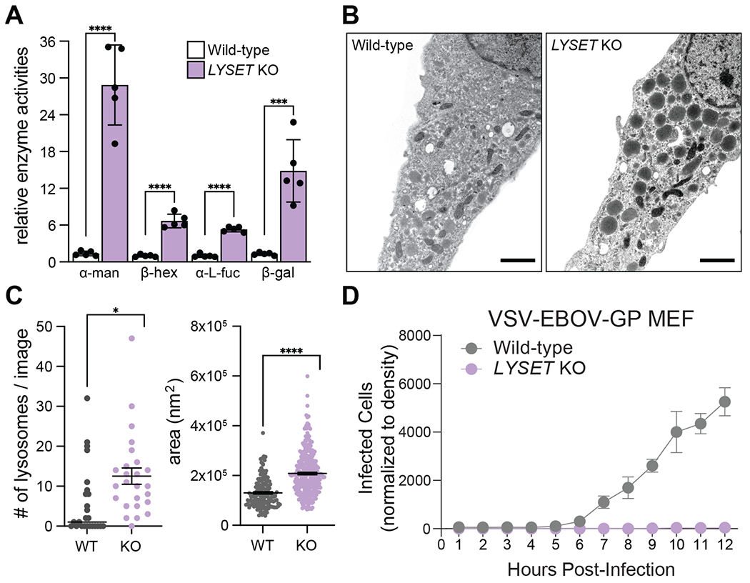
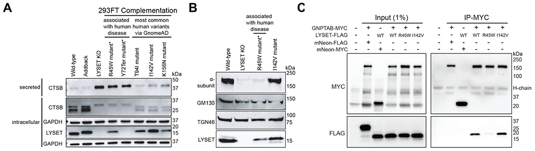
Lysosomes are key degradative compartments of the cell. Transport to lysosomes relies on GlcNAc-1-phosphotransferase-mediated tagging of soluble enzymes with mannose 6-phosphate (M6P). GlcNAc-1-phosphotransferase deficiency leads to the severe lysosomal storage disorder mucolipidosis II (MLII). Several viruses require lysosomal cathepsins to cleave structural proteins and thus depend on functional GlcNAc-1-phosphotransferase. We used genome-scale CRISPR screens to identify lysosomal enzyme trafficking factor (LYSET, also named TMEM251) as essential for infection by cathepsin-dependent viruses including severe acute respiratory syndrome coronavirus 2 (SARS-CoV-2). LYSET deficiency resulted in global loss of M6P tagging and mislocalization of GlcNAc-1-phosphotransferase from the Golgi complex to lysosomes. Lyset knockout mice exhibited MLII-like phenotypes, and human pathogenic LYSET alleles failed to restore lysosomal sorting defects. Thus, LYSET is required for correct functioning of the M6P trafficking machinery and mutations in LYSET can explain the phenotype of the associated disorder.


Conflict of interest statement

Competing interests: J.E.C. consulted for Janssen BioPharma on topics unrelated to this study. C.R.B. is a co-founder and Scientific Advisory Board member of Lycia Therapeutics, Palleon Pharmaceuticals, Enable Bioscience, Redwood Biosciences (a subsidiary of Catalent) and InterVenn Biosciences. C.A.B. reports compensation for consulting and/or SAB membership from Catamaran Bio, DeepCell Inc., Immunebridge, Sangamo Therapeutics, Bicycle Tx, and Revelation Biosciences on topics unrelated to this study. The remaining authors declare no competing interests.

Figures

Fig. 1.
Fig. 1.. LYSET is a critical host factor for multiple viruses that utilize activated cathepsins for entry.
(A) CRISPR screen for reovirus T3D (ReoT3D) host factors in U87MG cells. Significance of enrichment was determined via MAGeCK analysis (y-axis). All genes are plotted as bubbles on the x-axis, each representing a specific gene. A subset is color-coded according to function, labels show gene names. (B) Crystal violet staining after infection with ReoT3D representative of n=3 biologically independent replicates. “mock”: non-infected controls. (C) RT-qPCR quantification of ReoT3D RNA in infected U87MG and 293FT cells at MOI 1 at 72 hpi (mean ± SEM, n= 3) (D) U87MG or 293FT cells were infected with MOI of 1 ReoT3D virus. At 72 hpi, cells were lysed and viral titers determined via plaque assay (mean ± SEM, n = 3, ***p<0.001, ****p<0.0001; significance determined via one-way ANOVA with post-hoc Dunnett’s multiple comparisons test for (C) and (D)). (E) Time course of VSV-EBOV-GP infection of 293FT cells wild-type and LYSET KO (mean ± SEM, n = 3). (F) Bar-graph depicting independent infections of A549-ACE2 cells ± LYSET KO with SARS-CoV-2-mNeon using flow cytometry (mean ± SD, n = 3, ****p<0.0001; significance determined via unpaired, two-tailed student’s t test). (G) Infection of SARS-CoV-2 Delta and Omicron Spike reporter virus particles in Huh7.5.1 cell lines with or without LYSET KO. Cells were engineered to stably express ACE2 or ACE2 in combination with TMPRSS2, as indicated. Luciferase activity was measured at 72 hpi and normalized to wild-type cells (mean ± SEM, n = 6, ***p<0.001, ****p<0.0001; significance determined via significance was determined by unpaired, two-tailed student’s t-test with Welch’s correction).
Fig. 2.
Fig. 2.. LYSET is required for global lysosomal enzyme transport via M6P-tagging.
(A) Immunoblot analysis of cathepsin B (CTSB) in lysates (intracellular) and in extracellular medium (secreted) from wild-type cells and clonal 293FT cell lines containing knockout (KO) mutations in indicated genes. (B) Cathepsin activity in cells was determined by quantification of BMV-109 fluorescence signal in live 293FT wild-type and KO cells as the raw corrected total cellular fluorescence (CTCF) in arbitrary fluorescence units (AFU) (mean ± SD, n = 100 cells, **** p<0.0001; significance determined via one-way ANOVA with a post-hoc Dunnett’s multiple comparisons test). (C) Proteomic analysis of wild-type and LYSET KO MEFs by unbiased data independent acquisition (DIA). DIA was used for intracellular and secreted proteins. (D) Z-score analysis of individual peptides that contain the M6P moiety as determined using glycoproteomics. Peptides were derived from indicated lysosomal proteins in wild-type, LYSET KO and GNPTAB KO 293FT cells; n = 3 replicates for each cell line. (E) Immunoblot analysis of M6P-tagged proteins from 293FT wild-type, GNPTAB KO, S1P KO, and LYSET KO cells using an M6P-specific single-chain antibody fragment (M6P).
Fig. 3.
Fig. 3.. LYSET binds to GNPTAB and co-localizes with GNPTAB/GNPTG in Golgi apparatus cisternae.
(A) Immunofluorescence microscopy in HAP1 cells showing co-localization of LYSET with the cis-Golgi marker GM130, and GNPTAB. Scale bar, 10 μm (B) Transmission electron microscopy (TEM) immunogold staining (15 nm gold) shows LYSET in Golgi cisternae of SK-MEL-30 wild-type cells using ultrathin sections. (C) TEM double immunogold staining on ultrathin sections for LYSET (15 nm gold) and GNPTG (10 nm gold) in the Golgi apparatus of SK-MEL-30 wild-type cells. Arrowheads indicate co-localization. Scale bar, 200 nm. (D) Immunoprecipitation (IP) on lysates of cells expressing epitope-tagged proteins using anti-FLAG (left panel) or anti-MYC (right panel) magnetic beads, followed by immunoblot analysis with indicated antibodies. Input lysates are also analyzed.
Fig. 4.
Fig. 4.. LYSET KO triggers mislocalization of GNPTAB to the lysosome where it is degraded.
(A) Colocalization of GNPTAB with GM130 and LAMP2 in wild-type and LYSET KO cells and Manders colocalization quantification. Colocalization analysis was performed on at least n = 12 separate micrograph images. Scale bar, 10 μm (B) Immunoblot analysis of GNPTAB in 100K enriched ER/Golgi-fractions from wild-type, GNPTAB KO, and LYSET KO HAP1 cells. The antibody is specific for the α-subunit domain of GNPTAB. Golgi-marker (GM130) and ER-marker (PDI) proteins were used as loading controls. (C) Immunoblot analysis of 20K and 100K fractions from wild-type and LYSET KO HAP1 cells with or without bafilomycin A1 (BafA1) treatment. LAMP2, GM130 and GOLGIN-97 proteins were used as controls for preparation and loading.
Fig. 5.
Fig. 5.. Lyset KO mice display characteristics used to diagnose ML-II disease including elevated blood serum levels of lysosomal enzymes and aberrant lysosomes in isolated cells.
(A) Relative enzyme activities of α-mannosidase (α-man), β-hexosaminidase (β-hex), α-L-fucosidase (α-L-fuc) and β-galactosidase (β-gal) from blood sera of adult wild-type (WT) and Lyset KO mice; WT activities set to 1; mean ± SD, n = 5 mice, ***p<0.001, ****p<0.0001, unpaired student’s t test (two-tailed). (B) Electron micrographs of WT and Lyset KO MEFs. Scale bar, 1 μm. (C) Numbers of lysosomes and areas counted for 25 independent images for WT and Lyset KO (KO) MEFs; mean ± SEM; unpaired, two-tailed student’s t test with Welch’s correction; *P<0.05, ****P< 0.0001. (D) Time course of VSV-EBOV-GP infection of WT and KO MEFs (mean ± SD, n = 3)
Fig. 6.
Fig. 6.. Pathogenic LYSET mutations fail to restore lysosomal transport defects in LYSET KO cells.
(A) Immunoblot of 293FT lysates or supernatants from LYSET KO cells complemented with wild-type LYSET (Addback) or human variants. R45W and Y72Ter are patient-mutations (22); T94I, I142V and K156N variants are not disease-associated. (B) Immunoblots of 100K enriched-Golgi/ER fractions from 293FT KO cells complemented with indicated LYSET alleles. (C) Immunoprecipitation (IP) on lysates of cells expressing epitope-tagged proteins using anti-MYC magnetic beads followed by immunoblot analysis with indicated antibodies.

References

    1. Mercer J, Schelhaas M, Helenius A, Virus entry by endocytosis. Annual review of biochemistry 79, 803–833 (2010). - PubMed
    1. Mainou BA, Dermody TS, In search of cathepsins: how reovirus enters host cells. DNA and cell biology 31, 1646–1649 (2012). - PMC - PubMed
    1. Chandran K, Sullivan NJ, Felbor U, Whelan SP, Cunningham JM, Endosomal proteolysis of the Ebola virus glycoprotein is necessary for infection. Science (New York, N.Y.) 308, 1643–1645 (2005). - PMC - PubMed
    1. Shirato K, Kawase M, Matsuyama S, Middle East respiratory syndrome coronavirus infection mediated by the transmembrane serine protease TMPRSS2. Journal of virology 87, 12552–12561 (2013). - PMC - PubMed
    1. Hoffmann M et al., SARS-CoV-2 Cell Entry Depends on ACE2 and TMPRSS2 and Is Blocked by a Clinically Proven Protease Inhibitor. Cell 181, 271–280.e278 (2020). - PMC - PubMed