

Stromal Vascular Fraction Reverses the Age-Related Impairment in Revascularization following Injury
- PMID: 36075199
- PMCID: PMC9780192
- DOI: 10.1159/000526002
Stromal Vascular Fraction Reverses the Age-Related Impairment in Revascularization following Injury
Abstract
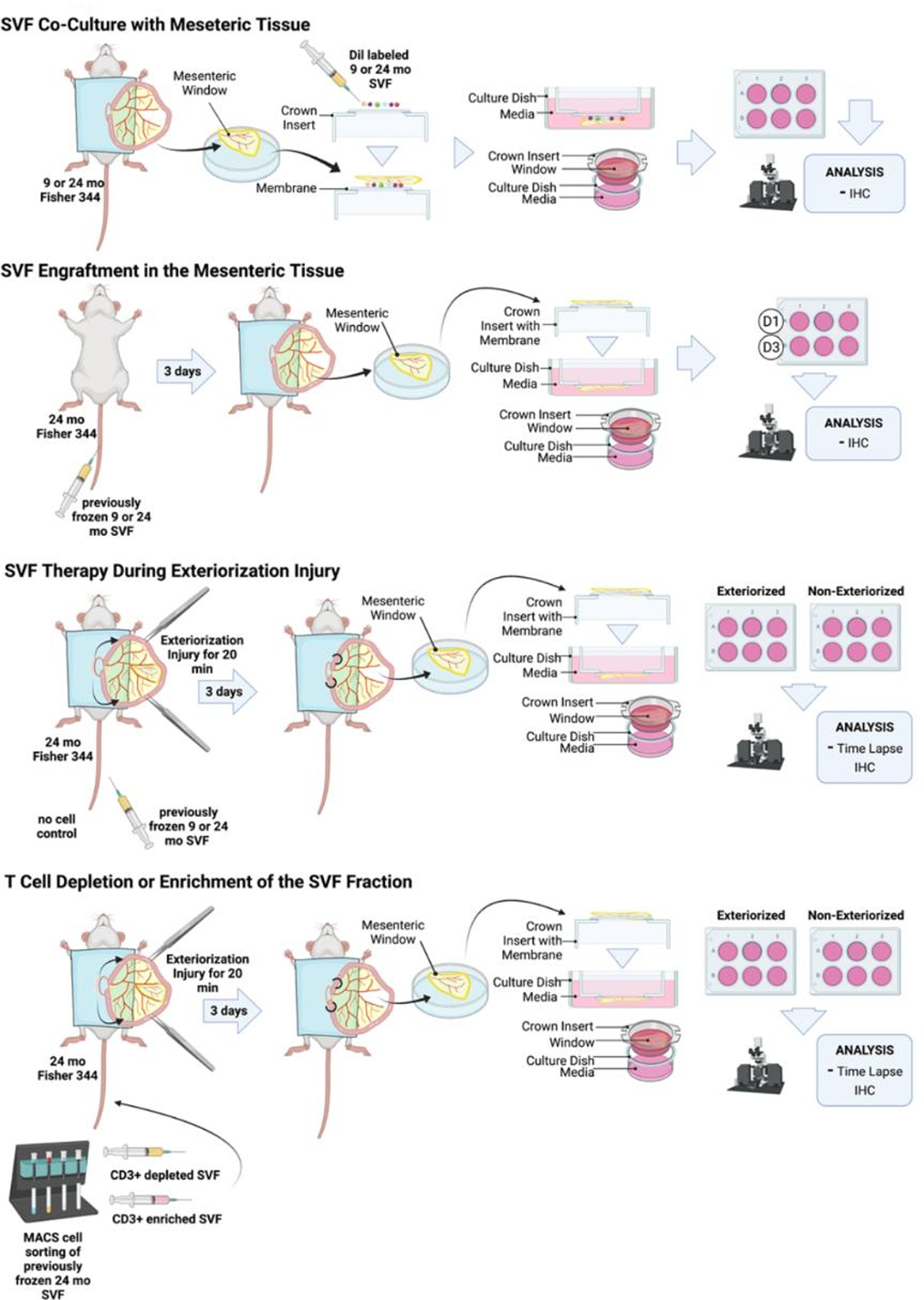
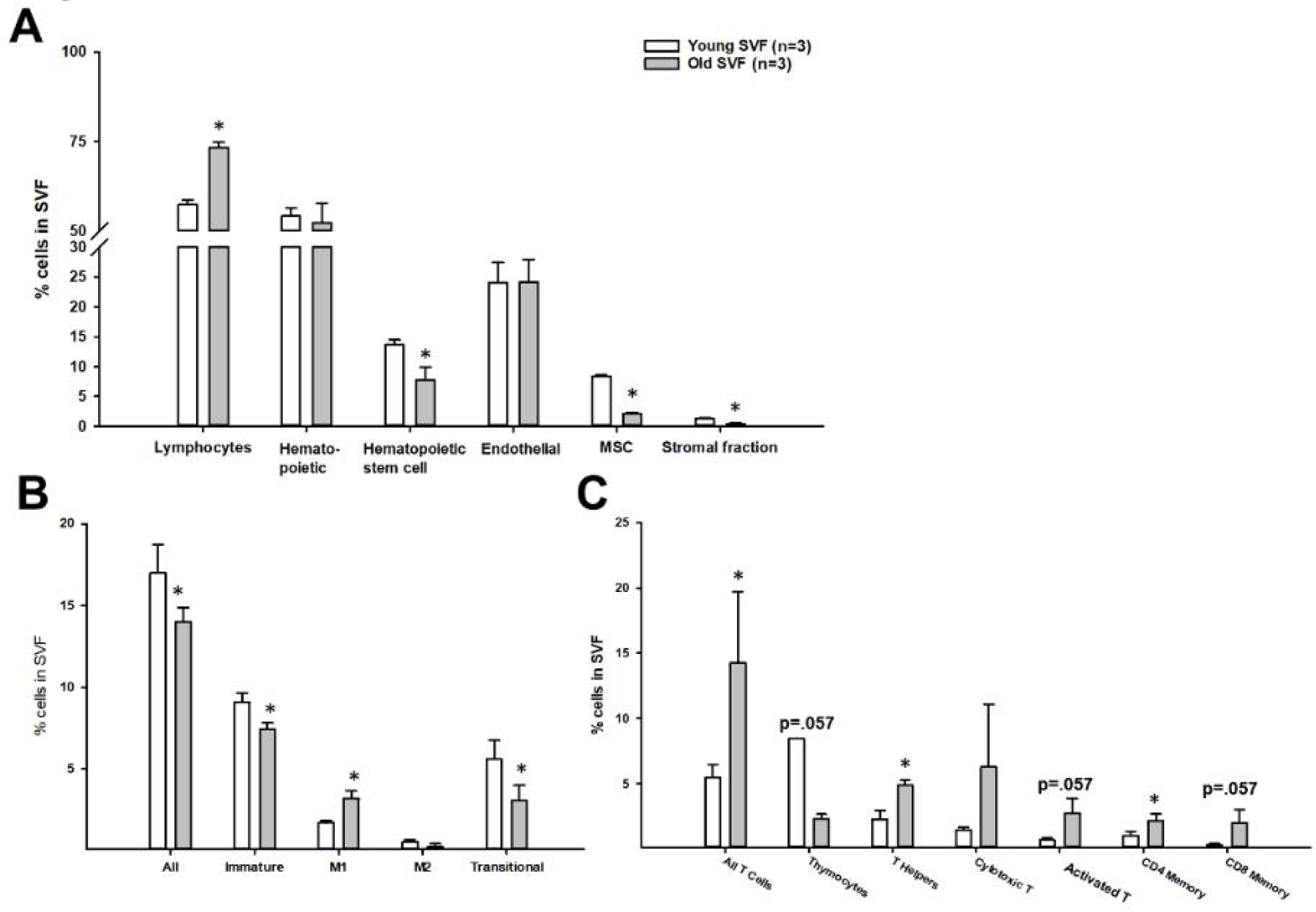
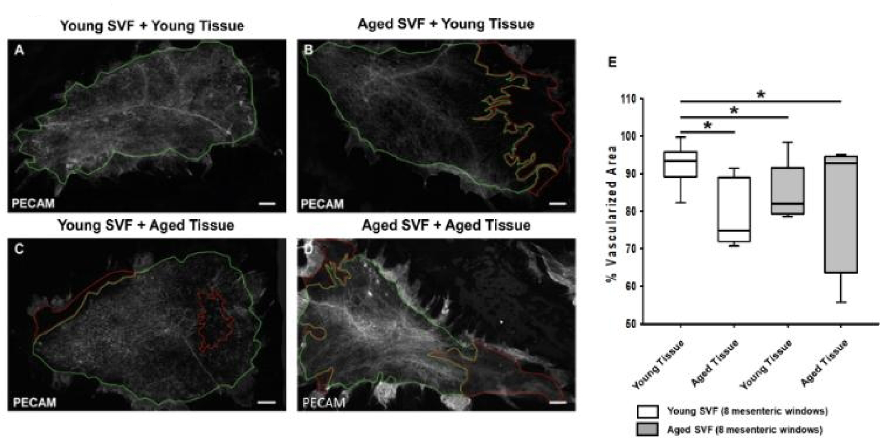
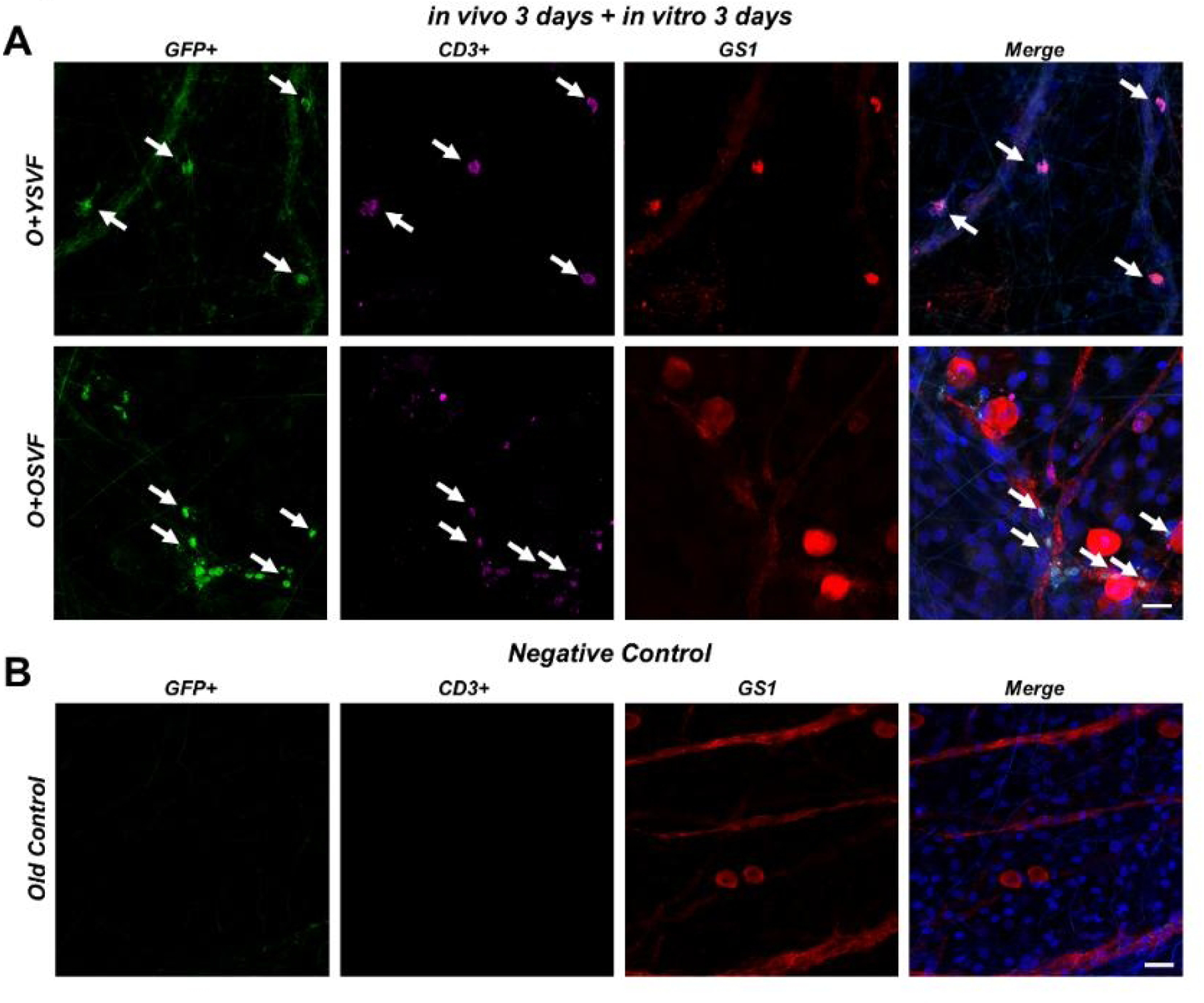
Adipose-derived stromal vascular fraction (SVF) has emerged as a potential regenerative therapy, but few studies utilize SVF in a setting of advanced age. Additionally, the specific cell population in SVF providing therapeutic benefit is unknown. We hypothesized that aging would alter the composition of cell populations present in SVF and its ability to promote angiogenesis following injury, a mechanism that is T cell-mediated. SVF isolated from young and old Fischer 344 rats was examined with flow cytometry for cell composition. Mesenteric windows from old rats were isolated following exteriorization-induced (EI) hypoxic injury and intravenous injection of one of four cell therapies: (1) SVF from young or (2) old donors, (3) SVF from old donors depleted of or (4) enriched for T cells. Advancing age increased the SVF T-cell population but reduced revascularization following injury. Both young and aged SVF incorporated throughout the host mesenteric microvessels, but only young SVF significantly increased vascular area following EI. This study highlights the effect of donor age on SVF angiogenic efficacy and demonstrates how the ex vivo mesenteric-window model can be used in conjunction with SVF therapy to investigate its contribution to angiogenesis.
Keywords: Aging; Angiogenesis; Cell therapy; Injury; Revascularization; T cells.
© 2022 S. Karger AG, Basel.
Conflict of interest statement
Conflicts of Interest
The authors have no conflicts of interest to declare.
Figures


Similar articles
-
Adipose-derived stromal vascular fraction cells isolated from old animals exhibit reduced capacity to support the formation of microvascular networks.Exp Gerontol. 2015 Mar;63:18-26. doi: 10.1016/j.exger.2015.01.044. Epub 2015 Jan 21. Exp Gerontol. 2015. PMID: 25617825 Free PMC article.
-
Adipose stromal vascular fraction reverses mitochondrial dysfunction and hyperfission in aging-induced coronary microvascular disease.Am J Physiol Heart Circ Physiol. 2022 Oct 1;323(4):H749-H762. doi: 10.1152/ajpheart.00311.2022. Epub 2022 Aug 26. Am J Physiol Heart Circ Physiol. 2022. PMID: 36018760 Free PMC article.
-
Enhanced beta-1 adrenergic receptor responsiveness in coronary arterioles following intravenous stromal vascular fraction therapy in aged rats.Aging (Albany NY). 2019 Jul 11;11(13):4561-4578. doi: 10.18632/aging.102069. Aging (Albany NY). 2019. PMID: 31296794 Free PMC article.
-
Adipose-Derived Stromal Vascular Fraction Cells: Update on Clinical Utility and Efficacy.Crit Rev Eukaryot Gene Expr. 2015;25(2):145-52. doi: 10.1615/critreveukaryotgeneexpr.2015013057. Crit Rev Eukaryot Gene Expr. 2015. PMID: 26080608 Review.
-
A systematic review of autologous adipose-derived stromal vascular fraction (SVF) for the treatment of acute cutaneous wounds.Arch Dermatol Res. 2022 Jul;314(5):417-425. doi: 10.1007/s00403-021-02242-x. Epub 2021 May 28. Arch Dermatol Res. 2022. PMID: 34047823
Cited by
-
Investigation of the stemness and wound-healing potential of long-term cryopreserved stromal vascular fraction cells.Regen Ther. 2025 Mar 13;29:128-139. doi: 10.1016/j.reth.2025.02.004. eCollection 2025 Jun. Regen Ther. 2025. PMID: 40162021 Free PMC article.
-
Stromal Vascular Fraction Restores Vasodilatory Function by Reducing Oxidative Stress in Aging-Induced Coronary Microvascular Disease.Antioxid Redox Signal. 2023 Feb;38(4-6):261-281. doi: 10.1089/ars.2021.0249. Epub 2022 Sep 28. Antioxid Redox Signal. 2023. PMID: 35950616 Free PMC article.
-
State of the Art in the Standardization of Stromal Vascular Fraction Processing.Biomolecules. 2025 Jan 30;15(2):199. doi: 10.3390/biom15020199. Biomolecules. 2025. PMID: 40001502 Free PMC article. Review.
-
Challenges in the clinical translation of stromal vascular fraction therapy in regenerative medicine.World J Stem Cells. 2025 Jun 26;17(6):103775. doi: 10.4252/wjsc.v17.i6.103775. World J Stem Cells. 2025. PMID: 40585955 Free PMC article. Review.
-
Analyzing the Clinical Potential of Stromal Vascular Fraction: A Comprehensive Literature Review.Medicina (Kaunas). 2024 Jan 27;60(2):221. doi: 10.3390/medicina60020221. Medicina (Kaunas). 2024. PMID: 38399509 Free PMC article.
References
-
- Lopez MJ and Spencer ND, In vitro adult rat adipose tissue-derived stromal cell isolation and differentiation. Methods Mol Biol, 2011.. 702: p. 37–46. - PubMed
