. 2022 Oct;298(10):102465.
doi: 10.1016/j.jbc.2022.102465. Epub 2022 Sep 6.

The iron-sulfur cluster assembly (ISC) protein Iba57 executes a tetrahydrofolate-independent function in mitochondrial [4Fe-4S] protein maturation

Affiliations

The iron-sulfur cluster assembly (ISC) protein Iba57 executes a tetrahydrofolate-independent function in mitochondrial [4Fe-4S] protein maturation

Ulrich Mühlenhoff et al. J Biol Chem. 2022 Oct.

Abstract

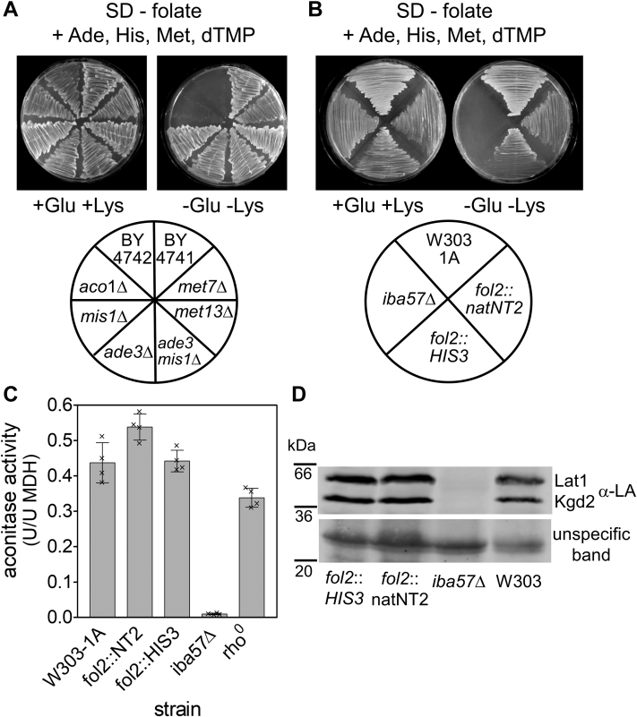
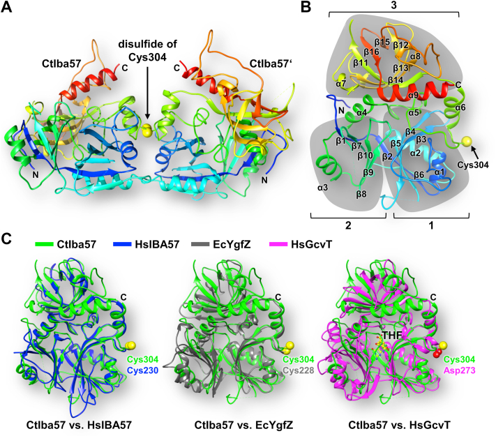
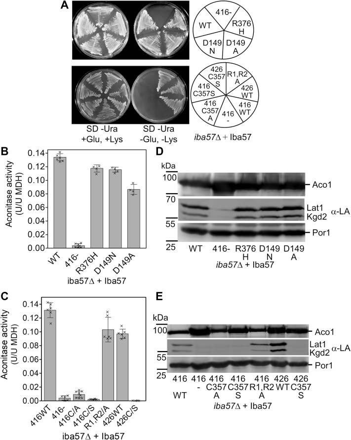
Mitochondria harbor the bacteria-inherited iron-sulfur cluster assembly (ISC) machinery to generate [2Fe-2S; iron-sulfur (Fe-S)] and [4Fe-4S] proteins. In yeast, assembly of [4Fe-4S] proteins specifically involves the ISC proteins Isa1, Isa2, Iba57, Bol3, and Nfu1. Functional defects in their human equivalents cause the multiple mitochondrial dysfunction syndromes, severe disorders with a broad clinical spectrum. The bacterial Iba57 ancestor YgfZ was described to require tetrahydrofolate (THF) for its function in the maturation of selected [4Fe-4S] proteins. Both YgfZ and Iba57 are structurally related to an enzyme family catalyzing THF-dependent one-carbon transfer reactions including GcvT of the glycine cleavage system. On this basis, a universally conserved folate requirement in ISC-dependent [4Fe-4S] protein biogenesis was proposed. To test this idea for mitochondrial Iba57, we performed genetic and biochemical studies in Saccharomyces cerevisiae, and we solved the crystal structure of Iba57 from the thermophilic fungus Chaetomium thermophilum. We provide three lines of evidence for the THF independence of the Iba57-catalyzed [4Fe-4S] protein assembly pathway. First, yeast mutants lacking folate show no defect in mitochondrial [4Fe-4S] protein maturation. Second, the 3D structure of Iba57 lacks many of the side-chain contacts to THF as defined in GcvT, and the THF-binding pocket is constricted. Third, mutations in conserved Iba57 residues that are essential for THF-dependent catalysis in GcvT do not impair Iba57 function in vivo, in contrast to an exchange of the invariant, surface-exposed cysteine residue. We conclude that mitochondrial Iba57, despite structural similarities to both YgfZ and THF-binding proteins, does not utilize folate for its function.

Keywords: crystallography; folate; iron–sulfur cluster assembly; iron–sulfur protein; mitochondria; mitochondrial disease.


Conflict of interest statement

Conflict of interest The authors declare that they have no conflicts of interest with the contents of this article.

Figures

Figure 1
Figure 1
Folate is not required formitochondrial [4Fe–4S] protein biogenesis in Saccharomyces cerevisiae.A, the indicated yeast strains (BY4741 background; MATa his3Δ leu2Δ met15Δ ura3Δ) with defects in the utilization or modification of folate were cultivated on folate-free synthetic defined (SD) minimal medium containing the four folate-dependent supplements Ade, His, Met, and dTMP in the presence or the absence of Lys and Glu. BY4742 (MATα his3Δ leu2Δ lys2Δ ura3Δ) and aco1Δ (W303-1A background) requiring supplementation with either Lys or Glu served as control. B, two independent folate-auxotroph fol2Δ deletion strains (W303-1A background) were streaked onto folate-free SD minimal medium agar plates plus Ade, His, Met, and dTMP in the presence or the absence of Lys and Glu. C and D, cells cultivated in folate-free SD minimal medium plus Ade, His, Met, and dTMP were assayed for (C) aconitase enzyme activities (relative to malate dehydrogenase [MDH]) in cell extracts and (D) the lipoylation (LA) of the E2 subunits of pyruvate (Lat1) and 2-ketoglutarate dehydrogenases (Kgd2) by immunostaining of isolated mitochondrial extracts. An unspecific band crossreacting with the anti-LA antibody served as a loading control. W303-1A wildtype and rho0 cells as well as iba57Δ were used as controls. Error bars indicate the SD (n ≥ 4). dTMP, deoxythymidine monophosphate.
Figure 2
Figure 2
Escherichia coli YgfZ does not complement Saccharomyces cerevisiae iba57Δ cells.A, strain iba57Δ (W303-1A background) was transformed with the indicated combinations of plasmids (Table S3) that allow the expression of E. coli YgfZ and IscA in mitochondria under the control of the TDH3 promoter. Cells were cultivated on synthetic defined (SD) minimal medium agar plates in the presence or the absence of Lys and Glu. B, cells were cultivated in SD minimal medium, and cell extracts were assayed for aconitase activities. iba57Δ expressing yeast IBA57 from a plasmid and cells with empty vector served as controls. Error bars indicate the standard deviation (n ≥ 4). C, iba57Δ cells harboring low-copy (p416) or high-copy (p426) vectors for the expression of EcYgfZ with a C-terminal Myc tag under the control of the TDH3 promoter were fractionated into mitochondria (Mit) and postmitochondrial supernatant (PMS) fractions. iba57Δ cells with the empty vector (p416) served as a control. Fractions were analyzed for the presence of EcYgfZ by immunostaining with α-Myc antibodies. The asterisks (∗) likely indicate the noncleaved EcYgfZ-Myc precursor. Stains for mitochondrial Aco1 and cytosolic Dre2 serve to document the quality of the fractionation. An unspecific protein stained with Fast Green FCF served as a loading control. EcYgfZ, Escherichia coli YgfZ.
Figure 3
Figure 3
Crystal structure of Chaetomium thermophilum Iba57 at 2.4 Å resolution.A, disulfide-bridged (via Cys304) dimer of CtIba57 as found in the crystal. Residues are rainbow colored from N (blue) to C termini (red). B, cartoon representation of CtIba57 monomer (coloring as in A). The three individual domains are highlighted in gray (1, 2, 3). The sulfur of the essential Cys304 is depicted as a yellow sphere. C, superposition of CtIba57 (green) with HsIBA57 (blue, left; Protein Data Bank [PDB] code: 6QE4), EcYgfZ (gray, middle; PDB code: 1NRK), and HsGcvT (magenta, right; PDB code: 1WSV). The conserved Cys residues of Iba57 (Ct and Hs) and EcYgfZ as well as the structural equivalent Asp273 of HsGcvT are depicted as spheres. THF of HsGcvT is shown in yellow (for enlargement, see Fig. 4A). CtIba57, CtIba57, Chaetomium thermophilum Iba57; EcYgfZ, Escherichia coli YgfZ; HsGcvT, human GcvT; HsIBA57, human IBA57; THF, tetrahydrofolate.
Figure 4
Figure 4
The structure of CtIba57 is incompatible with THF binding.A, superposition of CtIba57 (green) and HsGcvT (magenta; Protein Data Bank [PDB] code: 1WSV) highlighting the THF (yellow)-binding region of HsGcvT. The loops β9–10 and α5 that would interfere with THF binding in CtIba57 are marked. Blue arrows indicate the distances of the structural rearrangements of the respective loops. (For a comparison including HsIBA57 and EcYgfZ, refer to Fig. S4). B, the cavity of HsGcvT (red) encompassing its THF-binding site (THF pocket) and its lipoyl entrance tunnel was calculated using PyMOL. Side chains of residues in β9–10 and α5 loops of CtIba57 (green; shown as balls and sticks) protrude into the THF pocket of HsGcvT. C, comparison of residues involved in THF (yellow) binding to various proteins. Left, the canonical THF-binding pocket as found in HsGcvT (magenta), TrmE (cyan; PDB code: 1XZQ), and DMGO (sienna; PDB code: 1PJ6). The residue numbers are indicated in parenthesis for HsGcvT. Right, comparison of the THF-binding residues of HsGcvT (as shown left) with the structurally equivalent residues of CtIba57 (green), HsIBA57 (blue, PDB code: 6QE4), and EcYgfZ (gray, PDB code: 1NRK). The residue numbers are indicated in parenthesis for CtIba57. Only the side chains are depicted as sticks. Contacts are indicated by black lines. The catalytically important N10 position of THF is marked. CtIba57, Chaetomium thermophilum Iba57; EcYgfZ, Escherichia coli YgfZ; HsGcvT, human GcvT; HsIBA57, human IBA57; THF, tetrahydrofolate.
Figure 5
Figure 5
Cavities and surfaces of Iba57 and EcYgfZ exclude productive THF binding.A, cavities inside the cartoon ribbon representation were calculated for the indicated proteins using PyMOL. In HsGcvT, the cavity forms both the tunnel for the lipoyl arm of GCSH and the binding pocket for THF (yellow). B, the cavities extracted from A with attached human GCSH protein (location taken from the GCSH–HsGcvT complex (Protein Data Bank [PDB] code: 3A8I). The dihydrolipoyl moiety attached to GCSH protein is depicted in light green. THF (yellow) is shown as found in HsGcvT. C, surface charges (calculated by APBS biomolecular solvation software suite (80)) of the indicated proteins (blue: positive; red, negative). The β9–10- and α5-loops and the C terminus are indicated in CtIba57. The potential THF-binding pocket is indicated in EcYgfZ (yellow arrow). THF in HsGcvT is shown in yellow, and the poly-Glu binding site is outlined by a yellow dotted line. CtIba57, Chaetomium thermophilum Iba57; EcYgfZ, Escherichia coli YgfZ; HsGcvT, human GcvT; THF, tetrahydrofolate.
Figure 6
Figure 6
Potential THF-interacting residues are dispensable for in vivo function of ScIba57.A, yeast iba57Δ cells expressing either wildtype (WT) ScIba57 or the indicated point mutation variants from a centromeric plasmid (p416) under the control of the endogenous IBA57 promoter were cultivated on solid synthetic defined (SD) minimal medium in the presence or the absence of Lys and Glu. iba57Δ with an empty vector (416) served as control. Overproduction of the ScIba57–C357S variant in iba57Δ cells from a high-copy vector (p426) did not lead to high-copy suppression. B and C, cells cultivated in SD minimal medium plus Lys and Glu were assayed for aconitase enzyme activities (relative to malate dehydrogenase [MDH]). D and E, the presence of aconitase and the lipoylation (LA) of the E2 subunits of pyruvate (Lat1) and 2-ketoglutarate dehydrogenases (Kgd2) was determined by immunostaining of isolated mitochondrial extracts. A stain for mitochondrial Por1 served as a loading control. Error bars indicate the standard deviation (n ≥ 4). ScIba57, Saccharomyces cerevisiae Iba57; THF, tetrahydrofolate.

References

    1. Zanello P. Structure and electrochemistry of proteins harboring iron-sulfur clusters of different nuclearities. Part IV. Canonical, non-canonical and hybrid iron-sulfur proteins. J. Struct. Biol. 2019;205:103–120. - PubMed
    1. Braymer J.J., Freibert S.A., Rakwalska-Bange M., Lill R. Mechanistic concepts of iron-sulfur protein biogenesis in biology. Biochim. Biophys. Acta Mol. Cell Res. 2021;1868 - PubMed
    1. Lill R., Freibert S.A. Mechanisms of mitochondrial iron-sulfur protein biogenesis. Annu. Rev. Biochem. 2020;89:471–499. - PubMed
    1. Maio N., Jain A., Rouault T.A. Mammalian iron-sulfur cluster biogenesis: recent insights into the roles of frataxin, acyl carrier protein and ATPase-mediated transfer to recipient proteins. Curr. Opin. Chem. Biol. 2020;55:34–44. - PMC - PubMed
    1. Ciofi-Baffoni S., Nasta V., Banci L. Protein networks in the maturation of human iron-sulfur proteins. Metallomics. 2018;10:49–72. - PubMed

Publication types

MeSH terms

LinkOut - more resources