

Recombinant human erythropoietin induces neuroprotection, activates MAPK/CREB pathway, and rescues fear memory after traumatic brain injury with delayed hypoxemia in mice
- PMID: 36075467
- PMCID: PMC10515732
- DOI: 10.1016/j.brainres.2022.148074
Recombinant human erythropoietin induces neuroprotection, activates MAPK/CREB pathway, and rescues fear memory after traumatic brain injury with delayed hypoxemia in mice
Abstract
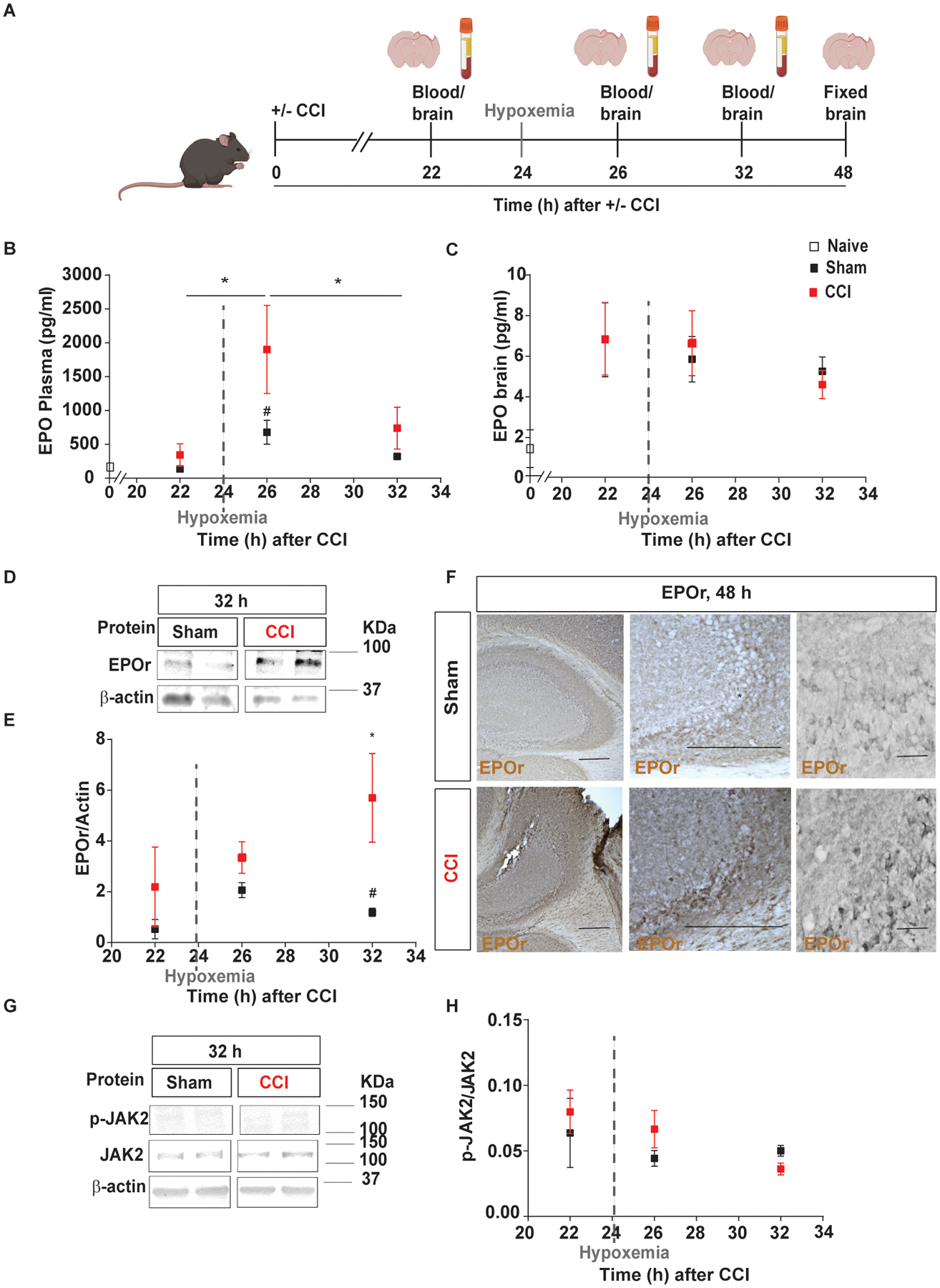
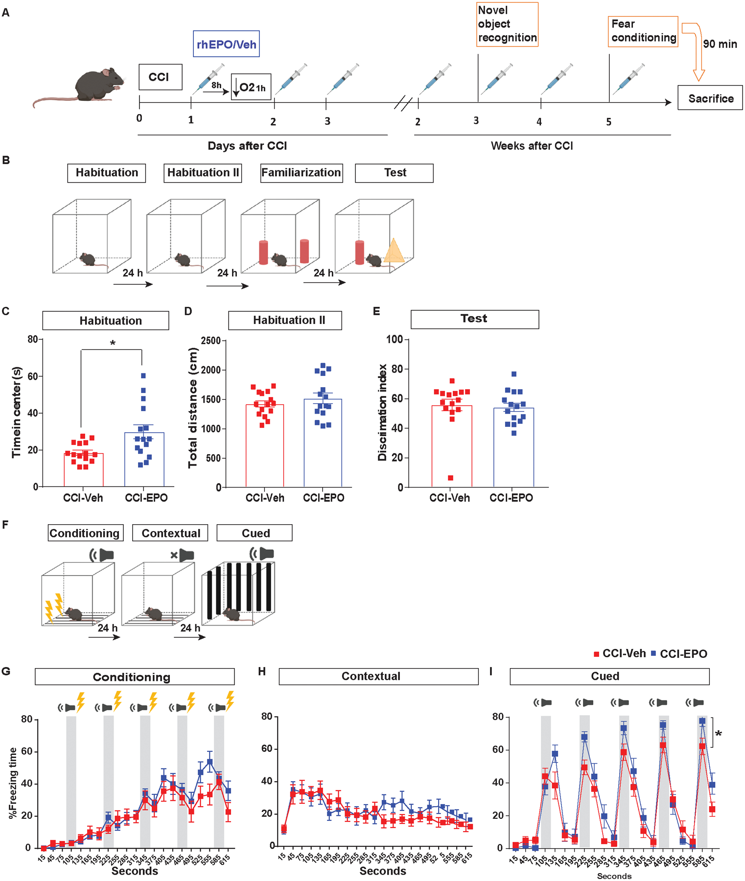
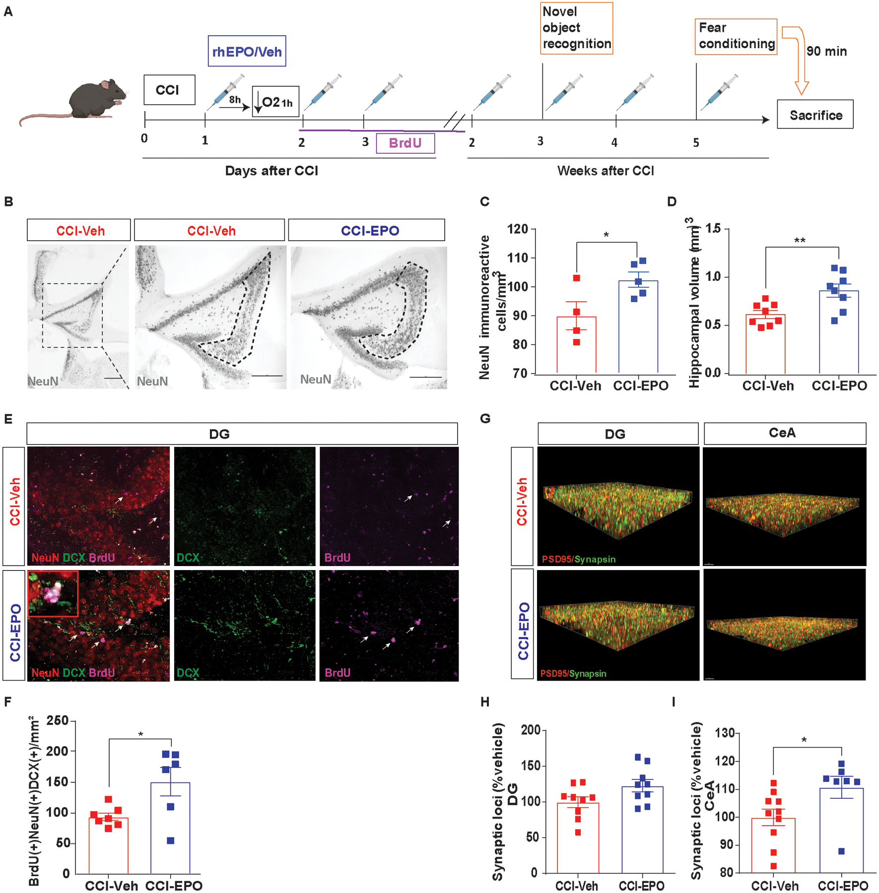
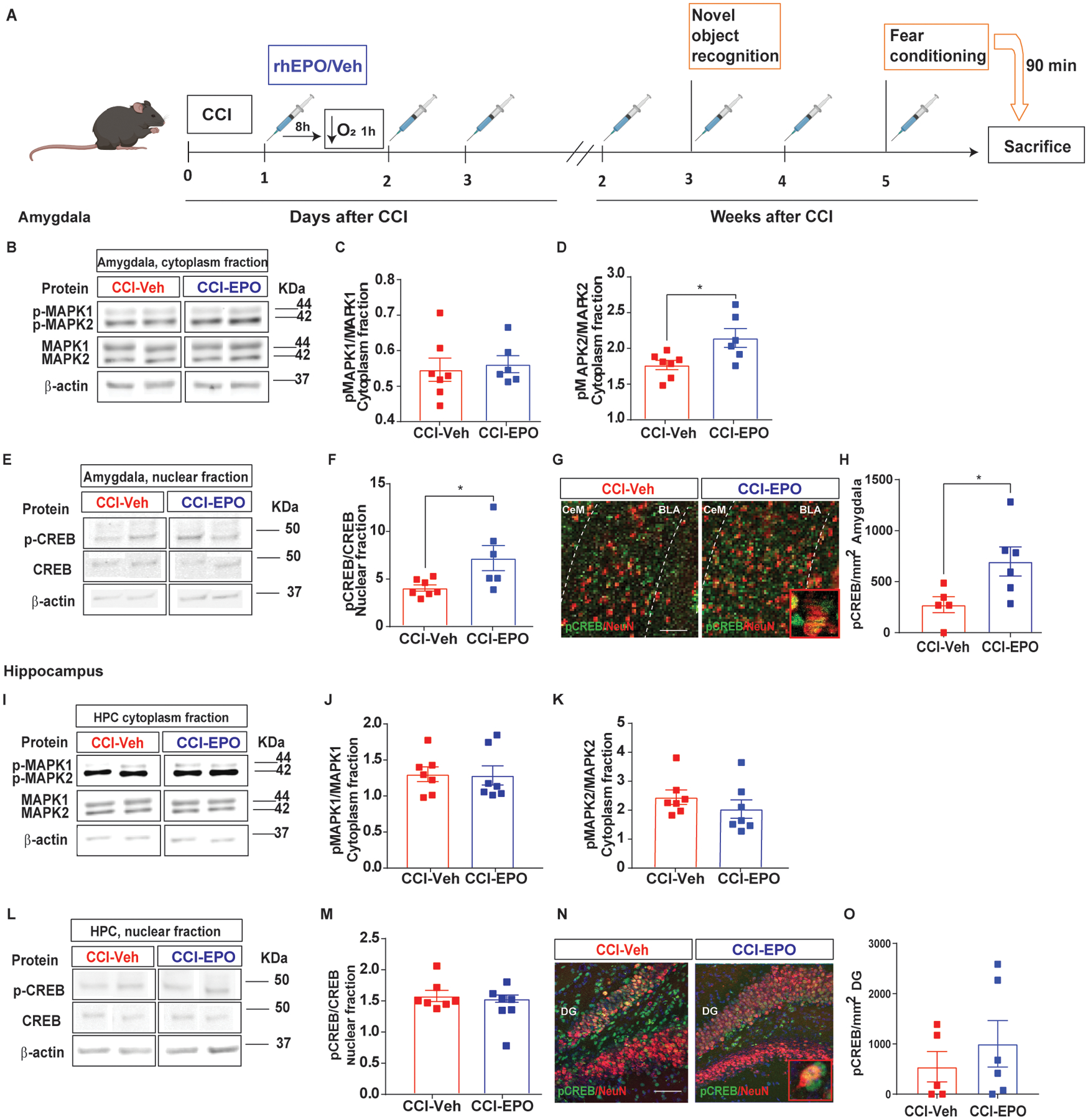
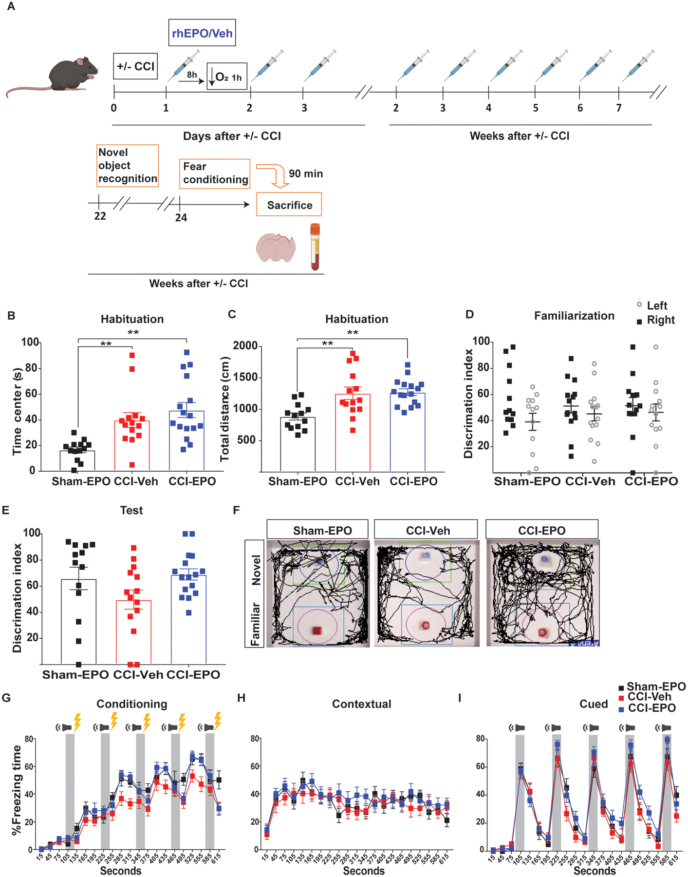
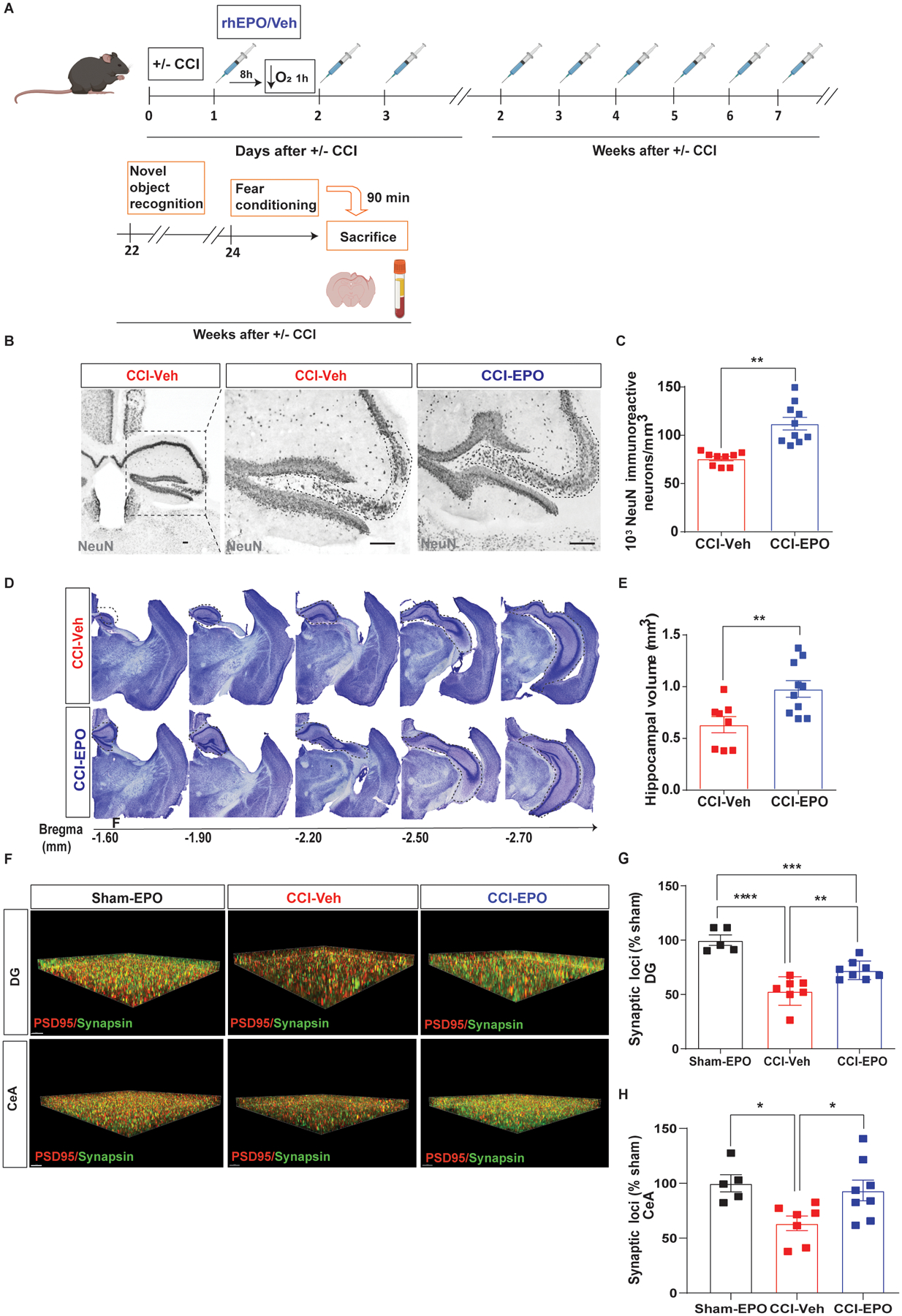
Therapeutic interventions targeting secondary insults, such as delayed hypoxemia, provide a unique opportunity for treatment in severe traumatic brain injury (TBI). Erythropoietin (EPO) is a hypoxia-responsive cytokine with important roles in neurodevelopment, neuroprotection and neuromodulation. We hypothesized that recombinant human erythropoietin (rhEPO) administration would mitigate injury in a combined injury model of TBI and delayed hypoxemia. Utilizing a clinically relevant murine model of TBI and delayed hypoxemia, we characterized how ongoing rhEPO administration influenced neurogenesis, neuroprotection, synaptic density and, behavioral outcomes early after TBI, and the impact on long-lasting outcomes 6 months after injury. We employed novel object recognition (NOR) and fear conditioning to assess long-term memory. At 1-month post-injury, we observed a significant increase in cued-fear memory response in the rhEPO-injured mice compared with vehicle-injured mice. This was associated with neuroprotection and neurogenesis in the hippocampus and mitogen-activated protein kinase (MAPK)/cAMP response element-binding protein (CREB) signaling activation and increased of excitatory synaptic density in the amygdala. Early rhEPO treatment after injury reduced neurodegeneration and increased excitatory synaptic density in the hippocampus and amygdala at 6 months post-injury. However at 6 months post-injury (4 months after discontinuation of rhEPO), we did not observe changes in behavioral assessments nor MAPK/CREB pathway activation. In summary, these data demonstrate that ongoing rhEPO treatment initiated at a clinically feasible time point improves neurological, cognitive, and histological outcomes after TBI in the setting of secondary hypoxemic insults.
Keywords: Erythropoietin; Fear conditioning; Hypoxemia; Neurogenesis; Neuroprotection; Traumatic brain injury.
Copyright © 2022 Elsevier B.V. All rights reserved.
Conflict of interest statement
Declaration of Competing Interest The authors declare that they have no known competing financial interests or personal relationships that could have appeared to influence the work reported in this paper.
Figures
Similar articles
-
Recombinant Erythropoietin Induces Oligodendrocyte Progenitor Cell Proliferation After Traumatic Brain Injury and Delayed Hypoxemia.Neurotherapeutics. 2023 Oct;20(6):1859-1874. doi: 10.1007/s13311-023-01443-8. Epub 2023 Sep 28. Neurotherapeutics. 2023. PMID: 37768487 Free PMC article.
-
Chemogenetic inhibition of amygdala excitatory neurons impairs rhEPO-enhanced contextual fear memory after TBI.Neurosci Lett. 2023 May 1;804:137216. doi: 10.1016/j.neulet.2023.137216. Epub 2023 Mar 28. Neurosci Lett. 2023. PMID: 36997018 Free PMC article.
-
N-acetylcysteine reduces brain injury after delayed hypoxemia following traumatic brain injury.Exp Neurol. 2021 Jan;335:113507. doi: 10.1016/j.expneurol.2020.113507. Epub 2020 Oct 13. Exp Neurol. 2021. PMID: 33065076 Free PMC article.
-
Therapeutic effect of erythropoietin in patients with traumatic brain injury: a meta-analysis of randomized controlled trials.J Neurosurg. 2017 Jul;127(1):8-15. doi: 10.3171/2016.4.JNS152909. Epub 2016 Jul 1. J Neurosurg. 2017. PMID: 27367243
-
Erythropoietin: emerging role of erythropoietin in neonatal neuroprotection.Pediatr Neurol. 2014 Oct;51(4):481-8. doi: 10.1016/j.pediatrneurol.2014.06.008. Epub 2014 Jun 24. Pediatr Neurol. 2014. PMID: 25266611 Free PMC article. Review.
Cited by
-
Mechanistic insights into Nrf2-driven pathogenesis and therapeutic targeting in spinal cord injury.Front Immunol. 2025 Jul 10;16:1574834. doi: 10.3389/fimmu.2025.1574834. eCollection 2025. Front Immunol. 2025. PMID: 40709178 Free PMC article. Review.
-
Microglial polarization pathways and therapeutic drugs targeting activated microglia in traumatic brain injury.Neural Regen Res. 2026 Jan 1;21(1):39-56. doi: 10.4103/NRR.NRR-D-24-00810. Epub 2024 Dec 7. Neural Regen Res. 2026. PMID: 39665832 Free PMC article.
-
Therapy of traumatic brain injury by modern agents and traditional Chinese medicine.Chin Med. 2023 Mar 11;18(1):25. doi: 10.1186/s13020-023-00731-x. Chin Med. 2023. PMID: 36906602 Free PMC article. Review.
-
Recombinant Erythropoietin Induces Oligodendrocyte Progenitor Cell Proliferation After Traumatic Brain Injury and Delayed Hypoxemia.Neurotherapeutics. 2023 Oct;20(6):1859-1874. doi: 10.1007/s13311-023-01443-8. Epub 2023 Sep 28. Neurotherapeutics. 2023. PMID: 37768487 Free PMC article.
-
Chemogenetic inhibition of amygdala excitatory neurons impairs rhEPO-enhanced contextual fear memory after TBI.Neurosci Lett. 2023 May 1;804:137216. doi: 10.1016/j.neulet.2023.137216. Epub 2023 Mar 28. Neurosci Lett. 2023. PMID: 36997018 Free PMC article.
References
-
- Adamcio B, Sargin D, Stradomska A, Medrihan L, Gertler C, Theis F, Zhang M, Muller M, Hassouna I, Hannke K, Sperling S, Radyushkin K, El-Kordi A, Schulze L, Ronnenberg A, Wolf F, Brose N, Rhee JS, Zhang W, Ehrenreich H, 2008. Erythropoietin enhances hippocampal long-term potentiation and memory. BMC Biol. 6, 37. - PMC - PubMed
-
- Arango MF, Bainbridge D, 2008. Magnesium for acute traumatic brain injury. Cochrane Database Syst Rev. CD005400. - PubMed
-
- Brines M, Cerami A, 2005. Emerging biological roles for erythropoietin in the nervous system. Nat Rev Neurosci. 6, 484–94. - PubMed
-
- Celorrio M, Abellanas MA, Rhodes J, Goodwin V, Moritz J, Vadivelu S, Wang L, Rodgers R, Xiao S, Anabayan I, Payne C, Perry AM, Baldridge MT, Aymerich MS, Steed A, Friess SH, 2021a. Gut microbial dysbiosis after traumatic brain injury modulates the immune response and impairs neurogenesis. Acta Neuropathol Commun. 9, 40. - PMC - PubMed
Publication types
MeSH terms
Substances
Grants and funding
LinkOut - more resources
Full Text Sources
Medical
Research Materials
