Review
. 2022 Sep 8;19(1):223.
doi: 10.1186/s12974-022-02584-x.

Neuroinflammation represents a common theme amongst genetic and environmental risk factors for Alzheimer and Parkinson diseases

Affiliations
Review

Neuroinflammation represents a common theme amongst genetic and environmental risk factors for Alzheimer and Parkinson diseases

Rachel J Boyd et al. J Neuroinflammation. .

Abstract

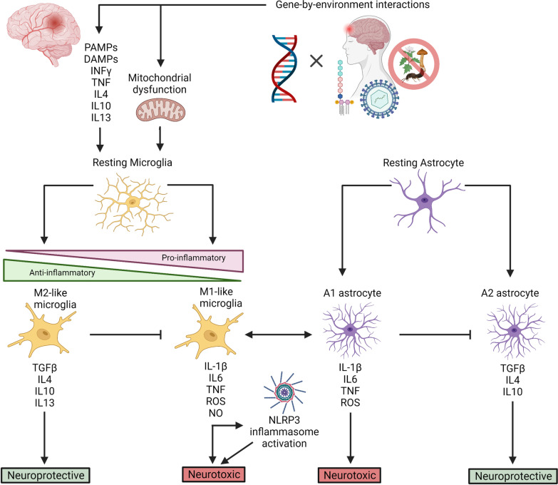
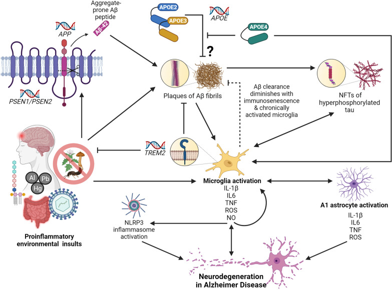
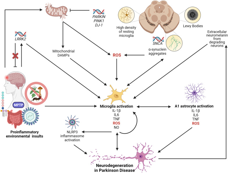
Multifactorial diseases are characterized by inter-individual variation in etiology, age of onset, and penetrance. These diseases tend to be relatively common and arise from the combined action of genetic and environmental factors; however, parsing the convoluted mechanisms underlying these gene-by-environment interactions presents a significant challenge to their study and management. For neurodegenerative disorders, resolving this challenge is imperative, given the enormous health and societal burdens they impose. The mechanisms by which genetic and environmental effects may act in concert to destabilize homeostasis and elevate risk has become a major research focus in the study of common disease. Emphasis is further being placed on determining the extent to which a unifying biological principle may account for the progressively diminishing capacity of a system to buffer disease phenotypes, as risk for disease increases. Data emerging from studies of common, neurodegenerative diseases are providing insights to pragmatically connect mechanisms of genetic and environmental risk that previously seemed disparate. In this review, we discuss evidence positing inflammation as a unifying biological principle of homeostatic destabilization affecting the risk, onset, and progression of neurodegenerative diseases. Specifically, we discuss how genetic variation associated with Alzheimer disease and Parkinson disease may contribute to pro-inflammatory responses, how such underlying predisposition may be exacerbated by environmental insults, and how this common theme is being leveraged in the ongoing search for effective therapeutic interventions.

Keywords: Alzheimer disease; Chronic inflammation; Common genetic disease; Gene-by-environment interactions; Inflammatory response; Multifactorial phenotypes; Neurodegeneration; Neuroinflammation; Parkinson disease.


Conflict of interest statement

The authors declare no competing interests.

Figures

Fig. 1
Fig. 1
The neuroinflammatory response. Upon infection, insult, or injury to brain tissue and/or organelles, resting microglia are stimulated by PAMPs, DAMPs, reactive oxygen species (ROS), and cytokines. Activated microglia exist along a spectrum of pro-inflammatory (M1-like; neurotoxic) and anti-inflammatory (M2-like; neuroprotective) states, where pro-inflammatory cytokines (i.e., IL-1β, IL6, TNF), reactive oxygen species (ROS), free radicals (NO), and anti-inflammatory cytokines (i.e., TGFB, IL4, IL10, IL13) inform various neurotoxic and/or neuroprotective pathways. Pro-inflammatory cytokines, particularly IL-1β, can activate the NLRP3 inflammasome, which further modulates neurotoxic pathways. Cytokine release is also a means by which microglia communicate with one another and with astrocytes. Resting astrocytes are activated through such cellular communication with microglia, and exist as either A1 (pro-inflammatory) or A2 (anti-inflammatory) astrocytes, where cytokine release can cross-talk with microglia and further activate neurotoxic or neuroprotective pathways
Fig. 2
Fig. 2
The role of neuroinflammation in Alzheimer disease (AD). Microglia activation, and the subsequent release of pro-inflammatory cytokines, ROS, and NO, is a central event in the onset of neurodegeneration in AD. Pro-inflammatory microglia release cytokines that cause a feed-forward loop of microglia activation, activate the NLRP3 inflammasome, and facilitate cross-talk with A1 astrocytes, all of which coalesce on neurodegenerative pathways. In AD, microglia can be activated upon detection of PAMPs, DAMPs, and other pro-inflammatory molecules as a result of environmental insult—including gut dysbiosis and sustained exposure to LPS endotoxin, viral infection, and TBI. These pro-inflammatory environmental insults are also associated with accelerated amyloid β (Aβ) plaque deposition and the overexpression of APP and β-secretase (PSEN1/PSEN2). Mutations in PSEN1 and PSEN2 can result in the cleavage of APP to produce 42-residue, aggregate-prone peptides, and mutations in APP can lead to overproduction, misfolding, and formation of Aβ fibrils. Microglia become activated upon sensing DAMPs from Aβ plaques, and are sequestered to clear them. However, chronically activated microglia and immunosenescence diminish the efficacy of this ability over time. TREM2 modulates microglia activity and survival. Mutations in TREM2 can impact the ability of microglia to modulate cytokine production, phagocytose bacteria, and clear neural debris. When Aβ plaques are not cleared, they, and the chronically activated microglia, can induce the formation of NFTs. Hyperphosphorylated tau protein can further activate microglia. Although their exact mechanism of action is unknown, the APOE2 and APOE3 isoforms are protective against AD and are thought to play a role in clearing Aβ plaques. The APOE4 isoform, however, is unable to clear Aβ plaques and forms neurotoxic fragments that can activate microglia
Fig. 3
Fig. 3
The role of neuroinflammation in Parkinson disease (PD). In PD, the SN is preferentially vulnerable to neuron loss. The high density of resting microglia in this region (12% of cells—the highest in the brain) is thought to render the SN vulnerable to the effects of a robust inflammatory response. Furthermore, neurodegeneration in the SN can cause a feed-forward loop, where, upon neuronal degradation, the high concentration of neuromelanin in the SN is released into the extracellular space and is cleared by activated microglia. Microglia activation, and the subsequent release of pro-inflammatory cytokines, ROS, and NO, is also a central event in the onset of neurodegeneration in PD. Like in AD, pro-inflammatory microglia release cytokines that cause a feed-forward loop of chronic microglia activation, activate the NLRP3 inflammasome, and facilitate cross-talk with A1 astrocytes, all of which coalesce on neurodegenerative pathways. In PD, microglia can be activated upon detection of PAMPs, DAMPs, and other pro-inflammatory molecules as a result of environmental insult—including gut dysbiosis and sustained exposure to LPS endotoxin, viral infection, TBI, MPTP, and pesticides. These pro-inflammatory insults can result in the overexpression of LRRK2, which is also pro-inflammatory and can modulate cytokine production. Mutations in LRRK2 can modulate PD risk, but gene knockouts impair microglia activation and are protective against pro-inflammatory environmental insults. Environmental insults further activate microglia through mitochondrial-mediated toxicity. Pesticides inhibit NADH dehydrogenase, which can lead to the production of ROS and mitochondrial DAMPs, both of which activate microglia. PARKIN, PINK1, and DJ-1 play roles in the clearance of damaged mitochondria, and mutations in these genes can prevent proper clearance and the production of ROS and DAMPs. Pathological hallmarks of PD are aggregates of SNCA surrounded by Lewy Bodies. SNCA mutations, amplification, and overexpression, can result in the formation of these protein aggregates, which are identified and cleared by DAMP-sensing microglia. SNCA can also produce ROS, which interacts with the P2X7 receptor on microglia

References

    1. Antonarakis SE, Chakravarti A, Cohen JC, Hardy J. Mendelian disorders and multifactorial traits: the big divide or one for all? Nat Rev Genet. 2010;11(5):380–384. doi: 10.1038/nrg2793. - DOI - PubMed
    1. Reeve A, Simcox E, Turnbull D. Ageing and Parkinson’s disease: why is advancing age the biggest risk factor? Ageing Res Rev. 2014;14(1):19–30. doi: 10.1016/j.arr.2014.01.004. - DOI - PMC - PubMed
    1. Alzheimer’s Association 2021 Alzheimer’s disease facts and figures. Alzheimer’s Dement. 2021;17(3):327–406. doi: 10.1002/alz.12328. - DOI - PubMed
    1. Nalls MA, Blauwendraat C, Vallerga CL, Heilbron K, Bandres-Ciga S, Chang D, et al. Identification of novel risk loci, causal insights, and heritable risk for Parkinson’s disease: a meta-analysis of genome-wide association studies. Lancet Neurol. 2019;18(12):1091–1102. doi: 10.1016/S1474-4422(19)30320-5. - DOI - PMC - PubMed
    1. Wightman DP, Jansen IE, Savage JE, Shadrin AA, Bahrami S, Holland D, et al. A genome-wide association study with 1,126,563 individuals identifies new risk loci for Alzheimer’s disease. Nat Genet. 2021;53(9):1276–1282. doi: 10.1038/s41588-021-00921-z. - DOI - PMC - PubMed