. 2022 Sep 8;13(1):467.
doi: 10.1186/s13287-022-03166-7.

YAP and TAZ play a crucial role in human erythrocyte maturation and enucleation

Affiliations

YAP and TAZ play a crucial role in human erythrocyte maturation and enucleation

Nattaya Damkham et al. Stem Cell Res Ther. .

Abstract

Background: Yes-associated protein (YAP) and WW domain-containing transcription regulator protein 1 (WWTR1, also known as TAZ) are two key transcription co-activators of the Hippo pathway. Both were originally characterized as organ size and cell proliferation regulators. Later studies demonstrated that the Hippo pathway may play a role in Drosophila and mammal hematopoiesis. However, the role of the Hippo pathway in human erythropoiesis has not yet been fully elucidated.

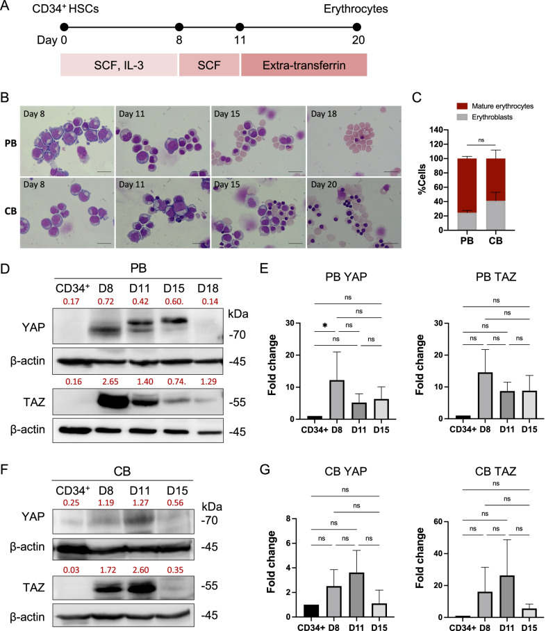
Methods: The role of YAP and TAZ was studied in human erythropoiesis and hematopoietic stem cell (HSC) lineage determination by using mobilized peripheral blood (PB) and cord blood (CB)-derived HSC as a model. HSCs were isolated and cultured in an erythroid differentiation medium for erythroid differentiation and culture in methylcellulose assay for HSC lineage determination study.

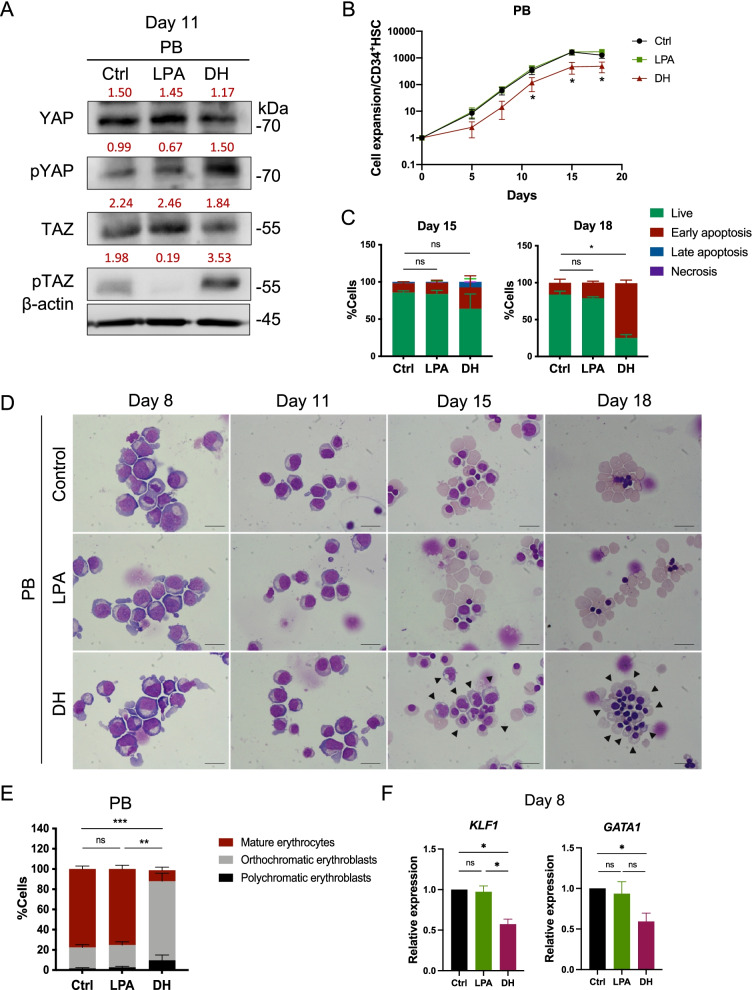
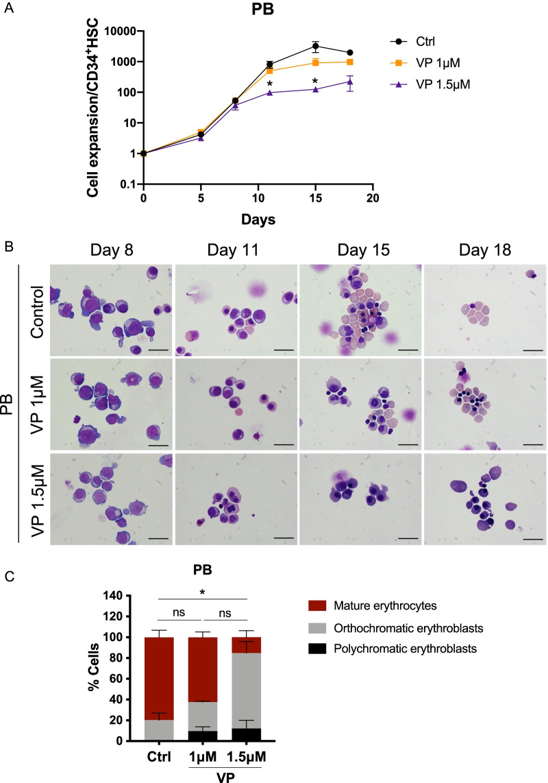
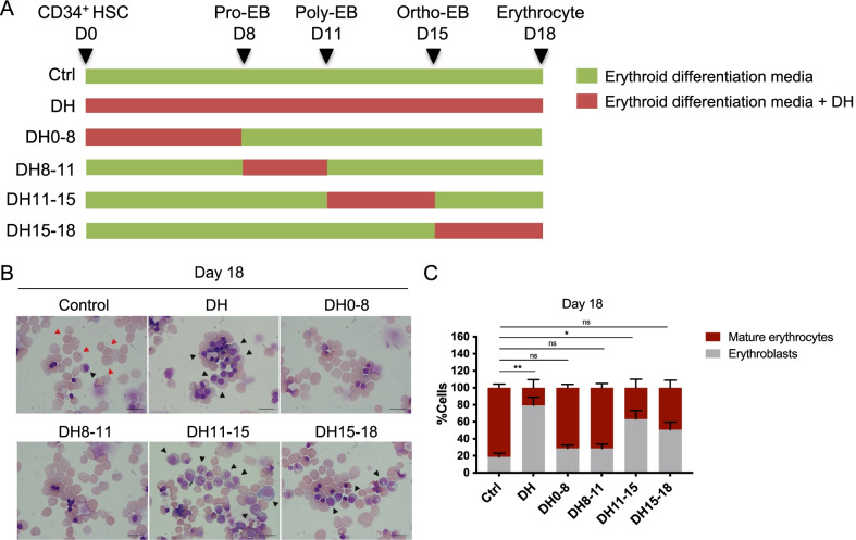
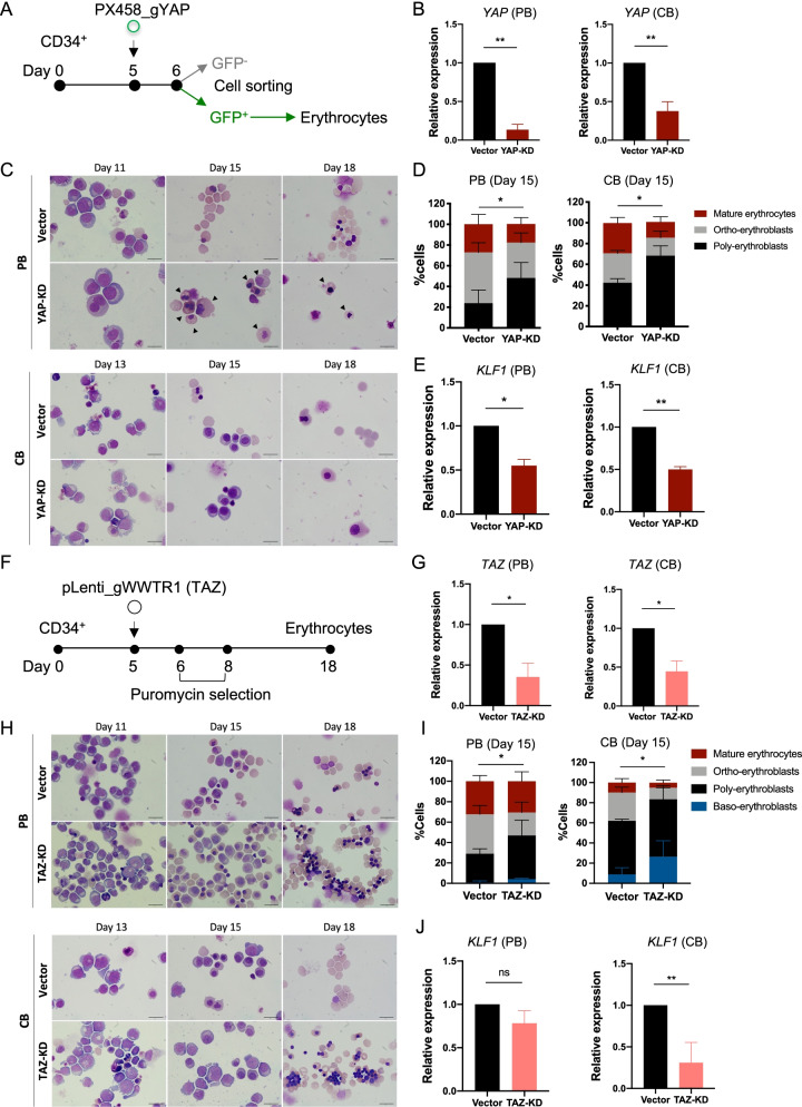
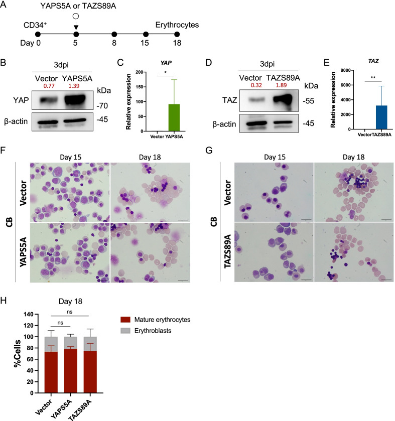
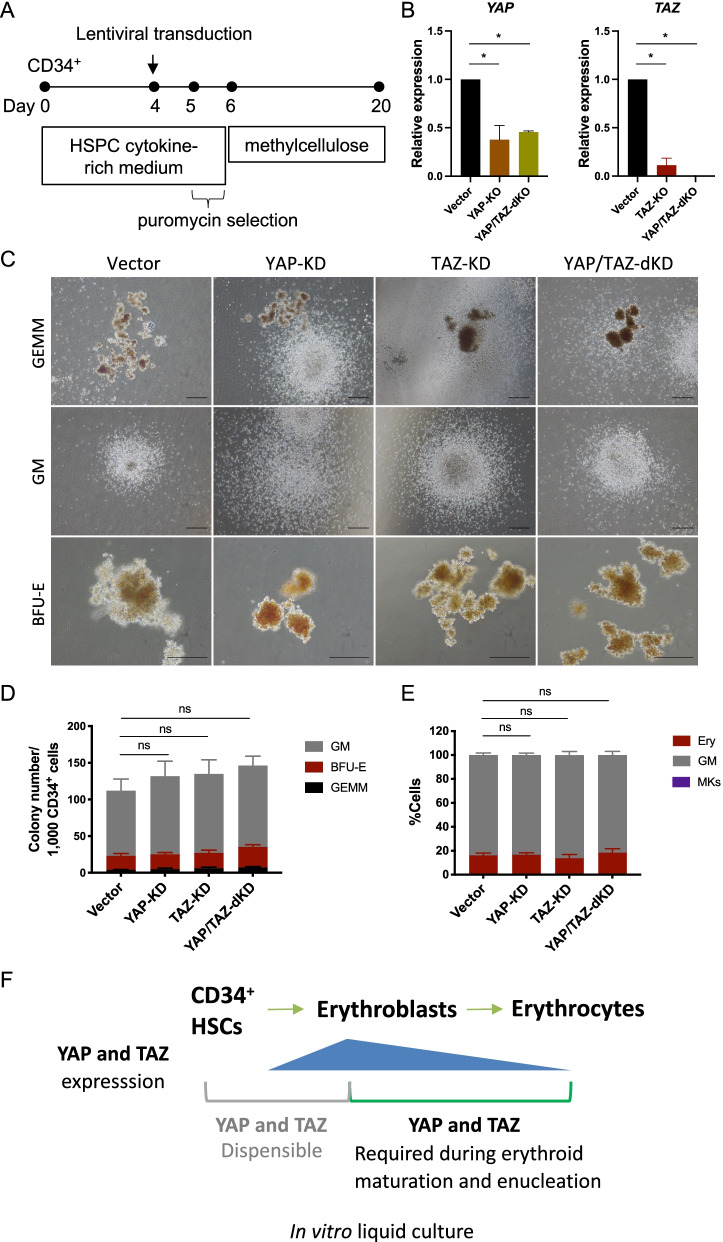
Results: YAP and TAZ were barely detectable in human HSCs, but became highly expressed in pro-erythroblasts and erythroblasts. Depletion or knockdown of YAP and/or TAZ did not affect the ability of HSC lineage specification to erythroid lineage in either methylcellulose assay or liquid culture. However, depletion of YAP and TAZ did impair erythroblast terminal differentiation to erythrocytes and their enucleation. Moreover, ectopic expression of YAP and TAZ in pro-erythroblasts did not exert an apparent effect on erythroid differentiation, expansion, or morphology.

Conclusions: This study demonstrated that YAP/TAZ plays important role in erythroid maturation and enucleation but is dispensable for lineage determination of human HSCs.

Keywords: Erythroid differentiation; Hematopoietic stem cells; Hippo pathway; TAZ; YAP.


Conflict of interest statement

All authors declare no personal or professional conflicts of interest relating to any aspect of this study.

Figures

Fig. 1
Fig. 1
Expression of YAP and TAZ in CD34+ HSC-derived erythroblasts. A Schematic of erythroid differentiation from human CD34+ HSCs in the three-stage erythroid culture system. B Wright’s staining of erythroid cells during differentiation of mobilized-peripheral blood (PB)- and cord blood (CB)-CD34+ HSCs showing the morphologic change from CD34+ HSC to pro-erythroblasts on day 8, erythroblasts on days 11–15, and mature erythrocytes on days 18 and 20 in in vitro culture, respectively. C Percentage of mature erythrocytes derived from PB- and CB-CD34+ HSCs in the three-stage erythroid culture system at the end of culture. At least 500 cells were counted in each group (n = 4). D, F Western blot analysis of total YAP and TAZ expression at days (D) 8, 11, 15, and 18 of erythroid-differentiated cells derived from PB- and CB-CD34+ HSCs, respectively. Relative expression levels of YAP and TAZ to β-actin as measured by Image J software were labeled in red (PB and CB, n = 5, 3, respectively). E, G Fold intensity of YAP and TAZ were analyzed compared to CD34+ cells. Data represent the mean ± standard error of the mean (SEM), *p < 0.5. Scale bar, 20 μm
Fig. 2
Fig. 2
Depletion of YAP/TAZ protein by DH-impaired erythroid cell proliferation and maturation. A Expression of YAP and TAZ after 10 μM lysophosphatidic acid (LPA; a YAP/TAZ activator) and dobutamine hydrochloride (DH; a YAP/TAZ inhibitor) treatment of PB-CD34+ HSC-derived erythroblasts for 11 days. Relative levels to β-actin were labeled in red. B Fold increase of cells during erythroid differentiation from PB-CD34+ HSCs after treatment with LPA (green) and DH (red) when compared to control (black) (n = 5). C Cell apoptosis of PB-CD34+ HSC-derived erythroid cells at days 15, and 18 after LPA and DH treatment, as analyzed by Annexin V and 7-AAD staining (n = 3). D Representative cell morphology during erythroid differentiation from PB-CD34+ HSCs showed morphological delay around day 15, and most of the remaining cells were erythroblasts (black arrow) at day 18 after DH treatment. E Percentage of mature erythrocytes and erythroblasts at the terminal stage of differentiation (day 18). At least 500 cells were counted in each group (n = 9). F Expression of the erythroid-specific genes KLF1 and GATA1, after LPA and DH treatment for 8 days (n = 3). Data represent the mean ± SEM, *p < 0.5, **p < 0.01, ***p < 0.001. Scale bar, 20 μm
Fig. 3
Fig. 3
Inhibition of YAP/TAZ-TEAD interaction by verteporfin impaired erythroid differentiation. A Fold increase of PB-CD34+ HSC-derived erythroid cells after treatment with verteporfin (VP) at 1.0 μM (orange) and 1.5 μM (purple) compared to control (black) (n = 3). B Representative cell morphology during erythroid differentiation after VP treatment. C Percentages of mature erythrocytes and erythroblasts on day 18. At least 500 cells were counted in each group (n = 3). Data represent the mean ± SEM, *p < 0.5. Scale bar, 20 μm
Fig. 4
Fig. 4
DH treatment specifically impaired erythroid maturation and enucleation from erythroblasts to erythrocytes. A Schematic of DH treatment at various time points during erythroid differentiation. B Representative of erythroid cell morphology of differentiated cells at day 18 following treatment with DH at the various time points described above, showing mature erythrocytes (red arrows) and erythroblasts (black arrows). C Percentages of mature erythrocytes and erythroblasts after timed DH treatment during differentiation at the terminal differentiation stage (day 18). At least 500 cells were counted in each group (n = 4). Data represent the mean ± SEM, *p < 0.5, **p < 0.01. Scale bar, 20 μm
Fig. 5
Fig. 5
YAP and TAZ knockdown delayed erythroid maturation and enucleation. A Schematic of YAP knockdown during erythroid differentiation using the CRISPR/Cas9 PX458-GFP_gYAP plasmid. B YAP mRNA expression of PB- and CB-CD34+ HSC-derived erythroblasts at 3 days post-nucleofection, as analyzed by qRT-PCR (n = 4). C A representative of erythroid cell morphology during differentiation after YAP-KD showed delayed differentiation and more immature cells (black arrow) compared to control. D Percentages of cells after YAP-KD of PB- and CB-CD34+ HSCs counted at day 15 of differentiation. At least 200 cells were counted in each group (n = 4). E KLF1 mRNA expression of sorted GFP+ cells at 3 days after nucleofection (n = 3). F Schematic of TAZ knockdown during erythroid differentiation using CRISPR/Cas9 pLenti_gWWTR1 (TAZ). G TAZ mRNA expression at 3 days post-nucleofection (n = 3, 4, respectively). H Representative erythroid cell morphology derived from PB- and CB-CD34+ HSCs, after TAZ-KD. I Percentages of cells after TAZ-KD at day 15 of differentiation. At least 200 cells were counted in each group (n = 3, 4, respectively). J KLF1 mRNA expression after TAZ-KD analyzed 3 days after nucleofection (n = 3). Data represent the mean ± SEM. *p < 0.05, **p < 0.01. Scale bar, 20 μm
Fig. 6
Fig. 6
Overexpression of YAP/TAZ had no apparent effect on erythroid differentiation. A Schematic of YAP and TAZ overexpression in erythroid cells using constitutively active YAP and TAZ plasmids named YAPS5A and TAZS89A, respectively. B YAP protein expression analyzed by Western blotting at 3 days post-nucleofection and relative expression to β-actin were labeled in red. C YAP mRNA expression as analyzed by qRT-PCR at 3 days post-nucleofection (n = 4). D TAZ protein expression as analyzed by Western blotting at 3 days post-nucleofection and relative expression to β-actin were labeled in red. E TAZ mRNA expression was analyzed by qRT-PCR at 3 days post-nucleofection (n = 5). F, G Representative cell morphology after YAP and TAZ overexpression. H Percentages of cells after YAP and TAZ overexpression at day 18 of differentiation. At least 200 cells were counted in each group (n = 3). Data represented in the mean ± SEM. *p < 0.05, **p < 0.01. Scale bar, 20 μm
Fig. 7
Fig. 7
Knockdown of YAP or TAZ in CD34+ HSCs did not alter HSC commitment to erythroid or myeloid lineages. A Schematic of YAP and TAZ knockdown in CD34+ HSCs using CRISPR/Cas9 lentiviral system. B YAP and TAZ mRNA expression as analyzed by qRT-PCR at 2 days after lentiviral transduction (n = 4, 3 respectively). C Representative of colony-forming unit (CFU) assay of YAP-KD, TAZ-KD, and YAP/TAZ-double (d) KD in PB-CD34+ HSCs after being cultured in methylcellulose for 14 days showing colony-forming unit granulocyte/erythroid/macrophage/megakaryocyte (CFU-GEMM), colony-forming unit granulocyte/macrophage (CFU-GM) and Burst-forming unit erythroid (BFU-E). D The colony number of YAP-KD, TAZ-KD, and YAP/TAZ-dKD derived from PB-CD34+ HSCs counted on day 14 of methylcellulose culture (n = 3). E Percentage of erythroid cells (Ery) and granulocytes/macrophages (GM) after YAP-KD, TAZ-KD, and YAP/TAZ-dKD of cells from methylcellulose (n = 3). F Summary of this study. Data represent the mean ± SEM, *p < 0.05. Scale bar, 200 μM

Similar articles

Cited by

References

    1. Trakarnsanga K, Griffiths RE, Wilson MC, Blair A, Satchwell TJ, Meinders M, et al. An immortalized adult human erythroid line facilitates sustainable and scalable generation of functional red cells. Nat Commun. 2017;8:14750. doi: 10.1038/ncomms14750. - DOI - PMC - PubMed
    1. Ingley E. Integrating novel signaling pathways involved in erythropoiesis. IUBMB Life. 2012;64(5):402–410. doi: 10.1002/iub.1024. - DOI - PubMed
    1. Xu T, Wang W, Zhang S, Stewart RA, Yu W. Identifying tumor suppressors in genetic mosaics: the Drosophila lats gene encodes a putative protein kinase. Development. 1995;121(4):1053–1063. doi: 10.1242/dev.121.4.1053. - DOI - PubMed
    1. Justice RW, Zilian O, Woods DF, Noll M, Bryant PJ. The Drosophila tumor suppressor gene warts encodes a homolog of human myotonic dystrophy kinase and is required for the control of cell shape and proliferation. Genes Dev. 1995;9(5):534–546. doi: 10.1101/gad.9.5.534. - DOI - PubMed
    1. Wu S, Huang J, Dong J, Pan D. Hippo encodes a Ste-20 family protein kinase that restricts cell proliferation and promotes apoptosis in conjunction with salvador and warts. Cell. 2003;114(4):445–456. doi: 10.1016/S0092-8674(03)00549-X. - DOI - PubMed

MeSH terms

Substances