Review
. 2022 Dec;149(14):1842-1861.
doi: 10.1017/S0031182022001160. Epub 2022 Aug 16.

Parasites and the neuroendocrine control of fish intestinal function: an ancient struggle between pathogens and host

Affiliations
Review

Parasites and the neuroendocrine control of fish intestinal function: an ancient struggle between pathogens and host

Giampaolo Bosi et al. Parasitology. 2022 Dec.

Abstract

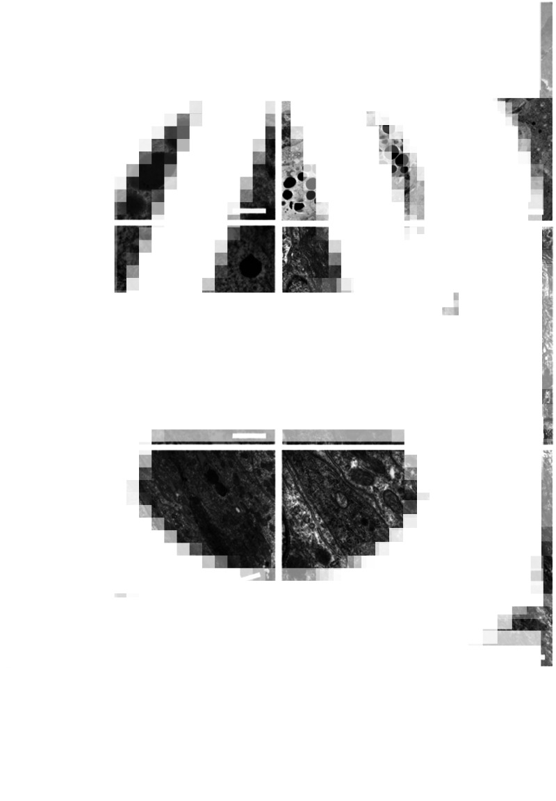
Most individual fish in wild and farmed populations can be infected with parasites. Fish intestines can harbour protozoans, myxozoans and helminths, which include several species of digeneans, cestodes, nematodes and acanthocephalans. Enteric parasites often induce inflammation of the intestine; the pathogen provokes changes in the host physiology, which will be genetically selected for if they benefit the parasite. The host response to intestinal parasites involves neural, endocrine and immune systems and interaction among these systems is coordinated by hormones, chemokines, cytokines and neurotransmitters including peptides. Intestinal fish parasites have effects on the components of the enteric nervous and endocrine systems; mechanical/chemical changes impair the activity of these systems, including gut motility and digestion. Investigations on the role of the neuroendocrine system in response to fish intestinal parasites are very few. This paper provides immunohistochemical and ultrastructural data on effects of parasites on the enteric nervous system and the enteric endocrine system in several fish–parasite systems. Emphasis is on the occurrence of 21 molecules including cholecystokinin-8, neuropeptide Y, enkephalins, galanin, vasoactive intestinal peptide and serotonin in infected tissues.

Keywords: Enteric endocrine system; enteric nervous system; gut physiology; intestinal parasites; teleost.


Conflict of interest statement

The authors declare having no conflict of interest.

Figures

None
Graphical abstract
Fig. 1.
Fig. 1.
Schematic view of the intestinal wall of fish with attached acanthocephalan. The intestinal mucosa is lined by a simple columnar epithelium consisting of typical epithelial cells with a sparse intermingling of mucus cells and ECs. Note the presence of the myenteric plexus with neurons between 2 muscle layers.
Fig. 2.
Fig. 2.
Components of the enteric nervous system and EES in several fish–helminth systems. (A) Salmo trutta infected with Pomphorhynchus laevis (out of microscope field), showing neurons of the myenteric plexus (thick arrows) and nerve fibres (thin arrows) immunoreactive to the anti-SP between the inner and outer intestinal muscle layers. The curved arrows indicate ECs in the epithelium. In the tunica propria-submucosa, numerous SP-positive mast cells are evident (arrowheads). CML, LML, circular and longitudinal muscle layer of tunica muscularis; TM, tunica mucosa; TPS, tunica propria-submucosa; scale bar: 100 μm. (B) Oncorhynchus mykiss parasitized with Eubothrium crassum (out of microscope field) showing 2 open-type ECs immunoreactive to anti-CCK8 in the rainbow trout intestinal epithelium; 1 of them is near a mucus cell (asterisk). Note the neuropod (thin arrow) of the cell on the right; scale bar: 10 μm. (C) Esox lucius harbouring Acanthocephalus lucii (out of microscope field), showing 2 closed-type ECs immunoreactive to anti-somatostatin-14 in the gastric epithelium of the pike. The neuropod is evident in the top left EC (thin arrow); scale bar: 20 μm.
Fig. 3.
Fig. 3.
Close relationship between ECs and epithelial components of the innate defence against parasites. Both images are from Silurus glanis intestine infected with the cestode Glanitaenia osculata (not shown). (A) ECs (curved arrows) immunoreactive to anti-serotonin close to the epithelial mucus cells (thin arrows). Immunohistochemistry counterstained with the Alcian blue/periodic acid Schiff method (AB/PAS). The AB/PAS stain reveals mucus cells in blue, magenta or violet depending on their content of acidic, neutral or mixed (acidic + neutral) mucins; scale bar: 50 μm. (B) ECs (curved arrows) immunoreactive to anti-met-enkephalin near intraepithelial mast cells (thick arrows). Mast cells are magenta due to periodic acid Schiff. Immunohistochemistry counterstained with the AB/PAS stain; scale bar: 50 μm.
Fig. 4.
Fig. 4.
Fish infected with intestinal digeneans. (A) The digenean Helicometra fasciata (arrow) attached with its sucker (arrowheads) to an intestinal fold (curved arrow) of the European eel. Azan Mallory stained; scale bar: 100 μm. (B) ECs (curved arrows) immunoreactive to anti-met-enkephalin in the intestinal epithelium of the Squalius cephalus infected with a digenean (d); scale bar: 100 μm. (C) Chelon saliens parasitized with Dicrogaster contractus (out of microscope field). Pituitary adenylate cyclase-activating polypeptide-immunoreactive ECs (curved arrows), and neurons and nerve fibres (thin arrows) in the infected mullet intestine; scale bar: 100 μm.
Fig. 5.
Fig. 5.
EES in fish infected with intestinal cestodes. (A) Silurus glanis parasitized with G. osculata (arrow). IF, intestinal fold. Haematoxylin–eosin stain; scale bar: 50 μm. (B) Salmo trutta infected with Cyathocephalus truncatus (arrow). ECs (curved arrows) immunoreactive to anti-glucagon in the intestinal epithelium are evident; scale bar: 100 μm. (C) Numerous ECs (curved arrows) immunoreactive to anti-CCK8 in the intestine of O. mykiss infected with the cestode E. crassum (arrow). The asterisk indicates a pyloric caecum. M, muscle layer; scale bar: 500 μm. (D) High number of ECs (curved arrows) in the intestinal epithelium of the stickleback Gasterosteus aculeatus parasitized with cestodes (not shown); scale bar: 50 μm.
Fig. 6.
Fig. 6.
Enteric nervous system and EES in fish infected with intestinal cestodes. (A) Salmo trutta infected with C. truncatus (arrowhead). Epithelial ECs (curved arrows) and nerve fibres (thick arrows) in the inner circular muscle layer, both immunoreactive to anti-met-enkephalin; scale bar: 100 μm. (B) Anguilla anguilla harbouring Proteocephalus macrocephalus (not shown). High magnification of neurons and nerve fibres (thick arrows) immunoreactive to pituitary adenylate cyclase-activating polypeptide in the myenteric plexus; scale bar: 100 μm.
Fig. 7.
Fig. 7.
Fish intestine infected with acanthocephalans. (A) Salmo trutta parasitized with Dentitruncus truttae, parasite proboscis (arrow) reached tunica propria-submucosa (asterisk). AB/PAS; scale bar: 100 μm. (B) Squalius cephalus harbouring P. laevis, showing anti-galanin immunoreactive neurons and nerve fibres (thick arrows) in the myenteric plexus of the parasitized chub. The thin arrows show subtle nerve fibres in the connective capsule (asterisk) around the bulb of the acanthocephalan (not shown); scale bar: 50 μm. (C) Salmo trutta infected with P. laevis. High number of nerve endings (arrows) immunoreactive to anti-VIP in inner circular muscle layer of fish intestine. The asterisk indicates the acanthocephalan proboscis; scale bar: 50 μm. (D) Salmo truttaP. laevis system, showing neurons and nerve fibres (thick arrows) of the myenteric plexus immunoreactive to anti-VIP. Note the occurrence of numerous nerve endings in longitudinal muscle layers (thin arrows). The asterisk shows the bulb of worm surrounded by a connective capsule (cc); scale bar: 50 μm. (E) Coregonus lavaretus infected with D. truttae (not shown), several ECs (curved arrows) immunoreactive to anti-SP in the intestinal epithelium are evident. The ECs are close to mucus cells secreting mainly mixed mucins (acid + neutral, deep violet stain, AB/PAS); scale bar: 50 μm. (F) Squalius cephalusP. laevis system. In the intestinal epithelium of the infected chub, a high number of ECs (curved arrows) immunoreactive to anti-met-enkephalin are visible. The asterisk indicates the acanthocephalan; scale bar: 100 μm.
Fig. 8.
Fig. 8.
Fish intestine parasitized with nematodes. (A) Nematode (arrow) inside the C. lavaretus pyloric caecum is evident. Haematoxylin–eosin stain; scale bar: 200 μm. (B) Anguilla anguilla intestine is infected with Contracaecum rudolphii larva (asterisk) between the tunica propria-submucosa (TS) and tunica muscularis (TM). Haematoxylin–eosin stain; scale bar: 200 μm. (C) Microphotograph shows C. rudolphii larvae inside a cyst (asterisk) on the outer surface of the eel gut. In the myenteric plexus, neurons and nerve fibres (arrows) are immunoreactive to the anti-met-enkephalin; scale bar: 200 μm. (D) Some C. rudolphii larvae in 2 cysts on external surface of the eel gut, neurons and nerve fibres (arrows) of the myenteric plexus immunoreactive to anti-leu-enkephalin are visible; scale bar: 200 μm.
Fig. 9.
Fig. 9.
SB of A. anguilla infected with the nematode Anguillicoloides crassus (not shown). (A) Nerve fibres (thick arrows) immunoreactive to the anti-CGRP are numerous in the infected SB. Many subtle nerve fibres (thin arrow) are near blood vessels; scale bar: 50 μm. (B) The arrows indicate several nerve fibres immunoreactive to anti-VIP in a region rich in blood vessels (asterisks); scale bar: 50 μm. (C) Large ganglia with numerous neurons immunoreactive to anti-n-NOS in the tunica muscularis (TM) of the infected eel SB. E, epithelium; TPS, tunica propria-submucosa; scale bar: 100 μm. (D) Numerous anti-NPY immunoreactive ECs (curved arrows) in the epithelium of the infected eel SB. The blood vessels are ectatic and filled with erythrocytes (asterisks). The red thick arrows indicate 2 parasite larvae; scale bar: 100 μm.
Fig. 10.
Fig. 10.
Transmission electron micrographs of neuroendocrine cells in the intestine of different fish species infected with helminths. In all neuroendocrine cells, numerous small electron-dense granules in cytoplasm are evident. (A) Intestine of Chelon ramada infected with digeneans; within the epithelium a neuroendocrine cell (arrow) is attached to a mast cell (asterisk); scale bar: 0.80 μm. (B) A neuroendocrine cell (arrow) and a mast cell within the tunica propria-submucosa of the intestine of C. lavaretus harbouring a nematode; scale bar: 3 μm. (C) A neuroendocrine cell (arrow) between nuclei of 2 intestinal epithelial cells of E. lucius parasitized with an acanthocephalan; scale bar: 1.4 μm. (D) Micrograph shows a neuroendocrine cell (arrow) within the epithelium of A. anguilla intestine infected with digeneans; scale bar: 2 μm. (E) A neuroendocrine cell (arrow) within the intestinal epithelium of S. glanis harbouring a cestode; scale bar: 3 μm. (F) In a higher magnification of Fig. 2, several small electron-dense granules are visible near the rough endoplasmic reticulum (arrows) in the cytoplasm; scale bar: 0.6 μm.

Similar articles

Cited by

References

    1. Adamo SA (2002) Modulating the modulators: parasites, neuromodulators and host behavioural change. Brain Behavior and Evolution 60, 370–377. - PubMed
    1. Ahlman H and Nilsson O (2001) The gut as the largest endocrine organ in the body. Annals of Oncology 12, S63–S68. - PubMed
    1. Aldegunde M and Mancebo M (2006) Effects of neuropeptide Y on food intake and brain biogenic amines in the rainbow trout (Oncorhynchus mykiss). Peptides 27, 719–727. - PubMed
    1. Aldman G and Holmgren S (1995) Intraduodenal fat and amino acids activate gallbladder motility in the rainbow trout, Oncorhynchus mykiss. General and Comparative Endocrinology 100, 27–32. - PubMed
    1. Anderson RC (2000) Nematode Parasites of Vertebrates: Their Development and Transmission. Wallingford, UK: CABI Publishing.

Publication types