. 2022 Aug 23;23(17):9524.
doi: 10.3390/ijms23179524.

Mutant WDR45 Leads to Altered Ferritinophagy and Ferroptosis in β-Propeller Protein-Associated Neurodegeneration

Affiliations

Mutant WDR45 Leads to Altered Ferritinophagy and Ferroptosis in β-Propeller Protein-Associated Neurodegeneration

Sokhna Haissatou Diaw et al. Int J Mol Sci. .

Abstract

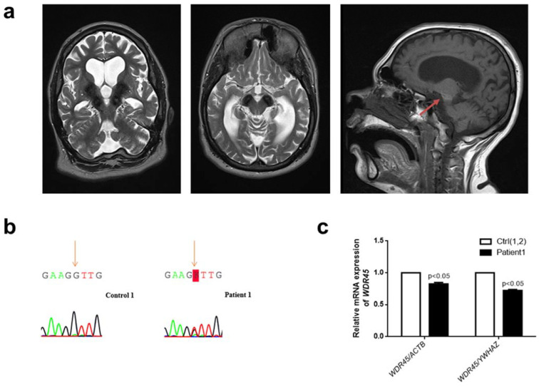
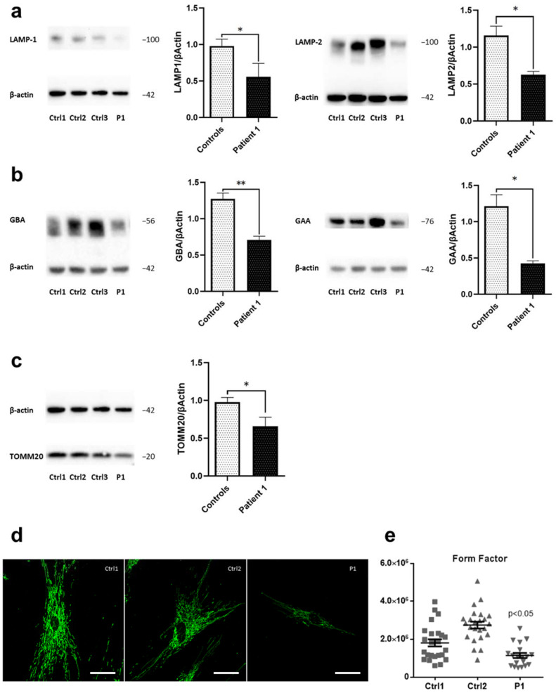
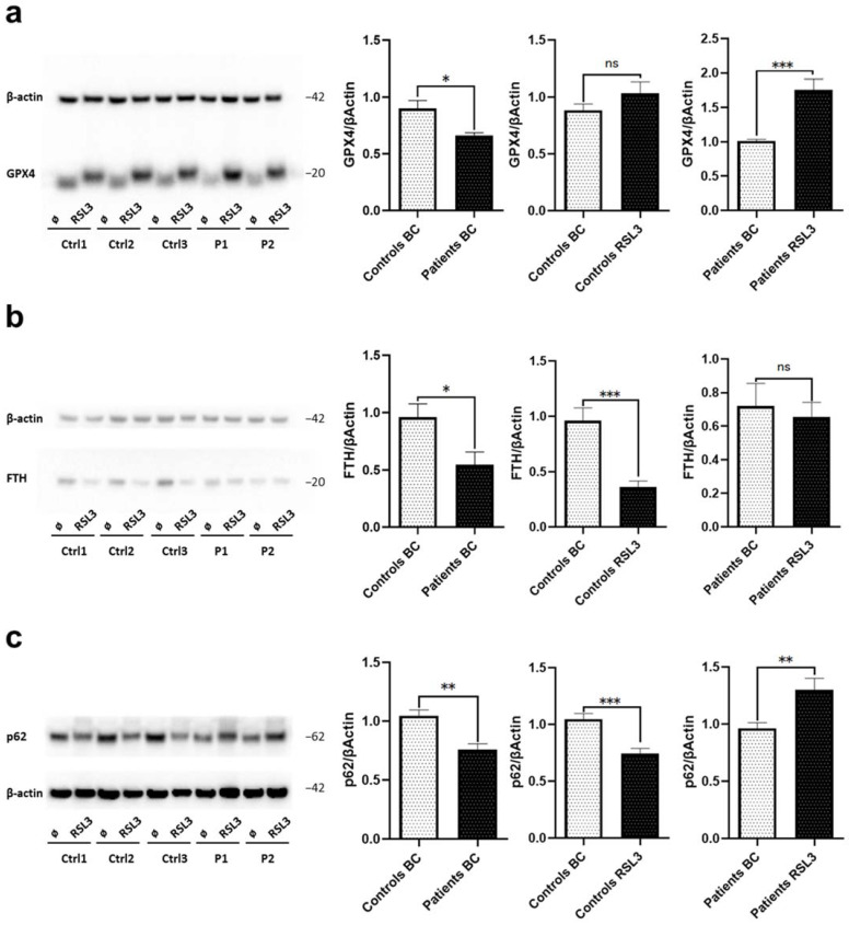
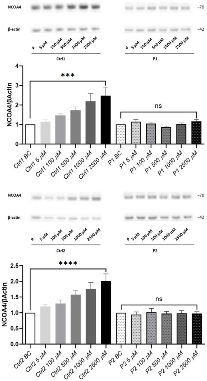
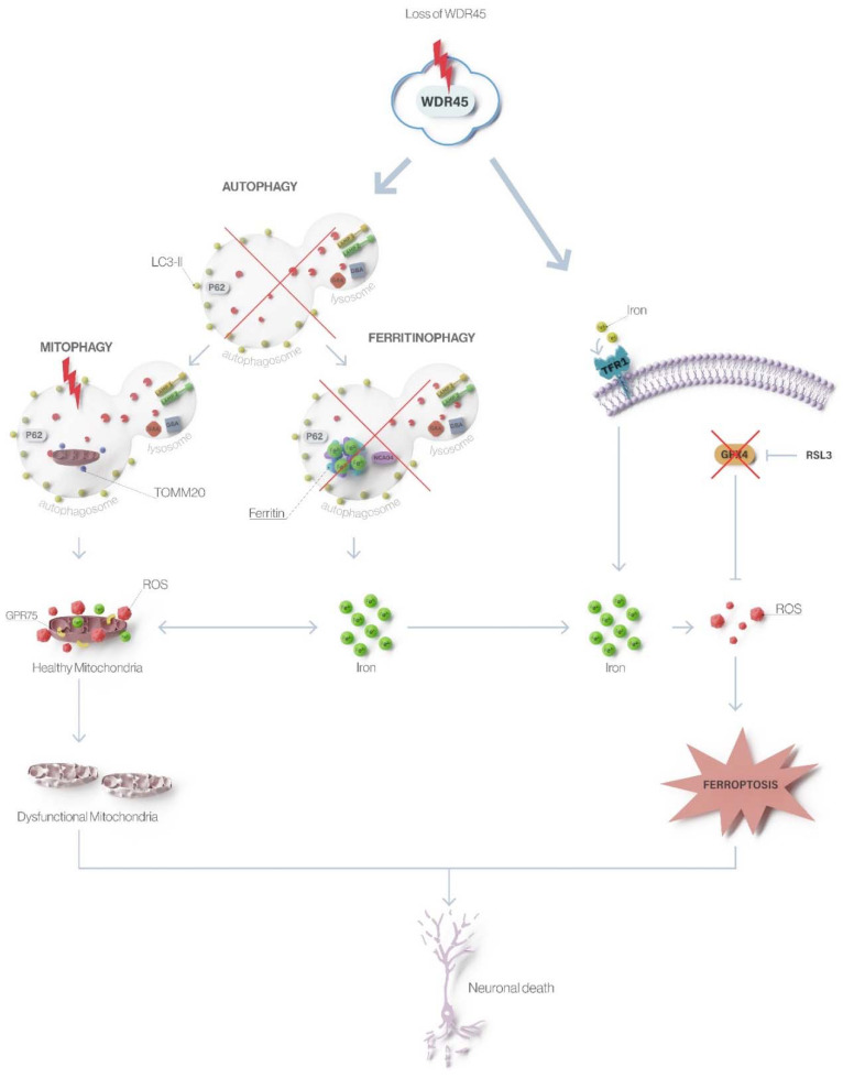
Beta-propeller protein-associated neurodegeneration (BPAN) is a subtype of neurodegeneration with brain iron accumulation (NBIA) caused by loss-of-function variants in WDR45. The underlying mechanism of iron accumulation in WDR45 deficiency remains elusive. We established a primary skin fibroblast culture of a new BPAN patient with a missense variant p.(Asn61Lys) in WDR45 (NM_007075.3: c.183C>A). The female patient has generalized dystonia, anarthria, parkinsonism, spasticity, stereotypies, and a distinctive cranial MRI with generalized brain atrophy, predominantly of the cerebellum. For the functional characterization of this variant and to provide a molecular link of WDR45 and iron accumulation, we looked for disease- and variant-related changes in the patient’s fibroblasts by qPCR, immunoblotting and immunofluorescence comparing to three controls and a previously reported WDR45 patient. We demonstrated molecular changes in mutant cells comprising an impaired mitochondrial network, decreased levels of lysosomal proteins and enzymes, and altered autophagy, confirming the pathogenicity of the variant. Compared to increased levels of the ferritinophagy marker Nuclear Coactivator 4 (NCOA4) in control cells upon iron treatment, patients’ cells revealed unchanged NCOA4 protein levels, indicating disturbed ferritinophagy. Additionally, we observed abnormal protein levels of markers of the iron-dependent cell death ferroptosis in patients’ cells. Altogether, our data suggests that WDR45 deficiency affects ferritinophagy and ferroptosis, consequentially disturbing iron recycling.

Keywords: BPAN; GBA; GPX4; NCOA4; WDR45; autophagy; ferritinophagy; ferroptosis.


Conflict of interest statement

The authors declare no conflict of interest.

Figures

Figure 1
Figure 1
Clinical and genetic analysis of a novel female patient (Patient 1) with a missense WDR45 variant. (a) Brain MRI from Patient 1. The globus pallidus and the substantia nigra are extensively hypointense bilaterally on axial T2 sequences indicating high levels of iron deposition. The characteristic BPAN T1-hyperintense halo surrounding the substantia nigra is also noted (red arrow). (b) Electropherograms of Sanger sequencing of complementary DNA (cDNA) from cultured fibroblasts from Patient 1 (right panel) and a healthy individual (Control 1) (left panel) in the reverse direction. The variant is highlighted: “K” represents “G” or “T.” (c) The quantification of WDR45 mRNA levels in fibroblasts from Patient 1 compared to fibroblasts from two healthy individuals (Control 1 and Control 2—Ctrl(1,2)). Means and standard error of the mean (SEM) are indicated. Quantification is shown with the mean control level set as 1. The results are based on the mean ratios of WDR45 expression compared to two reference genes, ACTB and YWHAZ. Statistical significance was analyzed by one-way ANOVA (p < 0.05 refers to a significant difference between Patient 1 and two healthy controls (Ctrl(1,2)).
Figure 2
Figure 2
Disrupted lysosomal and mitochondrial integrity in WDR45-mutant fibroblasts. Western blot analysis of total protein extract from: (a) fibroblasts from WDR45 mutation carrier (P1—Patient 1) and three healthy controls (Ctrl1, Ctrl2, Ctrl3) with antibodies against the lysosomal proteins LAMP-1 (left panel) and LAMP-2 (right panel), and β-actin (loading control), (b) with antibodies against lysosomal enzyme GBA and β-actin (loading control) (left panel) and with antibodies against lysosomal enzyme GAA and β-actin (loading control (right panel). (c) Patient 1’s fibroblasts and three healthy controls with antibodies against the mitochondrial protein TOMM20 and β-actin (loading control). Data analysis was carried out for all Western blots with Ctrl1 set as 1. The error bars indicate standard error of the mean of n ≥ 3 independent experiments. Statistical significance was analyzed by Unpaired t-test (* p < 0.05; ** p < 0.01). (d) The mitochondrial network was visualized by confocal microscopy in fixed cells immuno-stained with anti-GRP75 (green). The scale bar corresponds to 100 μm. (e) A mean form factor was calculated as a measure of mitochondrial interconnectivity by using ImageJ (NIH software). Each dot represents the value from a single cell (15–20 cells per cell culture), and the mean and the error bar (SEM) per individual are indicated. Statistical significance was analyzed by one-way ANOVA (p < 0.05 refers to a significantly decreased form factor in Patient 1’s fibroblasts compared to healthy controls).
Figure 3
Figure 3
WDR45-mutant fibroblasts exhibit altered autophagy. Western blot analysis of total protein extract from fibroblasts from: (a) WDR45 mutation carrier (P1-Patient 1) and three healthy controls (Ctrl1, Ctrl2, Ctrl3) with antibody against the autophagosome marker LC3-II (membrane-bound form), and β-actin (loading control) under basal conditions, (b) and upon treatment with 10 nM of Bafilomycin A1 for 6 h. (c) Patient 1’s fibroblasts (P1) and three healthy controls (Ctrl1, Ctrl2, Ctrl3) with antibodies against Beclin 1 and β-actin (loading control). Data analysis was carried out for all Western Blots with Ctrl1 set as 1. The error bars indicate the standard error of the mean of n ≥ 3 independent experiments. Statistical significance was analyzed by Unpaired t-test (ns refers to p ≥ 0.05; ** p < 0.01).
Figure 4
Figure 4
Loss of WDR45 is linked to disrupted iron recycling via ferroptosis. Western blot analysis of total protein extract from fibroblasts from: (a) Patients’ fibroblasts (P1, P2) and three healthy controls (Ctrl1, Ctrl2, Ctrl3) with antibodies against GPX4 and β-actin (loading control) under basal conditions and upon treatment with 1.5 µM of RSL3 for 6 h. (b) WDR45 mutation carriers (Patients, P1 and P2) and three healthy controls (Ctrl1, Ctrl2, Ctrl3) with antibody against FTH and β-actin (loading control) under basal conditions and upon treatment with 1.5 µM of RSL3 for 6 h. (c) Patients’ fibroblasts and three healthy controls with antibodies against p62 and β-actin (loading control) under basal conditions (BC) and upon treatment with 1.5 µM of RSL3 for 6 h. Data analysis was carried out for all Western blots with Ctrl1 in basal conditions set as 1 (first and second diagrams) or P1 in basal conditions (third diagram). The error bars indicate the standard error of the mean of n ≥ 3 independent experiments. Statistical significance was analyzed by Unpaired t-test (ns refers to p ≥ 0.05 (not significant); * p < 0.05; ** p < 0.01; *** p < 0.001).
Figure 5
Figure 5
Loss of WDR45 is linked to disrupted iron recycling via ferritinophagy. Western blot analysis of total protein extract from fibroblasts from: WDR45 mutation carriers (P1 and P2) and two healthy controls (Ctrl1 and Ctrl2) with antibody against NCOA4 and β-actin (loading control) under basal conditions (BC) and upon treatment with 5, 100, 500, 1000 and 2500 µM of FAC for 24 h. Data analysis was carried out for all Western blots with Ctrl1 BC and P1 (for the upper panel) and Ctrl2 BC and P2 (for the lower panel) set as 1. The error bars indicate the standard error of the mean of n ≥ 3 independent experiments. One-way ANOVA analyzed statistical significance with post-hoc Turkey test (ns refers to p ≥ 0.05 (not significant); *** p < 0.001 and **** p < 0.0001 refers to significantly increased NCOA4 protein levels in fibroblasts treated with 2500 µM of FAC compared to the same but untreated cells (BC)).
Figure 6
Figure 6
Loss of WDR45 leads to altered autophagy, ferritinophagy, and ferroptosis in β-propeller protein-associated neurodegeneration. Mutations in WDR45 cause impaired autophagy as the primary defect that further leads to disturbed degradation of iron-rich ferritin (affecting ferritinophagy) and iron-containing organelles like mitochondria (affecting mitophagy). Lack of ferritinophagy leads to increased total cellular iron levels, which, through ROS formation and oxidative stress, triggers ferroptosis resulting in neuronal death.

References

    1. Gregory A., Hayflick S. Neurodegeneration with Brain Iron Accumulation Disorders Overview. In: Adam M.P., Ardinger H.H., Pagon R.A., Wallace S.E., Bean L.J., Gripp K.W., Mirzaa G.M., Amemiya A., editors. GeneReviews®. University of Washington; Seattle, WA, USA: 1993. - PubMed
    1. Saitsu H., Nishimura T., Muramatsu K., Kodera H., Kumada S., Sugai K., Kasai-Yoshida E., Sawaura N., Nishida H., Hoshino A., et al. De Novo Mutations in the Autophagy Gene WDR45 Cause Static Encephalopathy of Childhood with Neurodegeneration in Adulthood. Nat. Genet. 2013;45:445–449. doi: 10.1038/ng.2562. - DOI - PubMed
    1. Haack T.B., Hogarth P., Kruer M.C., Gregory A., Wieland T., Schwarzmayr T., Graf E., Sanford L., Meyer E., Kara E., et al. Exome Sequencing Reveals De Novo WDR45 Mutations Causing a Phenotypically Distinct, X-Linked Dominant Form of NBIA. Am. J. Hum. Genet. 2012;91:1144–1149. doi: 10.1016/j.ajhg.2012.10.019. - DOI - PMC - PubMed
    1. Hayflick S.J., Kruer M.C., Gregory A., Haack T.B., Kurian M.A., Houlden H.H., Anderson J., Boddaert N., Sanford L., Harik S.I., et al. Beta-Propeller Protein-Associated Neurodegeneration: A New X-Linked Dominant Disorder with Brain Iron Accumulation. Brain. 2013;136:1708–1717. doi: 10.1093/brain/awt095. - DOI - PMC - PubMed
    1. Arber C.E., Li A., Houlden H., Wray S. Review: Insights into Molecular Mechanisms of Disease in Neurodegeneration with Brain Iron Accumulation: Unifying Theories: Mechanisms of Neurodegeneration with Brain Iron Accumulation. Neuropathol. Appl. Neurobiol. 2016;42:220–241. doi: 10.1111/nan.12242. - DOI - PMC - PubMed