. 2022 Aug 24;23(17):9579.
doi: 10.3390/ijms23179579.

Impact of Growth Conditions on Pseudomonas fluorescens Morphology Characterized by Atomic Force Microscopy

Affiliations

Impact of Growth Conditions on Pseudomonas fluorescens Morphology Characterized by Atomic Force Microscopy

Houssem Kahli et al. Int J Mol Sci. .

Abstract

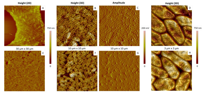
This work is dedicated to the characterization by Atomic Force Microscopy (AFM) of Pseudomonas fluorescens, bacteria having high potential in biotechnology. They were first studied first in optimal conditions in terms of culture medium and temperature. AFM revealed a more-or-less elongated morphology with typical dimensions in the micrometer range, and an organization of the outer membrane characterized by the presence of long and randomly distributed ripples, which are likely related to the organization of lipopolysaccharides (LPS). The outer membrane also presents invaginations, some of them showing a reorganization of ripples, which could be the first sign of a bacterial stress response. In a second step, bacteria grown under unfavorable conditions were characterized. The choice of the medium appeared to be more critical in the case of the second generation of cells, the less adapted medium inducing not only changes in the membrane organization but also larger damages in bacteria. An increased growth temperature affected both the usual "swollen" morphology and the organization of the outer membrane. Here also, LPS likely contribute to membrane remodelling, which makes them potential markers to track cell state changes.

Keywords: Atomic Force Microscopy; Gram-negative bacteria; Lipopolysaccharide; Pseudomonas fluorescens; bacterial surface; membrane; morphology; stress.


Conflict of interest statement

The authors declare no conflict of interest.

Figures

Figure 1
Figure 1
OM (A,C,E) and AFM height (B,D,F) images of P. fluorescens deposits made by simple deposition from suspensions at different O.D. values (O.D. = 1.0, images (A,B); O.D. = 0.5, images (C,D); O.D. = 0.3, images (E,F)).
Figure 2
Figure 2
AFM height or amplitude images of P. fluorescens deposits obtained by simple deposition (AD) or spin-coating (EH).
Figure 3
Figure 3
Morphological analysis of P. fluorescens cells. (AC) AFM 3D topography images of isolated cells; (D) AFM phase image corresponding to the red square in (C); (E) height profiles in length (blue) and width (red) of the cell shown in (C).
Figure 4
Figure 4
Histograms of P. fluorescens length (blue), width (red) and height (black). Measurements were performed on 120 aggregated cells (solid bars) and 40 isolated cells (hatched bars).
Figure 5
Figure 5
Membrane characteristics of P. fluorescens cells. (A,D) The 3D topographic AFM images of aggregated cells; (B,E) phase AFM images of areas in the red and orange squares in images (A) and (D), respectively; (C,F) phase AFM images of areas in the red and orange squares in images (B) and (E), respectively.
Figure 6
Figure 6
Relationship between membrane organization and invagination, shown by AFM images obtained in air and in tapping mode. (A) The 3D topographic images of three bacteria. Characteristics of cells numbered 1 and 2 are described in the main text; (B) corresponding phase image; (CF) phase images of areas in the red, grey, green and yellow squares in figure (B), respectively. White arrows indicate the bottom of the invagination.
Figure 7
Figure 7
Relationship between membrane organization and invaginations. (A,D,G) AFM 3D topographic images of three bacteria; (B,E,F) corresponding phase images; AFM images shown in (C,F,I) correspond to the areas in the squares indicated in (B,E,H), respectively. White arrows indicate the bottom of the invagination.
Figure 8
Figure 8
AFM amplitude images of P. fluorescens bacteria of first generation deposited by simple deposition from a culture in LB (A,B) and MH (C,D) media (15 h, 28 °C).
Figure 9
Figure 9
AFM amplitude images of P. fluorescens bacteria of second generation deposited by simple deposition from a culture in LB (A,B) and MH (C,D) media (15 h, 28 °C).
Figure 10
Figure 10
AFM images of P. fluorescens bacteria incubated for 15 h in LB medium at 37 °C (AD) and 28 °C (EH). AFM images are 3D height or topographic ones (A,C,E,G) and amplitude ones (B,D,F,H).
Figure 11
Figure 11
Effect of the incubation time on the morphology of aggregated P. fluorescens bacteria deposited by simple deposition. The bacteria were cultured in LB medium at 28 °C during different culture times: 12 h (A,B), 15h (C,D) and 24 h (E,F). AFM images are 3D height or topographic ones (A,C,E) and amplitude ones (B,D,F).
Figure 12
Figure 12
Effect of the culture time on the morphology of isolated P. fluorescens bacteria deposited by simple deposition. The bacteria were cultured in LB medium at 28 °C during different incubation times: 12 h (A,B), 15 h (C,D) and 24 h (E,F). AFM images are 3D height or topographic ones (A,C,E) and amplitude ones (B,D,F).

Similar articles

Cited by

References

    1. Lemanceau P. Effets bénéfiques de rhizobactéries sur les plantes: Exemple des Pseudomonas spp. fluorescents. Agronomie. 1992;12:413–437. doi: 10.1051/agro:19920601. - DOI
    1. Meyer J.-M. Pyoverdines: Pigments, siderophores and potential taxonomic markers of Fluorescent Pseudomonas Species. Arch. Microbiol. 2000;174:135–142. doi: 10.1007/s002030000188. - DOI - PubMed
    1. Baranski R., Klocke E., Nothnagel T. enhancing resistance of transgenic carrot to fungal pathogens by the expression of Pseudomonas Fluorescens Microbial Factor 3 (MF3) Gene. Physiol. Mol. Plant Pathol. 2007;71:88–95. doi: 10.1016/j.pmpp.2007.12.002. - DOI
    1. Wu B., He T., Wang Z., Qiao S., Wang Y., Xu F., Xu H. Insight into the mechanisms of plant growth promoting strain SNB6 on Enhancing the Phytoextraction in Cadmium Contaminated Soil. J. Hazard. Mater. 2020;385:121587. doi: 10.1016/j.jhazmat.2019.121587. - DOI - PubMed
    1. Bolwerk A., Lagopodi A.L., Wijfjes A.H.M., Lamers G.E.M., Chin-A-Woeng T.F.C., Lugtenberg B.J.J., Bloemberg G.V. Interactions in the Tomato Rhizosphere of Two Pseudomonas Biocontrol Strains with the Phytopathogenic Fungus Fusarium oxysporum f. Sp. Radicis-Lycopersici. MPMI. 2003;16:983–993. doi: 10.1094/MPMI.2003.16.11.983. - DOI - PubMed

MeSH terms

Substances

LinkOut - more resources