. 2022 Aug 27;23(17):9735.
doi: 10.3390/ijms23179735.

Balanced Free Essential Amino Acids and Resistance Exercise Training Synergistically Improve Dexamethasone-Induced Impairments in Muscle Strength, Endurance, and Insulin Sensitivity in Mice

Affiliations

Balanced Free Essential Amino Acids and Resistance Exercise Training Synergistically Improve Dexamethasone-Induced Impairments in Muscle Strength, Endurance, and Insulin Sensitivity in Mice

Jiwoong Jang et al. Int J Mol Sci. .

Abstract

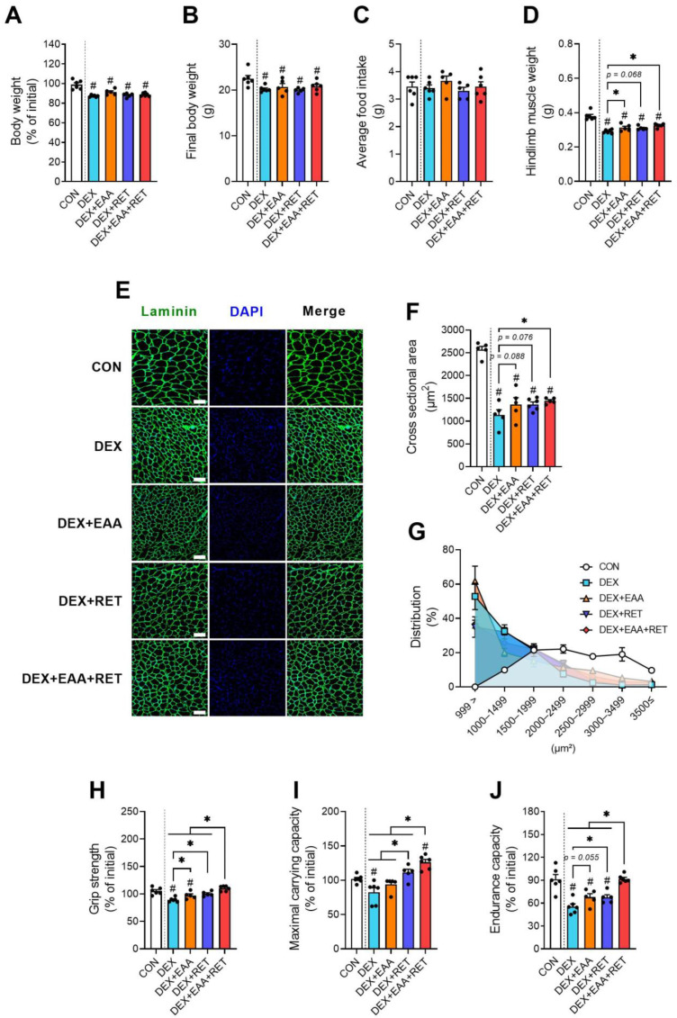
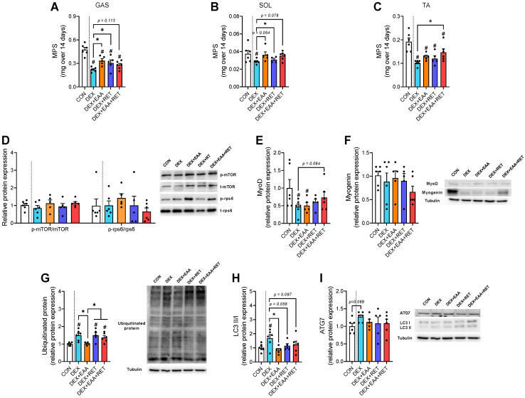
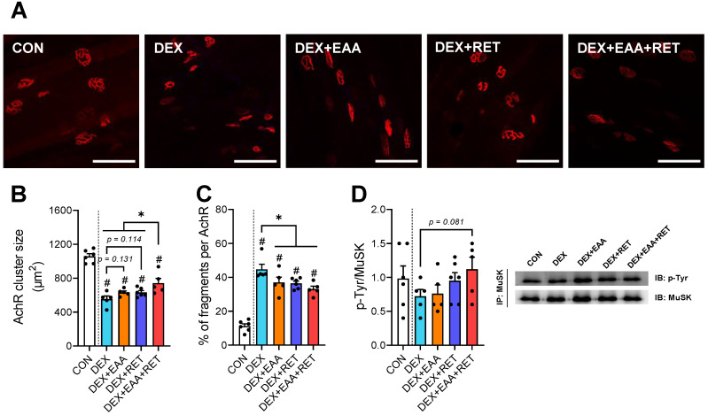
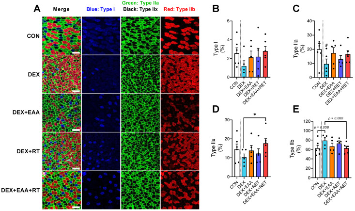
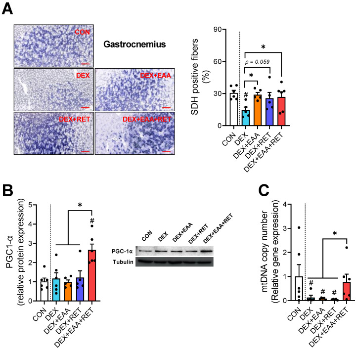
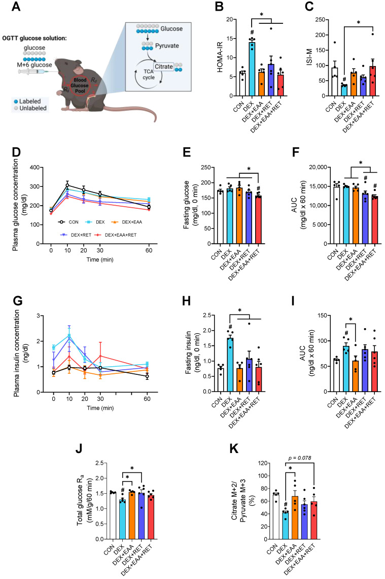
Our previous study shows that an essential amino acid (EAA)-enriched diet attenuates dexamethasone (DEX)-induced declines in muscle mass and strength, as well as insulin sensitivity, but does not affect endurance. In the present study, we hypothesized that the beneficial effects will be synergized by adding resistance exercise training (RET) to EAA, and diet-free EAA would improve endurance. To test hypotheses, mice were randomized into the following four groups: control, EAA, RET, and EAA+RET. All mice except the control were subjected to DEX treatment. We evaluated the cumulative rate of myofibrillar protein synthesis (MPS) using 2H2O labeling and mass spectrometry. Neuromuscular junction (NMJ) stability, mitochondrial contents, and molecular signaling were demonstrated in skeletal muscle. Insulin sensitivity and glucose metabolism using 13C6-glucose tracing during oral glucose tolerance tests were analyzed. We found that EAA and RET synergistically improve muscle mass and/or strength, and endurance capacity, as well as insulin sensitivity, and glucose metabolism in DEX-treated muscle. These improvements are accomplished, in part, through improvements in myofibrillar protein synthesis, NMJ, fiber type preservation, and/or mitochondrial biogenesis. In conclusion, free EAA supplementation, particularly when combined with RET, can serve as an effective means that counteracts the adverse effects on muscle of DEX that are found frequently in clinical settings.

Keywords: dexamethasone; essential amino acids; glucose metabolic flux; mitochondrial biogenesis; muscle atrophy; neuromuscular junction stability; physical performance; protein turnover; resistance exercise training.


Conflict of interest statement

I.-Y.K., S.H.P. and J.Y.J. are stockholders of Myocare. Inc., and Robert Wolfe is a shareholder in Essential Blends, LLC, and the Amino Company, LLC. All others have no potential conflict of interest.

Figures

Figure 1
Figure 1
EAA and RET synergistically improve DEX-induced declines in muscle strength and endurance. (A) The changes in body weight after 14 days of each treatment (n = 5–6). (B) The final body weight after 14 days of each treatment (n = 5–6). (C) Average food intake over 14 days (n = 5–6). (D) Total hindlimb muscle mass (soleus, plantaris, gastrocnemius, extensor digitorum longus, and tibialis anterior) (n = 5–6). (E) Representative image of a gastrocnemius muscle cross-section with laminin (green) and DAPI (blue) staining. Scale bar, 100 μm. (F) Cross-sectional area in gastrocnemius. (G) Myofiber size frequency distribution of gastrocnemius (n = 5–6). (H) Changes in four-limb grip strength over 14 days (n = 5–6). (I) Changes in maximal carrying capacity over 14 days (n = 5–6). (J) Changes in endurance capacity over 14 days (n = 5–6). Data are presented as mean ± S.E. # Significant difference from control group (p < 0.05). * Significant difference between labeled groups (p < 0.05). CON, sedentary control; DEX, dexamethasone; EAA, essential amino acids; RET, resistance exercise training.
Figure 2
Figure 2
EAA and/or RET stimulate muscle protein synthesis rate and suppression of autophagy activation in muscle of DEX-treated mice. Integrated myofibrillar protein synthesis rate in (A) gastrocnemius muscle (mixed muscle), (B) soleus muscle (slow oxidative muscle), and (C) tibialis anterior muscle (fast glycolytic muscle) for 14 days (n = 5–6 per muscle type). (D) Representative image and relative protein expression of mTOR and rps6 in gastrocnemius (n = 5–6). Representative image and relative protein expression of (E) MyoD and (F) myogenin in gastrocnemius (n = 5–6). (G) Representative image and relative protein expression of ubiquitinated-protein in gastrocnemius (n = 5–6). (H) Representative image and relative protein expression of the ratio of LC3B II/I and (I) ATG7 in gastrocnemius (n = 5–6). Data are presented as mean ± S.E. # Significant difference from control group (p < 0.05). * Significant difference between labeled groups (p < 0.05). CON, sedentary control; DEX, dexamethasone; EAA, essential amino acids; RET, resistance exercise training; MPS, myofibrillar protein synthesis; mTORC1, mammalian target of rapamycin complex 1; rps6, ribosomal protein s6; MyoD, myoblast determination protein 1; LC3, microtubule-association protein 1A/1B-light chain 3; ATG7, autophagy-related 7; GAS, gastrocnemius muscle; SOL, soleus muscle; TA, tibialis anterior muscle.
Figure 3
Figure 3
Combined treatment of EAA and RET restores a decrease in NMJ stability in skeletal muscle by DEX treatment. (A) Representative image of an acetylcholine receptor staining in plantaris. (n = 5–6). Scale bar, 100 μm. (B) Acetylcholine receptor cluster size in plantaris (n = 5–6). (C) Percentage of fragmented acetylcholine receptor in plantaris (n = 5–6). (D) Representative image and relative protein expression of Musk (n = 5–6). Data are presented as mean ± S.E. # Significant difference from control group (p < 0.05). * Significant difference between labeled groups (p < 0.05). CON, sedentary control; DEX, dexamethasone; EAAs, essential amino acids; RET, resistance exercise training; PLAN, plantaris muscle; AchR, acetylcholine receptor; MuSK, muscle-specific tyrosine kinase.
Figure 4
Figure 4
Combined treatment of EAA and RET prevents muscle fiber type shifting to fast glycolytic fiber due to DEX. (A) Representative image of immunofluorescence in gastrocnemius muscle for the triple-labeling with MHC type I (blue), MHC type IIa (green), MHC type IIx (black), and MHC type IIB (red). Scale bar, 100 μm (n = 5–6). (B) The proportion of MHC type-I-specific fibers (n = 5–6). (C) The proportion of MHC type-IIa-specific fibers (n = 5–6) (D) The proportion of MHC type-IIx-specific fibers (n = 5–6). (E) The proportion of MHC type-Iib-specific fibers (n = 5–6). Data are presented as mean ± S.E. # Significant difference from control group (p < 0.05). * Significant difference between labeled groups (p < 0.05). CON, sedentary control; DEX, dexamethasone; EAA, essential amino acids; RET, resistance exercise training; MHC, myosin heavy chain.
Figure 5
Figure 5
Combined treatment of EAA and RET completely restores the loss of mitochondrial biogenesis in skeletal muscle by DEX treatment. (A) Representative image and the proportion of SDH-positive muscle fibers in gastrocnemius (n = 5–6). Scale bar, 200 μm. (B) Representative image and relative protein expression of the PGC1-α in gastrocnemius (n = 5–6). (C) The relative mtDNA copy number with qPCR measurement in gastrocnemius (n = 5–6). Data are presented as mean ± S.E. # Significant difference from control group (p < 0.05). * Significant difference between labeled groups (p < 0.05). CON, sedentary control; DEX, dexamethasone; EAA, essential amino acids; RET, resistance exercise training; SDH, succinate dehydrogenase; PGC1-α, peroxisome proliferator-activated receptor gamma coactivator 1-alpha; mtDNA, mitochondrial DNA.
Figure 6
Figure 6
Combined treatment of EAA and RET completely restores impaired insulin sensitivity by DEX treatment. (A) Schematic showing glucose flux in vivo at systemic and myocellular levels using U-13C6 glucose tracer during OGTT. This figure was created with BioRender.com. (B) HOMA-IR index (n = 5–6). (C) Matsuda insulin sensitivity index (n = 5–6). (D) Plasma glucose concentration during the OGTT (n = 5–6). (E) Plasma glucose concentration at fasting state over 6 h (n = 5–6). (F) Area under the curve of plasma glucose concentration during OGTT (n = 5–6). (G) Plasma insulin concentration during the OGTT (n = 5–6). (H) Plasma insulin concentration at fasting state over 6 h (n = 5–6). (I) Area under the curve of plasma insulin concentration during OGTT (n = 5–6). (J) Total plasma glucose appearance during OGTT (n = 5–6). (K) Glucose flux into the Krebs cycle, reflected as the ratio of citrate M+2 to pyruvate M+3 in gastrocnemius muscle. Data are presented as mean ± S.E. # Significant difference from control group (p < 0.05). * Significant difference between labelled groups (p < 0.05). CON, sedentary control; DEX, dexamethasone; EAA, essential amino acids; RET, resistance exercise training; HOMA-IR, homeostasis model assessment of insulin resistance; ISI-M, Matsuda–DeFronzo insulin sensitivity index; AUC, area under the curve; glucose Ra, rate of plasma glucose appearance.

References

    1. Corssmit E.P., Dekkers O.M. Screening in adrenal tumors. Curr. Opin. Oncol. 2019;31:243–246. doi: 10.1097/CCO.0000000000000528. - DOI - PubMed
    1. Bromberg M.B., Carter O. Corticosteroid use in the treatment of neuromuscular disorders: Empirical and evidence-based data. Muscle Nerve. 2004;30:20–37. doi: 10.1002/mus.20075. - DOI - PubMed
    1. Lee H., Kim Y.I., Nirmala F.S., Jeong H.Y., Seo H.-D., Ha T.Y., Jung C.H., Ahn J. Chrysanthemum zawadskil Herbich attenuates dexamethasone-induced muscle atrophy through the regulation of proteostasis and mitochondrial function. Biomed. Pharmacother. 2021;136:111226. doi: 10.1016/j.biopha.2021.111226. - DOI - PubMed
    1. Kim Y., Park S., Lee J., Jang J., Jung J., Koh J.-H., Choi C.S., Wolfe R.R., Kim I.-Y. Essential Amino Acid-Enriched Diet Alleviates Dexamethasone-Induced Loss of Muscle Mass and Function through Stimulation of Myofibrillar Protein Synthesis and Improves Glucose Metabolism in Mice. Metabolites. 2022;12:84. doi: 10.3390/metabo12010084. - DOI - PMC - PubMed
    1. Sun L., Trausch-Azar J.S., Muglia L.J., Schwartz A.L. Glucocorticoids differentially regulate degradation of MyoD and Id1 by N-terminal ubiquitination to promote muscle protein catabolism. Proc. Natl. Acad. Sci. USA. 2008;105:3339–3344. doi: 10.1073/pnas.0800165105. - DOI - PMC - PubMed

LinkOut - more resources