Review
. 2022 Sep 1;23(17):9974.
doi: 10.3390/ijms23179974.

Nephritis-Associated Plasmin Receptor (NAPlr): An Essential Inducer of C3-Dominant Glomerular Injury and a Potential Key Diagnostic Biomarker of Infection-Related Glomerulonephritis (IRGN)

Affiliations
Review

Nephritis-Associated Plasmin Receptor (NAPlr): An Essential Inducer of C3-Dominant Glomerular Injury and a Potential Key Diagnostic Biomarker of Infection-Related Glomerulonephritis (IRGN)

Nobuyuki Yoshizawa et al. Int J Mol Sci. .

Abstract

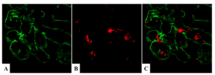
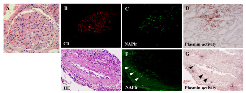
Nephritis-associated plasmin receptor (NAPlr) was originally isolated from the cytoplasmic fraction of group A Streptococci, and was found to be the same molecule as streptococcal glyceraldehyde-3-phosphate dehydrogenase (GAPDH) and plasmin receptor (Plr) on the basis of nucleotide and amino acid sequence homology. Its main functions include GAPDH activity, plasmin-binding capacity, and direct activation of the complement alternative pathway (A-P). Plasmin trapped by deposited NAPlr triggers the degradation of extracellular matrix proteins, such as glomerular basement membranes and mesangial matrix, and the accumulation of macrophages and neutrophils, leading to the induction of plasmin-related endocapillary glomerular inflammation. Deposited NAPlr at glomerular endocapillary site directly activates the complement A-P, and the endocapillary release of complement-related anaphylatoxins, C3a and C5a, amplify the in situ endocapillary glomerular inflammation. Subsequently, circulating and in situ-formed immune complexes participate in the glomerular injury resulting in NAPlr-mediated glomerulonephritis. The disease framework of infection-related glomerulonephritis (IRGN) has been further expanded. GAPDH of various bacteria other than Streptococci have been found to react with anti-NAPlr antibodies and to possess plasmin-binding activities, allowing glomerular NAPlr and plasmin activity to be utilized as key biomarkers of IRGN.

Keywords: C3-dominant glomerular injury; NAPlr (Bacterial GAPDH) mediated glomerulonephritis; immune complex-dominant glomerular injury; infection-related glomerulonephritis (IRGN); nephritis-associated plasmin receptor (NAPlr); poststreptococcal acute glomerulonephritis (PSAGN).


Conflict of interest statement

The authors declare no conflict of interest.

Figures

Figure 1
Figure 1
Graphical abstract of this review. NAPlr: nephritis-associated plasmin receptor, GAPDH: glyceraldehyde-3-phosphate dehydrogenase, MΦ: macrophage, PMN: polymorphonuclear neutrophil, GBM: glomerular basement membrane, GN: glomerulonephritis.
Figure 2
Figure 2
The amino acid and nucleotide sequences of NAPlr share high sequence identity with streptococcal Plr, as shown in the internal amino acid sequence of NAPlr. Because Plr and GAPDH are already established to be the same molecules, these three proteins are considered identical. NAPlr: nephritis-associated plasmin receptor, GAPDH: glyceraldehyde-3-phosphate dehydrogenase, Plr: plasmin receptor.
Figure 3
Figure 3
Two different patterns of glomerular C3 and IgG deposits in post-infectious glomerulonephritis. (A,B): C3 with IgG deposits, positive granular staining for C3 (A) and for IgG (B). (C,D): C3 without IgG deposits, positive granular staining for C3 (C) but completely negative staining for IgG (D). Original magnification: ×400. Photographs are cited from a previously published report [22].
Figure 4
Figure 4
Characteristic pathology in the early phase of PSAGN. (A): Light microscopy (HE); endocapillary proliferative glomerulonephritis. Original magnification: ×400. (B): C3 staining; fine granular deposits mainly on capillary wall, part of mesangium. Original magnification: ×400. (C): Electron microscopy; characteristic subepithelial dome-shaped humps (indicated by black arrows). PSAGN: post-streptococcal acute glomerulonephritis. Photographs are cited from a previously published textbook [23], where details of all the staining methods and magnifications have been described.
Figure 5
Figure 5
Difference in glomerular localization of NAPlr and immune complexes. (A): In the early phase, localization of NAPlr, which is presumed to be a free antigen, is primarily on the mesangium, part of the GBM, and infiltrating neutrophils. (B): In the early phase, C3 is localized mainly along the GBM (subepithelial), and partly on the mesangium. Original magnification: ×400. GBM: glomerular basement membrane, NAPlr: nephritis-associated plasmin receptor. Photographs are cited from a previously published report [2], where details of all the staining methods have been described.
Figure 6
Figure 6
Immunofluorescence staining with rabbit anti-NAPlr antibody in glomeruli from a PSAGN patient (10 days after onset) revealed glomerular NAPlr deposition mainly on the mesangium, the endocapillary side of GBM, and infiltrating neutrophils in a ring-like granular pattern. Original magnification: ×400. GBM: glomerular basement membrane, NAPlr: nephritis-associated plasmin receptor, PSAGN: post-streptococcal acute glomerulonephritis. This photograph is cited from a previously published report [24].
Figure 7
Figure 7
NAPlr localization relative to the glomerular basement membrane. Confocal microscopy images of double immunofluorescence staining for (A) α3 chain of collagen IV (FITC), (B) NAPlr (Alexa Fluor 594), and (C) the merged image in a post-streptococcal acute glomerulonephritis patient. NAPlr immunofluorescence is mostly seen on the inner side of glomerular tufts. Original magnification: ×400. NAPlr: nephritis-associated plasmin receptor. Photographs are cited from a previously published report [6], where details of all the staining methods have been described.
Figure 8
Figure 8
Reported mechanism for the tissue invasion of Streptococcus through direct binding pathway dependent on streptokinase and plasminogen. PLG: plasminogen, PLA: plasmin, SK: streptokinase, MMPs: matrix metalloproteases.
Figure 9
Figure 9
Schematic representation of the proposed mechanism for PSAGN. GAPDH: glyceraldehyde-3-phosphate dehydrogenase, GBM: glomerular basement membrane, END: endothelial cell, MES: mesangium, NAPlr: nephritis-associated plasmin receptor, MΦ: macrophage, PMN: polymorphonuclear neutrophil, PSAGN: post-streptococcal acute glomerulonephritis. This schema is modified and cited from a previously published paper [30].
Figure 10
Figure 10
In situ zymography for plasmin activity. (A) Positive NAPlr staining in the glomeruli of a PSAGN patient 18 days after onset. (B) Prominent plasmin activity was observed in a similar distribution to NAPlr deposition in the same glomeruli as shown in (A). This activity was completely inhibited by the addition of aprotinin (C). NAPlr: nephritis-associated plasmin receptor, PSAGN: post-streptococcal acute glomerulonephritis. Photographs are cited from a previously published report [11], where details of all the staining methods have been described. Original magnifications: ×200.
Figure 11
Figure 11
Cell-mediated immune response in PSAGN focused on NAPlr. Th: T helper/inducer cell, Ag-Ab complex: antigen-antibody complex, MΦ: macrophage, Neutro: neutrophil, END: endothelial cell. MES: mesangial cell, END: endothelial cell, GF/Cytok: growth factor/cytokines, NAPlr: nephritis-associated plasmin receptor, PSAGN: post-streptococcal acute glomerulonephritis.
Figure 12
Figure 12
(A) Immune complex-dominant glomerular injury. (B) NAPlr is fully saturated with IgG and complement components, however, IgG is not saturated with complement components. NAPlr: nephritis-associated plasmin receptor, NAPlr-Ab: NAPlr-antibody complex, NAPlr-Ab-C: NAPlr-antibody-complement complexes, MΦ: macrophage, PMN: polymorphonuclear neutrophil, C: complement, iC3b: inactivated C3b, NAPlr: nephritis-associated plasmin receptor.
Figure 13
Figure 13
Complement activation by NAPlr. (A) Immunoelectrophoresis showing conversion of C3 after incubation of NHS with NAPlr with or without Mg2⁺ and EGTA (middle). As a possible control, zymosan was added to NHS and indicates activated C3 (top). NHS not incubated with NAPlr shows a single arc of C3 (bottom). (B) Formation of iC3b from C3 in NHS incubated with various amounts of NAPlr. The plots use average values from triplicate assays. NAPlr: nephritis-associated plasmin receptor, EGTA: ethylene glycol tetraacetic acid, NHS: normal human serum.
Figure 14
Figure 14
(A): Representative light microscopy image of experimental glomerulonephritis induced by the administration of streptococcal nephritogenic protein in rabbits, 4 weeks after the start of injection. Moderate endothelial and mesangial proliferative changes with prominent infiltrating leucocytes are evident (H&E, original magnification ×400). (B): Immunofluorescence of glomeruli from representative rabbits in experiment, 4 weeks after the start of injections. C3 is intensely deposited along the glomerular basement membrane and mesangial areas (FITC-labelled goat anti-rabbit C3, original magnification ×200). Photographs are cited from a previously published report [45], where details of all the staining methods have been described.
Figure 15
Figure 15
Schematic representation of the complement activation through each pathway and NAPlr. Ag-Ab: antigen-antibody, MBL: mannose-binding lectin, MASP: mannose-binding lectin-associated serine proteases, C3(H₂O): hydrolyzed C3, MAC: membrane attack complex, NAPlr: nephritis-associated plasmin receptor.
Figure 16
Figure 16
A schematic diagram showing an approach to the classification of disease in a biopsy specimen, showing the morphological changes of glomerulonephritis with dominant C3. Post-infectious glomerulonephritis (GN) highlighted by red circle should be differentiated from C3 glomerulopathy. DDD: dense deposit disease, C3 GN: C3 glomerulonephritis.
Figure 17
Figure 17
(A): C3-dominant glomerular injury. (B): NAPlr is fully saturated with IgG and complement components, and IgG is also fully saturated with complement components. Ab: antibody, NAPlr-Ab: NAPlr-antibody, NAPlr-C: NAPlr-complement complex, NAPlr-Ab-C: NAPlr-antibody-complement complexes, C bar: activated complement, iC3b: inactivated C3b, A-P: alternative pathway, MΦ: macrophage, PMN; polymorphonuclear neutrophil, NAPlr: nephritis-associated plasmin receptor.
Figure 18
Figure 18
Kidney biopsy findings of C3 glomerulopathy with glomerular NAPlr deposition and associated plasmin activity. (AC): Double immunofluorescent staining for NAPlr (fluorescein isothiocyanate, green) and complement C3 (Alexa Fluor 594, red). NAPlr (A) and C3 (B) were both positive in the glomeruli, and their localization was essentially different in the merged images (C). (D): By in situ zymography, the plasmin activity was found to have a distribution similar to that of the NAPlr staining in the glomeruli. Original magnifications of (AD): ×200. (E): (Upper panel) Massive electron-dense deposits (arrow) were observed along the lamina densa in the glomerular basement membrane (GBM). (Lower panel) Subepithelial “hump”-shaped electron-dense deposits were also present in the GBM. Photographs are cited from a previously published report [71], where details of all the staining methods have been described.
Figure 19
Figure 19
Western blotting analysis of isolated bacterial protein. Protein staining of the blot of isolated protein from M. pneumoniae and Streptococcus pyogenes (left) and Western blotting using anti-NAPlr antibody (right). Western blotting revealed a specific band in the β-lane at around the predicted size of M. pneumoniae GAPDH (37 kDa: indicated by * with arrow), confirming the cross-reactivity of the anti-NAPlr antibody with M. pneumoniae GAPDH. The strong band in the α-lane with a molecular mass of 43 kDa, indicated by the arrow, is the NAPlr protein. Lanes: M1, prestained marker; α = isolated protein of S. pyogenes; β = isolated protein of M. pneumoniae; M2, Western 7 Protein Ladder [79].
Figure 20
Figure 20
Kidney biopsy findings for case 7 in Table 8. (A): Mesangial and endocapillary proliferation with double contours along the capillary walls and a cellular crescent (periodic acid-methenamine-silver stain, original magnification ×200). C3 (B) and NAPlr (C) were both positive in glomeruli, and their localization was essentially different. (D): In situ zymography showed that the glomerular distribution of plasmin activity was similar to that of NAPlr staining. Photographs are cited from a previously published report [83], where details of all the staining methods have been described.
Figure 21
Figure 21
Kidney biopsy findings for case 5 in Table 8. (A) Light microscopy showing diffuse proliferative and exudative glomerulonephritis with infiltrating neutrophils (periodic acid–Schiff, original magnification ×400). (B,C) C3 (B) and NAPlr (C) were both positive in glomeruli, and their localization was essentially different (B,C). (D) In situ zymography showed that the plasmin activity was similar to the NAPlr staining in the glomeruli in its distribution. Photographs are cited from a previously published report [89], where details of all the staining methods have been described.
Figure 22
Figure 22
Kidney biopsy findings for case 8 in Table 8. (A): Light microscopy showing endocapillary proliferation with massive eosinophil infiltration (H&E, original magnification ×400). (B): Immunostaining for C3. C3 was stained granularly along the glomerular tuft. NAPlr was positively stained in the glomeruli (C), and the distribution of plasmin activity (D) was similar to that of NAPlr staining in the glomeruli. (EG): Eosinophils were observed to infiltrate in the wall of the pulmonary artery, where NAPlr positive immunostaining and strong plasmin activity were detected (arrowheads) in addition to in the glomerulus. Photographs are cited from a previously published report [84], where details of all the staining methods have been described.

Similar articles

Cited by

References

    1. Yamakami K., Yoshizawa N., Wakabayashi K., Takeuchi A., Tadakuma T., Boyle M.D. The potential role for nephritis-associated plasmin receptor in acute poststreptococcal glomerulonephritis. Methods. 2000;21:185–197. doi: 10.1006/meth.2000.0990. - DOI - PubMed
    1. Yoshizawa N., Yamakami K., Fujino M., Oda T., Tamura K., Matsumoto K., Sugisaki T., Boyle M.D. Nephritis-associated plasmin receptor and acute poststreptococcal glomerulonephritis: Characterization of the antigen and associated immune response. J. Am. Soc. Nephrol. 2004;15:1785–1793. doi: 10.1097/01.ASN.0000130624.94920.6B. - DOI - PubMed
    1. Couser W.G., Johnson R.J. Post-infecitous glomerulonephritis. In: Neilson E.G., Couser W.G., editors. Immunologic Renal Diseases. Lippincott-Raven; Philadelphia, PA, USA: 1997. pp. 915–943.
    1. Nadasdy T., Silva F.G. Acute postinfectious glomerulonephritis and glomerulonephritis complicating persistent bacterial infection. In: Jennette J.C., Olson J.L., Schwartz M.M., Silva F.G., editors. Heptinstall’s Pathology of the Kidney. 6th ed. Lippincott Williams & Wilkins; Philadelphia, PA, USA: 2007. pp. 321–396.
    1. Nasr S.H., Fidler M.E., Valeri A.M., Cornell L.D., Sethi S., Zoller A., Stokes M.B., Markowitz G.S., D’Agati V.D. Postinfectious glomerulonephritis in the elderly. J. Am. Soc. Nephrol. 2011;22:187–195. doi: 10.1681/ASN.2010060611. - DOI - PubMed