. 2022 Sep 2;23(17):10022.
doi: 10.3390/ijms231710022.

Effect of Titanium and Zirconia Nanoparticles on Human Gingival Mesenchymal Stromal Cells

Affiliations

Effect of Titanium and Zirconia Nanoparticles on Human Gingival Mesenchymal Stromal Cells

Michael Nemec et al. Int J Mol Sci. .

Abstract

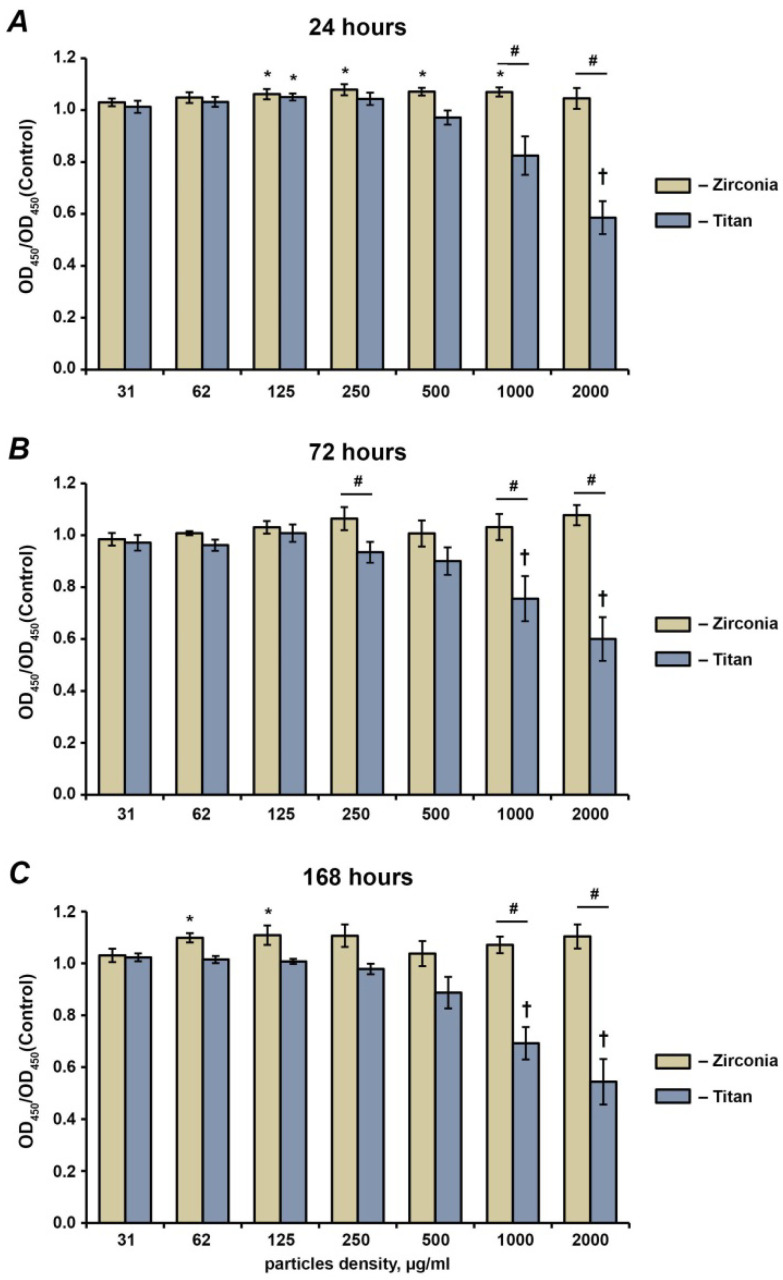
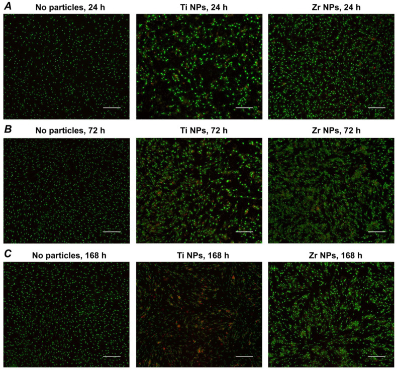
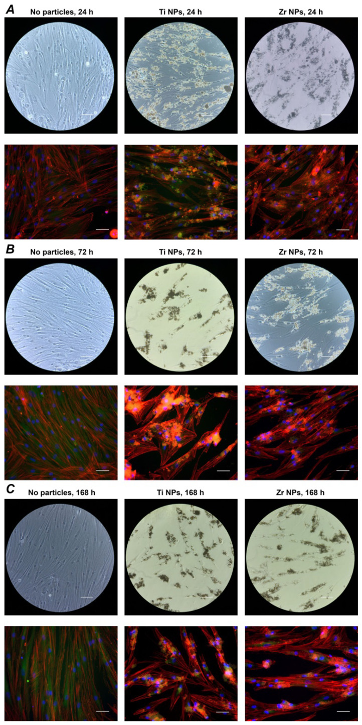
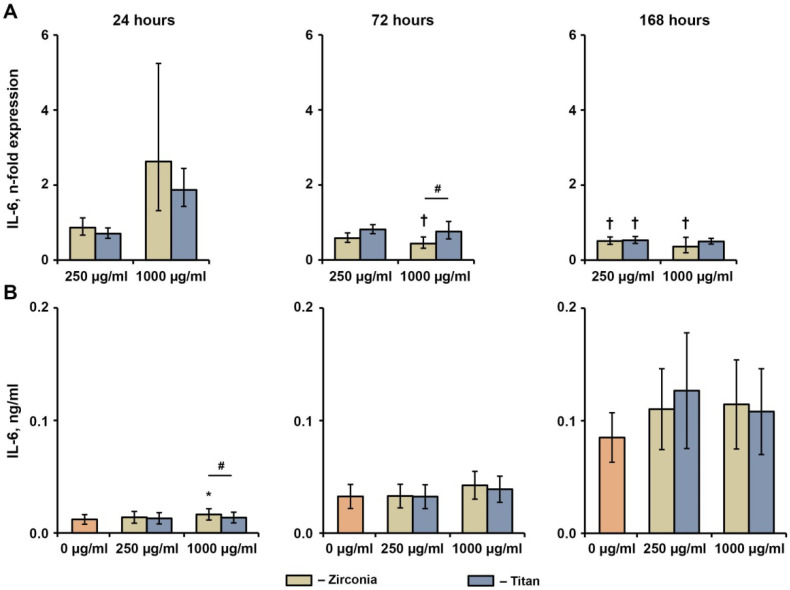
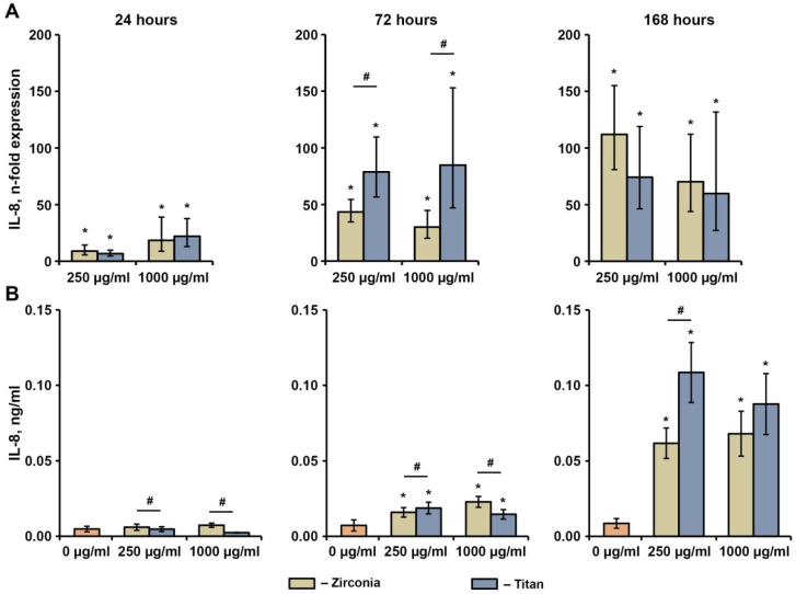
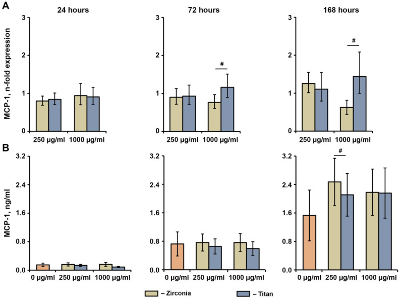
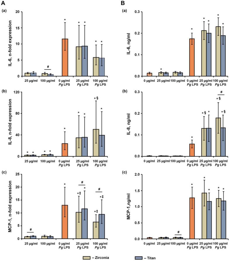
Nano- and microparticles are currently being discussed as potential risk factors for peri-implant disease. In the present study, we compared the responses of human gingival mesenchymal stromal cells (hG-MSCs) on titanium and zirconia nanoparticles (<100 nm) in the absence and presence of Porphyromonas gingivalis lipopolysaccharide (LPS). The primary hG-MSCs were treated with titanium and zirconia nanoparticles in concentrations up to 2.000 µg/mL for 24 h, 72 h, and 168 h. Additionally, the cells were treated with different nanoparticles (25−100 µg/mL) in the presence of P. gingivalis LPS for 24 h. The cell proliferation and viability assay and live−dead and focal adhesion stainings were performed, and the expression levels of interleukin (IL)-6, IL-8, and monocyte chemoattractant protein (MCP)-1 were measured. The cell proliferation and viability were inhibited by the titanium (>1000 µg/mL) but not the zirconia nanoparticles, which was accompanied by enhanced apoptosis. Both types of nanoparticles (>25 µg/mL) induced the significant expression of IL-8 in gingival MSCs, and a slightly higher effect was observed for titanium nanoparticles. Both nanoparticles substantially enhanced the P. gingivalis LPS-induced IL-8 production; a higher effect was observed for zirconia nanoparticles. The production of inflammatory mediators by hG-MSCs is affected by the nanoparticles. This effect depends on the nanoparticle material and the presence of inflammatory stimuli.

Keywords: dental implants; human gingival mesenchymal stromal cells; nanoparticles; titanium; zirconia.


Conflict of interest statement

The authors declare no conflict of interest.

Figures

Figure 1
Figure 1
Effect of different NPs on the proliferation and viability of hG-MSCs. The hG-MSCs were cultured in the absence or in the presence of titanium or zirconia NPs at different concentrations for 24 h (A), 72 h (B), or 168 h (C). The proliferation and viability were measured using the MTT method. The Y-axis represents the ratios of optical densities (OD) measured at 570 nm in hG-MSCs cultured with NPs to those in the control group (without NPs). Data are presented as means ± s.e.m. of six independent experiments with hG-MSCs isolated from six different donors. Note: *—significantly higher compared to control, p < 0.05; †—significantly lower compared to control, p < 0.05; #—significantly different between Ti NPs and Zr NPs, p < 0.05.
Figure 2
Figure 2
Live–dead staining of hG-MSCs cultured with or without different NPs. The hG-MSCs were cultured in the absence or in the presence of Ti NPs or Zr NPs at a concentration of 1000 µg/mL for 24 h (A), 72 h (B), or 168 h (C) and stained with a live–dead staining kit. Viable and dead cells are visualized in green and red, respectively. Images were taken from a representative experiment. The scale bar corresponds to 200 µm.
Figure 3
Figure 3
Focal adhesion staining of hG-MSCs cultured with or without NPs. The hG-MSCs were cultured in the absence or in the presence of Ti NPs or Zr NPs at a concentration of 250 µg/mL for 24 h (A), 72 h (B), or 168 h (C) and the cells were stained with a focal adhesion staining kit. F-actin was stained with TRITC-conjugated phalloidin (red), the focal adhesions with anti-vinculin and FITC-conjugated secondary antibody (green), and the nucleus with DAPI (blue). The scale bars correspond to 20 µm.
Figure 4
Figure 4
Effects of NPs on the basal IL-6 expression in hG-MSCs. The hG-MSCs were cultured in the absence or in the presence of different NPs for 24 h, 72 h, or 168 h. The resulting IL-6 gene expression (A) and IL-6 protein production (B) rates were measured via qPCR and ELISA, respectively. (A) Y-axis represents n-fold expression of IL-6 in hG-MSCs cultured with NPs compared to the unstimulated control (n-fold expression = 1). N-fold expression was calculated using the 2−ΔΔCt method, using GAPDH as the reference gene. (B) Y-axis shows the concentration of IL-6 in the conditioned media. Data are presented as means ± s.e.m. of six independent experiments performed with hG-MSCs isolated from six different donors. Note: *—significantly higher compared to control, p < 0.05; †—significantly lower compared to control, p < 0.05; #—significantly different between Ti NPs and Zr NPs, p < 0.05.
Figure 5
Figure 5
Effects of NPs on the basal IL-8 expression in hG-MSCs. The hG-MSCs were cultured in the absence or in the presence of different NPs for 24 h, 72 h, or 168 h. The resulting IL-8 gene expression (A) and IL-8 protein production (B) rates were measured via qPCR and ELISA, respectively. (A) Y-axis represents n-fold expression of IL-8 in hG-MSCs cultured with NPs compared to unstimulated control (n-fold expression = 1). N-fold expression was calculated using the 2−ΔΔCt method, using GAPDH as the reference gene. (B) Y-axis shows the concentration of IL-8 in the conditioned media Data are presented as means ± s.e.m. of six independent experiments performed with hG-MSCs isolated from six different donors. Note: *—significantly higher compared to control, p < 0.05; #—significantly different between Ti NPs and Zr NPs, p < 0.05.
Figure 6
Figure 6
Effects of NPs on the basal MCP-1 expression in hG-MSCs. The hG-MSCs were cultured in the absence or in the presence of different NPs for 24 h, 72 h, or 168 h. The resulting MCP-1 gene expression (A) and MCP-1 protein production (B) rates were measured via qPCR and ELISA, respectively. (A) Y-axis represents n-fold expression of MCP-1 in hG-MSCs cultured with NPs compared to unstimulated control (n-fold expression = 1). N-fold expression was calculated using the 2−ΔΔCt method, using GAPDH as the reference gene. (B) Y-axis shows the concentration of MCP-1 in the conditioned media Data are presented as means ± s.e.m. of six independent experiments performed with hG-MSCs isolated from six different donors. Note: #—significantly different between Ti NPs and Zr NPs, p < 0.05.
Figure 7
Figure 7
The effects of different NPs on the P. gingivalis LPS-induced response in hG-MSCs. The hG-MSCs were stimulated with P. gingivalis LPS (Pg LPS) in the absence or in the presence of different NPs for 24 h. The resulting gene expression (A) and protein production (B) rates for IL-6, IL-8, and MCP-1 were determined via qPCR and ELISA, respectively. (A) Y-axis represents n-fold expression of target genes in hG-MSCs cultured under various conditions in relation to cells cultured without NPs and Pg LPS, calculated using the 2−ΔΔCt method, using GAPDH as a reference gene. (B) Y-axis shows the concentrations of various proteins in the conditioned media measured. Data are presented as means ± s.e.m. of six independent experiments performed with hG-MSCs isolated from six different donors. Note: *—significantly higher compared to control, p < 0.05; #—significantly different between Ti NPs and Zr NPs, p < 0.05; §—significantly higher compared to Pg LPS without NPs, p < 0.05; ‡—significantly lower compared to Pg LPS without NPs, p < 0.05.

Similar articles

Cited by

References

    1. Howe M.-S., Keys W., Richards D. Long-term (10-year) dental implant survival: A systematic review and sensitivity meta-analysis. J. Dent. 2019;84:9–21. doi: 10.1016/j.jdent.2019.03.008. - DOI - PubMed
    1. Buser D., Sennerby L., De Bruyn H. Modern implant dentistry based on osseointegration: 50 years of progress, current trends and open questions. Periodontology 2000. 2017;73:7–21. doi: 10.1111/prd.12185. - DOI - PubMed
    1. Steinemann S.G. Titanium—The material of choice? Periodontology. 2000;17:7–21. doi: 10.1111/j.1600-0757.1998.tb00119.x. - DOI - PubMed
    1. Cionca N., Hashim D., Mombelli A. Zirconia dental implants: Where are we now, and where are we heading? Periodontology 2000. 2017;73:241–258. doi: 10.1111/prd.12180. - DOI - PubMed
    1. Özkurt Z., Kazazoğlu E. Zirconia Dental Implants: A Literature Review. J. Oral Implantol. 2011;37:367–376. doi: 10.1563/AAID-JOI-D-09-00079. - DOI - PubMed