. 2022 Sep 2;23(17):10039.
doi: 10.3390/ijms231710039.

Transcription Factors Runx1 and Runx3 Suppress Keratin Expression in Undifferentiated Keratinocytes

Affiliations

Transcription Factors Runx1 and Runx3 Suppress Keratin Expression in Undifferentiated Keratinocytes

Eisaku Ogawa et al. Int J Mol Sci. .

Abstract

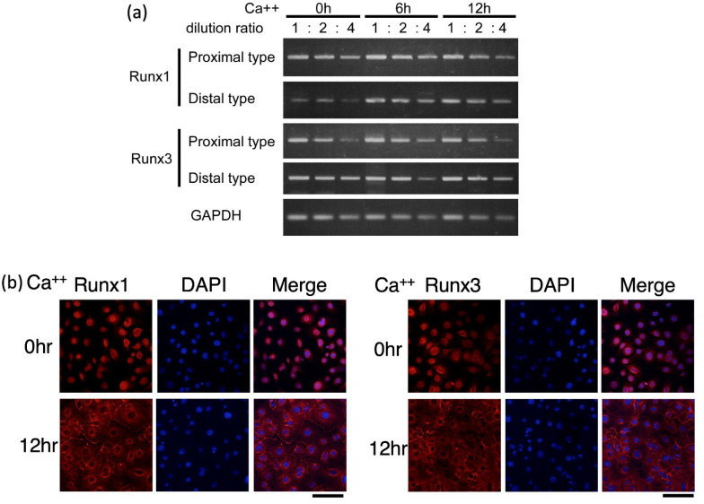
The Runt-related transcription factor (Runx) family has been suggested to play roles in stem cell regulation, tissue development, and oncogenesis in various tissues/organs. In this study, we investigated the possible functions of Runx1 and Runx3 in keratinocyte differentiation. Both Runx1 and Runx3 proteins were detected in primary cultures of mouse keratinocytes. Proteins were localized in the nuclei of undifferentiated keratinocytes but translocated to the cytoplasm of differentiated cells. The siRNA-mediated inhibition of Runx1 and Runx3 expression increased expression of keratin 1 and keratin 10, which are early differentiation markers of keratinocytes. In contrast, overexpression of Runx1 and Runx3 suppressed keratin 1 and keratin 10 expression. Endogenous Runx1 and Runx3 proteins were associated with the promoter sequences of keratin 1 and keratin 10 genes in undifferentiated but not differentiated keratinocytes. In mouse skin, the inhibition of Runx1 and Runx3 expression by keratinocyte-specific gene targeting increased the ratios of keratin 1- and keratin 10-positive cells in the basal layer of the epidermis. On the other hand, inhibition of Runx1 and Runx3 expression did not alter the proliferation capacity of cultured or epidermal keratinocytes. These results suggest that Runx1 and Runx3 likely function to directly inhibit differentiation-induced expression of keratin 1 and keratin 10 genes but are not involved in the regulation of keratinocyte proliferation.

Keywords: differentiation; keratinocyte; proliferation; runt-related transcription factor (Runx).


Conflict of interest statement

All authors have no conflict of interest to declare.

Figures

Figure 1
Figure 1
Expression of Runx1/Runx3 transcripts (a) and proteins (b) in cultured keratinocytes. The differentiation of keratinocytes was induced by increasing the calcium concentration in the medium for 0, 6, and 12 h of incubation as indicated. (a) RNA was extracted from the cells and processed for cDNA synthesis. Relative amounts of proximal and distal Runx1/Runx3 transcripts were evaluated by semi-quantitative PCR using the 1-, 2-, and 4-fold diluted cDNAs. GAPDH transcript was used as a control. The primers that were set to distinguish proximal versus distal type of transcripts yielded amplified products of rather long size. Since this made real time PCR experiments difficult to do, semi-quantitative PCR was chosen. Note that the purpose of experiments here was to confirm the presence of Runx transcripts but not try to measure the exact variation in transcript amounts. (b) Cells were fixed and immunostained for Runx1/Runx3 protein. DAPI was used to stain nuclei. Scale bar, 100 μm. In both (a,b) we performed three independent trials for each experimental set out and could obtain essentially similar results. In the figure, representative results are shown.
Figure 2
Figure 2
Effects of inhibition of Runx1/Runx3 expression on keratin 1/keratin 10 mRNA and protein expression in cultured keratinocytes. The cells were transfected with specific siRNA for Runx1 or Runx3, incubated for 4 h, induced to differentiate by raising the calcium concentration for 12 h, and harvested. Two different sequences were used for Runx1 and Runx3 siRNA, shown as siRNA1 and siRNA2. (a) Effects of Runx1/Runx3 siRNAs on Runx1/Runx3 expression. RNA was prepared from the cells and processed for RT-PCR. Proximal and distal Runx1/Runx3 transcripts were examined with GAPDH transcript as a control. (b) Effects of Runx1/Runx3 siRNAs on keratin 1 and keratin 10 protein expression. Protein lysates were prepared from the cells and processed for immunoblotting analysis of keratin 1, keratin 10, and β-actin (control). (c) Effects of Runx1/Runx3 siRNAs on keratin 1 and keratin 10 gene expression. RNA was prepared from the cells and processed for real-time RT-PCR analysis. The levels of keratin 1 and keratin 10 transcripts normalized relative to HPRT transcript are shown. In each experimental set out as above, we performed three independent trials and could obtain essentially similar results. In (a,b), representative results are shown, whereas in (c), mean ± standard deviation are shown, and statistically significant differences, if detected, are indicated by brackets (a student t-test; * p < 0.05, ** p < 0.01).
Figure 3
Figure 3
Effects of Runx1/Runx3 overexpression on keratin 1/keratin 10 transcript and protein levels in cultured keratinocytes. Cells were infected with adenovirus vectors carrying LacZ (control), Runx1, or Runx3 for 24 h and cell differentiation was induced by increasing the calcium concentration for 0, 6, and 12 h in (b) and for 12 h in (c). (a) Protein lysates were prepared from the cells and processed for immunoblotting analysis of Runx1, Runx3, and α-tubulin (control). (b) Protein lysates were prepared from the cells and processed for immunoblotting analysis of keratin 1, keratin 10, and α-tubulin (control). (c) RNA was prepared from the cells and processed for cDNA synthesis and real-time PCR analysis. The levels of keratin 1 and keratin 10 transcripts normalized relative to HPRT transcript are shown. In each experimental set out as above, we performed three independent trials and could obtain essentially similar results. In (a) and (b) representative results are shown, whereas in (c) mean ± standard deviation are shown and statistically significant differences, if detected, are indicated by brackets (a student t-test; * p < 0.05, ** p < 0.01). Note that the image in (a) is extensively magnified so that the endogenous Runx bands are visible. The bands indicated by an asterisk are due to a nonspecific reaction.
Figure 4
Figure 4
Chromatin immunoprecipitation (ChIP) of Runx1/Runx3 with keratin 1 and keratin 10 promoters. ChIP assay was performed to determine the binding of Runx1/Runx3 proteins to the keratin 1 and keratin 10 promoter sequences. The cells were cultured under low-calcium conditions (0 h) and high-calcium conditions (12 h), and cell lysates were prepared and processed for ChIP using anti-Runx1, anti-Runx3, or control IgG antibody. PCR was performed on the precipitated genomic DNA using primers flanking the Runx-binding consensus sites within the keratin 1 promoter and keratin 10 promoter. We performed three independent trials in the above experimental set out and could obtain essentially similar results. In the figure, representative results are shown.
Figure 5
Figure 5
Effects of Runx1/Runx3 knockout on keratin 1 and keratin 10 expression in keratinocytes. (a) Genotyping of the skin-specific Runx1 or Runx3 knockout mice. Genomic DNA was isolated from the tails of K5Cre-Runx1fl/fl mice, K5Cre-Runx3 fl/fl mice, and wild-type mice and processed for PCR. The alleles detected were K5-Cre transgene (top row), Runx1 allele (second row), Runx3 allele (third row), and β-actin (bottom row). (b) RT-PCR analysis of Runx1 or Runx3 transcripts in the epidermis-specific Runx1 or Runx3 knockout mice. RNA was isolated from the epidermis of K5Cre-Runx1fl/fl mice, K5Cre-Runx3 fl/fl mice, and wild-type mice and processed for RT-PCR. The detected transcripts were Runx1 allele (top row), Runx3 allele (second row), and β-actin (bottom row). Flox, floxed allele; wt, wild-type allele; Δ, deleted allele. (c,d) Immunostaining of keratin 1 and keratin 10 in the skin of epidermis-specific Runx1 or Runx3 knockout mice. Skin sections were prepared from K5Cre-Runx1fl/fl mice, K5Cre-Runx3 fl/fl mice, and wild-type mice and processed for staining with hematoxylin and eosin (H&E) and immunostaining for keratin 1 and keratin 10 (green fluorescence). Nuclei stained with DAPI are shown in blue. Arrows, skin surface; arrowheads, keratin 1-positive or keratin 10-positive basal keratinocytes; broken lines, dermal/epidermal junction. Scale bar, 50 μm. Six different visual fields were chosen randomly, using 40-fold magnification of optical lens and the numbers of keratin 1-positive and keratin 10-positive basal keratinocytes were counted (the total number of cells counted were roughly 1000). One of the authors (EO) prepared the tissue section and did immunostaining, whereas another author (TE), not informed of the mouse genotype of samples, counted the cell numbers through a microscope. The percentages of keratin 1-positive or keratin 10-positive cells per total epidermal cells are shown as the mean ± standard deviation. * p < 0.05. In (c,d), two different individual mice were used for each genotype and essentially similar results were obtained. In figure, representative results are shown.

Similar articles

Cited by

References

    1. Roop D. Defects in the barrier. Science. 1995;267:474–475. doi: 10.1126/science.7529942. - DOI - PubMed
    1. Tomic-Canic M., Komine M., Freedberg I.M., Blumenberg M. Epidermal signal transduction and transcription factor activation in activated keratinocytes. J. Dermatol. Sci. 1998;17:167–181. doi: 10.1016/S0923-1811(98)00016-4. - DOI - PubMed
    1. Ghadially R. 25 years of epidermal stem cell research. J. Investig. Dermatol. 2012;132:797–810. doi: 10.1038/jid.2011.434. - DOI - PMC - PubMed
    1. Ogawa E., Sato Y., Minagawa A., Okuyama R. Pathogenesis of psoriasis and development of treatment. J. Dermatol. 2018;45:264–272. doi: 10.1111/1346-8138.14139. - DOI - PubMed
    1. Ratushny V., Gober M.D., Hick R., Ridky T.W., Seykora J.T. From keratinocyte to cancer: The pathogenesis and modeling of cutaneous squamous cell carcinoma. J. Clin. Investig. 2012;122:464–472. doi: 10.1172/JCI57415. - DOI - PMC - PubMed