. 2022 Sep 2;23(17):10066.
doi: 10.3390/ijms231710066.

Selective Loading and Variations in the miRNA Profile of Extracellular Vesicles from Endothelial-like Cells Cultivated under Normoxia and Hypoxia

Affiliations

Selective Loading and Variations in the miRNA Profile of Extracellular Vesicles from Endothelial-like Cells Cultivated under Normoxia and Hypoxia

Anny Waloski Robert et al. Int J Mol Sci. .

Abstract

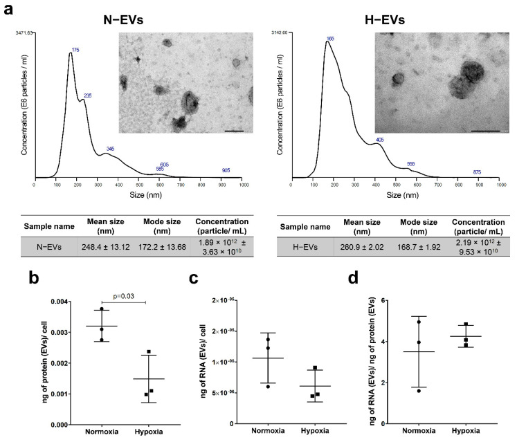
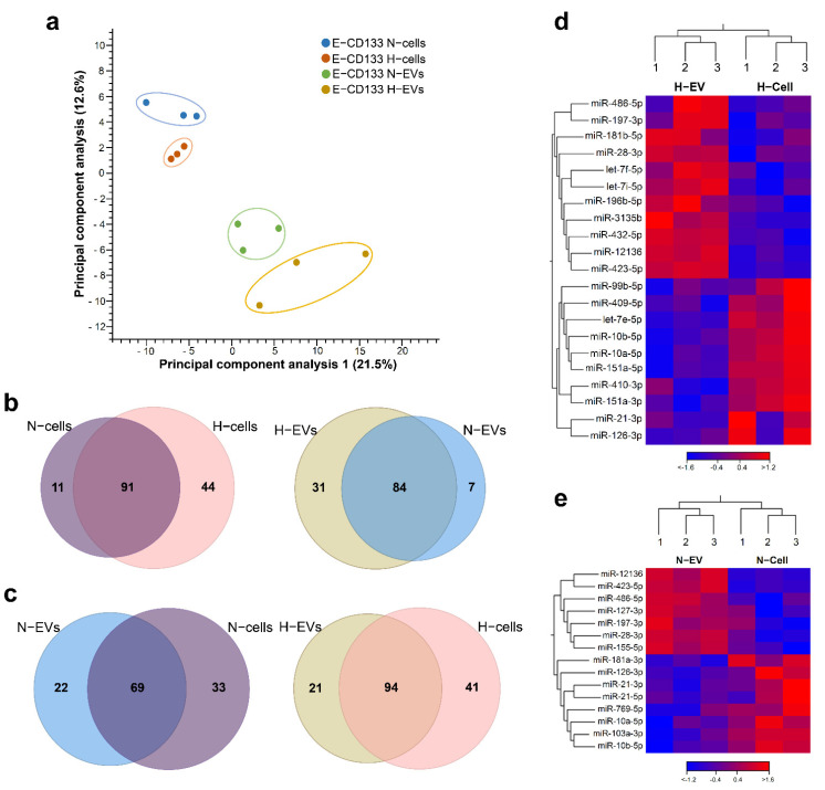
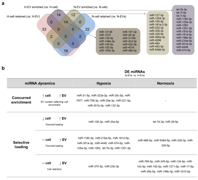
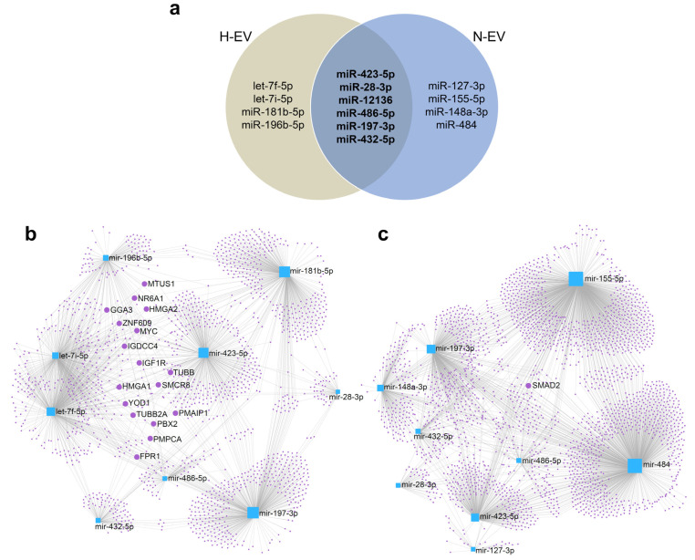
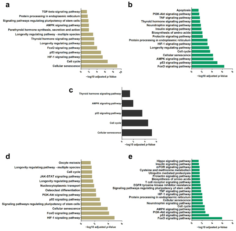
Endothelial-like cells may be obtained from CD133+ mononuclear cells isolated from human umbilical cord blood (hUCB) and expanded using endothelial-inducing medium (E-CD133 cells). Their use in regenerative medicine has been explored by the potential not only to form vessels but also by the secretion of bioactive elements. Extracellular vesicles (EVs) are prominent messengers of this paracrine activity, transporting bioactive molecules that may guide cellular response under different conditions. Using RNA-Seq, we characterized the miRNA content of EVs derived from E-CD133 cells cultivated under normoxia (N-EVs) and hypoxia (H-EVs) and observed that changing the O2 status led to variations in the selective loading of miRNAs in the EVs. In silico analysis showed that among the targets of differentially loaded miRNAs, there are transcripts involved in pathways related to cell growth and survival, such as FoxO and HIF-1 pathways. The data obtained reinforce the pro-regenerative potential of EVs obtained from E-CD133 cells and shows that fine tuning of their properties may be regulated by culture conditions.

Keywords: CD133+ cells; endothelial-like cells; extracellular vesicles; hypoxia; miRNA.


Conflict of interest statement

The authors do not declare any conflicts of interest. The funders had no role in the design of the study; in the collection, analyses, or interpretation of data; in the writing of the manuscript; or in the decision to publish the results.

Figures

Figure 1
Figure 1
E-CD133 cell and EVs under normoxia and hypoxia. (a) Transmission electron microscopy and NTA showing the size distribution of E-CD133 N-EVs and H-EVs (n = 3 technical replicates). Scale bar = 200 nm. Analysis of (b) the ratio of the protein content of the E-CD133 EVs/cell, (c) the ratio of the RNA content of the E-CD133 EVs/cell, and (d) the ratio of the RNA content/protein content of the E-CD133 EVs obtained from cells cultivated under normoxia and hypoxia. (Unpaired student t-test).
Figure 2
Figure 2
Comparison of miRNAs identified in normoxia and hypoxia E-CD133 cells and EVs. (a) Principal component analysis of miRNAs identified in the samples. (b) Venn diagrams comparing the identified miRNAs in E-CD133 cells (left) or E-CD133 EVs (right) in normoxia versus hypoxia culture conditions. (c) Venn diagrams comparing the identified miRNAs in E-CD133 cells versus E-CD133 EVs in normoxia (left) or hypoxia (right) culture conditions. (d) Heatmap of miRNAs differentially expressed (FDR ≤ 10%, log2(FC) < −1 and >1) among E-CD133 cells and E-CD133 EV cultured in hypoxia. (e) Heatmap of miRNAs differentially expressed (FDR ≤ 10%, log2(FC) < −1 and >1) among E-CD133 cells and E-CD133 EV cultured in normoxia.
Figure 3
Figure 3
Analysis of miRNAs enriched in E-CD133 EVs or retained in E-CD133. (a) Venn chart analysis of the miRNAs found as enriched in the EVs or retained in the cells during hypoxia and normoxia. (b) Analysis of the miRNA dynamics that may lead to a DE of miRNAs in H-EVs vs. N-EVs (up arrow = augmentation in miRNA expression/enrichment; down arrow = reduction in miRNA expression/enrichment; dash = no change in miRNA expression/enrichment).
Figure 4
Figure 4
Identification of higher expressed miRNA targets. (a) Venn diagram comparing the miRNAs enriched in N-EVs and/or H-EVs (vs. cells) with higher CPM. The miRNAs presented in the two EVs are highlighted in bold. (b) Network of top 10 miRNAs enriched in H-EVs (vs. H-cells) and its target mRNAs. (c) Network of top 10 miRNAs enriched in N-EVs (vs. N-cells) and its target mRNAs. The highlighted genes are targets of 4 or more miRNAs.
Figure 5
Figure 5
Analysis of miRNA targets and their regulated pathways. (a) KEGG analysis of mRNA targets of top 10 miRNAs enriched in H-EVs (vs. H-cells). (b) KEGG analysis of mRNA targets of top 10 miRNAs enriched in N-EVs (vs. N-cells). (c) KEGG analysis of mRNA targets of the six miRNAs more expressed in H- and N-EVs in comparison with cells. (d) KEGG analysis of mRNA targets of the four miRNAs more expressed in E-CD133 EVs in comparison with E-CD133 only in hypoxia condition. (e) KEGG analysis of mRNA targets of the four miRNAs more expressed in E-CD133 EVs in comparison with E-CD133 only in normoxia condition.

Similar articles

Cited by

References

    1. Théry C., Witwer K.W., Aikawa E., Alcaraz M.J., Anderson J.D., Andriantsitohaina R., Antoniou A., Arab T., Archer F., Atkin-Smith G.K., et al. Minimal Information for Studies of Extracellular Vesicles 2018 (MISEV2018): A Position Statement of the International Society for Extracellular Vesicles and Update of the MISEV2014 Guidelines. J. Extracell. Vesicles. 2018;7:1535750. doi: 10.1080/20013078.2018.1535750. - DOI - PMC - PubMed
    1. Cocucci E., Meldolesi J. Ectosomes and Exosomes: Shedding the Confusion between Extracellular Vesicles. Trends Cell Biol. 2015;25:364–372. doi: 10.1016/j.tcb.2015.01.004. - DOI - PubMed
    1. Van Niel G., Carter D.R.F., Clayton A., Lambert D.W., Raposo G., Vader P. Challenges and Directions in Studying Cell–Cell Communication by Extracellular Vesicles. Nat. Rev. Mol. Cell Biol. 2022;23:369–382. doi: 10.1038/s41580-022-00460-3. - DOI - PubMed
    1. Debbi L., Guo S., Safina D., Levenberg S. Boosting Extracellular Vesicle Secretion. Biotechnol. Adv. 2022;59:107983. doi: 10.1016/j.biotechadv.2022.107983. - DOI - PMC - PubMed
    1. Leitolis A., Suss P.H., Roderjan J.G., Angulski A.B.B., da Costa F.D.A., Stimamiglio M.A., Correa A. Human Heart Explant-Derived Extracellular Vesicles: Characterization and Effects on the in Vitro Recellularization of Decellularized Heart Valves. Int. J. Mol. Sci. 2019;20:1279. doi: 10.3390/ijms20061279. - DOI - PMC - PubMed