Review
. 2022 Sep 3;23(17):10068.
doi: 10.3390/ijms231710068.

Lipid-Based Nanoparticles as a Pivotal Delivery Approach in Triple Negative Breast Cancer (TNBC) Therapy

Affiliations
Review

Lipid-Based Nanoparticles as a Pivotal Delivery Approach in Triple Negative Breast Cancer (TNBC) Therapy

Aiswarya Chaudhuri et al. Int J Mol Sci. .

Abstract

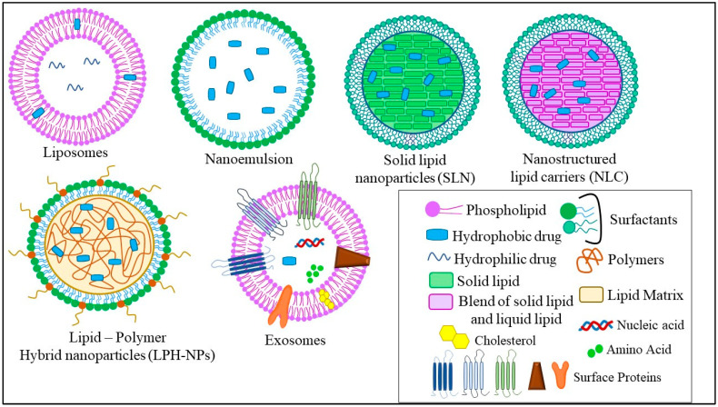
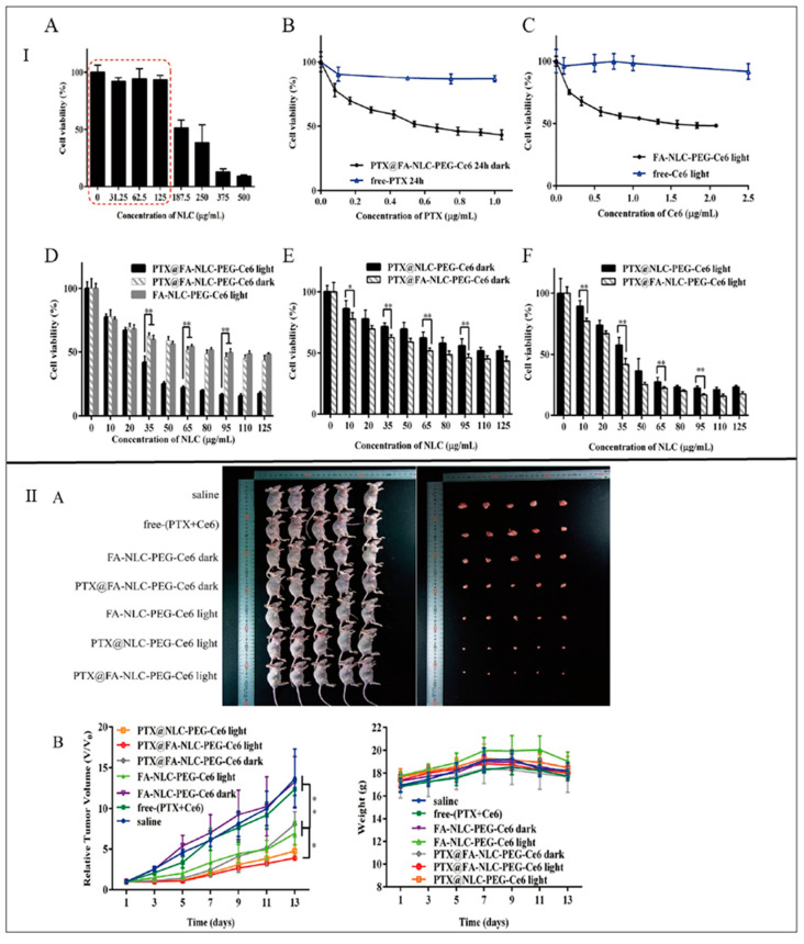
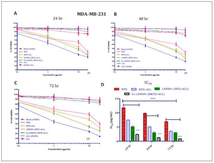
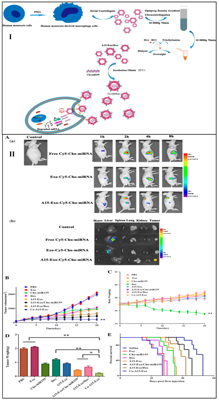
Triple-negative breast cancer is considered the most aggressive type of breast cancer among women and the lack of expressed receptors has made treatment options substantially limited. Recently, various types of nanoparticles have emerged as a therapeutic option against TNBC, to elevate the therapeutic efficacy of the existing chemotherapeutics. Among the various nanoparticles, lipid-based nanoparticles (LNPs) viz. liposomes, nanoemulsions, solid lipid nanoparticles, nanostructured lipid nanocarriers, and lipid-polymer hybrid nanoparticles are developed for cancer treatment which is well confirmed and documented. LNPs include various therapeutic advantages as compared to conventional therapy and other nanoparticles, including increased loading capacity, enhanced temporal and thermal stability, decreased therapeutic dose and associated toxicity, and limited drug resistance. In addition to these, LNPs overcome physiological barriers which provide increased accumulation of therapeutics at the target site. Extensive efforts by the scientific community could make some of the liposomal formulations the clinical reality; however, the relatively high cost, problems in scaling up the formulations, and delivery in a more targetable fashion are some of the major issues that need to be addressed. In the present review, we have compiled the state of the art about different types of LNPs with the latest advances reported for the treatment of TNBC in recent years, along with their clinical status and toxicity in detail.

Keywords: lipid–polymer hybrid nanoparticles; liposomes; nanoemulsion; nanostructured lipid carriers; solid lipid nanoparticles; targeted therapy; triple-negative breast cancer.


Conflict of interest statement

The authors declare no conflict of interest.

Figures

Figure 1
Figure 1
The mechanism of LNPs uptake into the lymphatic circulation: (A) Uptake of drug-loaded LNPs by Peyer’s patch into the lymphatic system: The drug-loaded LNPs are taken up by the M-cells of the enterocytes which are then taken up by the dendritic cells followed by Peyer’s patch from where the drug-loaded LNPs enter into the lymphatic system via afferent lymphatic. (B) Uptake of drug-loaded LNPs by an intestinal wall into the lymphatic system: The drug-loaded LNPs enter the lymphatic system through the intestinal wall in fours ways—(1) transcellular transport, (2) paracellular transport, (3) by inhibiting P-gp glycoprotein and cytochrome P450, or (4) by the production of chylomicrons. Abbreviations: M cell: membranous cell, LNPs: lipid-based nanoparticles; P-gp: P-glycoprotein.
Figure 2
Figure 2
Different types of LNPs used for the treatment of TNBC.
Figure 3
Figure 3
(I) An illustration displaying the construction of PTX-ILips and the release of PTX and aCD47 from the liposome for effective chemotherapy and immunotherapy respectively against TNBC. (II) Anticancer efficacy study and recurrence inhibition study in vivo: (A) Individual tumor growth curves in different groups. (B) Tumor growth kinetics of MDA-MB-231 tumors in mice treated with different formulations. (C) Survival rates of animals in various groups. (D) H&E staining of tumor slices collected from mice after the treatment of 21 days. Scale bar = 100 μm. (E) Photographs of lung metastatic nodules and histological assessment of lung metastatic nodules via H&E staining. Scale bar = 100 μm. (F) Numbers of lung metastatic nodules from each group. (G) Schematic illustration of the establishment of tumor recurrence model and therapy with different formulations. (H) Representative IVIS images of MDA-MB-231 tumor-bearing mice in each group. (I) Tumor volume growth curves after tumor implantation, subsequent surgery, and therapy. (J) Survival of mice in different treatment groups. Data are displayed as the mean ± SD. ** p < 0.01; **** p < 0.0001. Reprinted (adapted) with permission from [54]. Copyright (2021) American Chemical Society.
Figure 4
Figure 4
(I) Schematic diagram of TME remodulation by targeted delivery of puerarin-loaded nanoemulsion. (II) Combination treatment of nanoPue and nanoPTX on 4T1 tumor model: (A) nanoPue and nanoPTX combination treatment scheme. (B) Tumor growth curves of 4T1 tumors in different treatment groups. (C) The tumor weight and the representative tumor image at the end of the experiment in different treatment groups. (D) TUNEL staining of differently treated 4T1 tumor tissues. (E) Comparison of Ki67 expression of 4T1 tumors in different treatment groups. Scale bar represents 20 μm * p < 0.05, ** p < 0.01, *** p < 0.001 and **** p < 0.0001. Reprinted from Biomaterials, 235, Xu, et al., Nano-puerarin regulates tumor microenvironment and facilitates chemo- and immunotherapy in murine triple negative breast cancer model, 1-12, Copyright (2020), with permission from Elsevier [63].
Figure 5
Figure 5
(I) In vitro cytotoxicity assays: (A) Cell viability of MDA-MB-231 cells after incubation with FA-NLC-PEG-Ce6 (dark) NPs of different concentrations. (B) Cytotoxicity evaluation of free-PTX and PTX@FA-NLC-PEG-Ce6 in dark in MDA-MB-231 cells by MTT. (C) Cytotoxicity evaluation of free-PTX and PTX@FA-NLC-PEG-Ce6 in light in MDA-MB-231 cells by MTT. (D) Cytotoxicity comprehensive evaluation of single drug NLCs and combination drug NLCs in MDA-MB-231 cells by MTT. (E) Cytotoxicity evaluation of PTX@NLC-PEG-Ce6 and PTX@FA-NLC-PEG-Ce6 in MDA-MB-231 cells with red laser by MTT. (F) Cytotoxicity evaluation of PTX@NLC-PEG-Ce6 and PTX@FA-NLC-PEG-Ce6 in MDA-MB-231 cells without red laser by MTT. * p < 0.05, ** p < 0.01. (II). In vivo anti-cancer activity of NLCs in tumor-bearing nude mice after intravenous administration of saline, free (PTX + Ce6) and different kinds of NLCs, with red laser after 24 h of injection (each mouse for 30 min): (A) Photographs of sacrificed nude mice and the tumor tissues collected from them. (B) Changes of relative tumor volumes in MDA-MB-231 tumor-bearing nude mice of each group. Note: * p < 0.05, ** p < 0.01 (n = 5). Reprinted from International Journal of Pharmaceutics, 569, Zhang, et al., Construction and in vitro and in vivo evaluation of folic acid-modified nanostructured lipid carriers loaded with paclitaxel and chlorin e6, 1-12, Copyright (2019), with permission from Elsevier [82].
Figure 6
Figure 6
The in vitro anti-cancer activity of MTX- and ACL-based free and co-encapsulated LPHNPs after 24 (A), 48 (B), and 72 h (C) in MDA-MB-231 cell lines. The image (D) depicts the comparison of IC50 of different samples obtained after 24, 48, and 72 h (*** p < 0.05). The statistics were run to determine significance in IC50 by 2-way ANOVA, and data are presented as the mean of three independent experiments (SD, n = 6). Reprinted (adapted) with permission from [89]. Copyright (2017) American Chemical Society.
Figure 7
Figure 7
(I) Schematic diagram of isolation of exosomes, loading of Dox.Hcl and Cho-miR159 within the exosomes and release of Dox and Cho-miR159-loaded A15-Exo (Co-A15-Exo). (II) Biodistribution and antitumor efficacy of Co-A15-Exo in vivo: (A(a)). Images were taken 1 h, 2 h, 4 h, or 8 h after the administration of free Cy5-Cho-miRNA, Exo-Cy5-Cho-miRNA, or A15-Exo-Cy5-Cho-miRNA. (A(b)) Ex vivo imaging of tumor and organs collected at the end of the experiment (8 h post-injection). (B) Tumor growth curves of mice receiving different therapeutic regimens (n = 5, mean ± SD). (C) Body weight changes during treatment. Data are expressed as the mean ± SD (n = 5). ** p < 0.01, vs. PBS. (D). The weights of the excised tumor tissues from all groups. Data are expressed as the mean ± SD (n = 5). * p < 0.05 and ** p < 0.01 when compared with the indicated groups. (E) Survival rate of MDA-MB-231 tumor-bearing BALB/c nude mice [102].

References

    1. Gallo C. Triple Negative Breast Cancer Epidemiology. 2018. [(accessed on 1 April 2020)]. Available online: https://oncologynurse-ce.com/triple-negative-breast-cancer-epidemiology.
    1. Aqil F., Munagala R., Agrawal A.K., Jeyabalan J., Tyagi N., Rai S.N., Gupta R.C. Anthocyanidins Inhibit Growth and Chemosensitize Triple-Negative Breast Cancer via the NF-kappaB Signaling Pathway. Cancers. 2021;13:6428 - PMC - PubMed
    1. Cheng Z., Li M., Dey R., Chen Y. Nanomaterials for cancer therapy: Current progress and perspectives. J. Hematol. Oncol. 2021;14:85. - PMC - PubMed
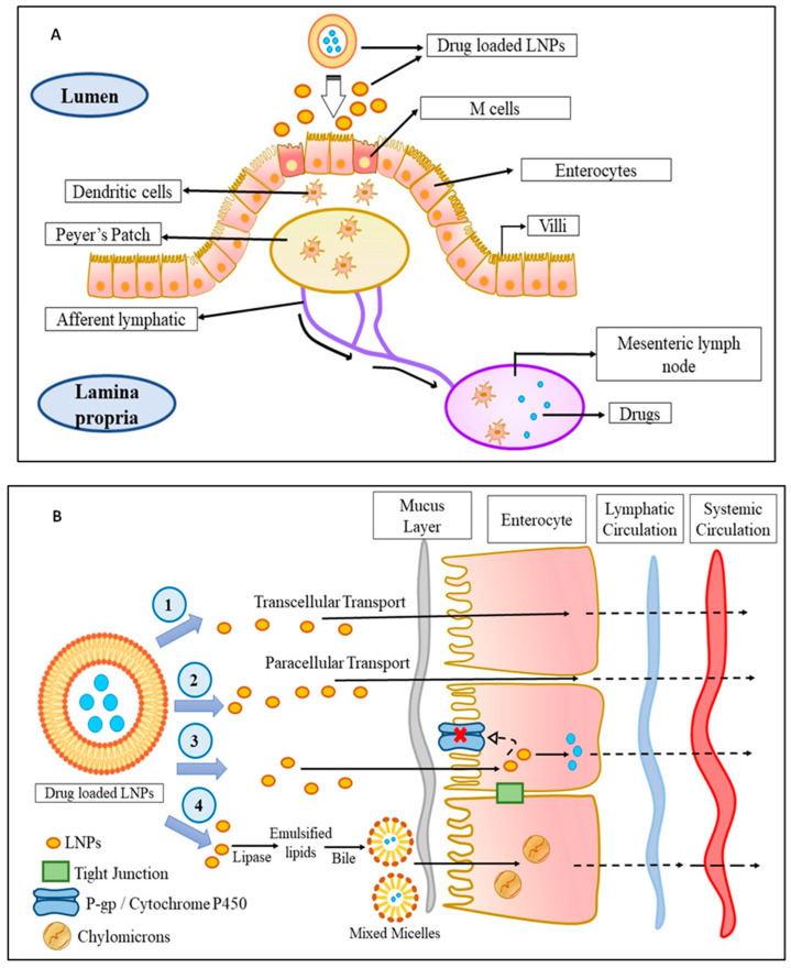
    1. El Moukhtari S.H., Rodriguez-Nogales C., Blanco-Prieto M.J. Oral lipid nanomedicines: Current status and future perspectives in cancer treatment. Adv. Drug Deliv. Rev. 2021;173:238–251. - PubMed
    1. Yan L., Shen J., Wang J., Yang X., Dong S., Lu S. Nanoparticle-Based Drug Delivery System: A Patient-Friendly Chemotherapy for Oncology. Dose-Response. 2020;18:1559325820936161. - PMC - PubMed

LinkOut - more resources