. 2022 Sep 3;23(17):10097.
doi: 10.3390/ijms231710097.

TNF-α Plus IL-1β Induces Opposite Regulation of Cx43 Hemichannels and Gap Junctions in Mesangial Cells through a RhoA/ROCK-Dependent Pathway

Affiliations

TNF-α Plus IL-1β Induces Opposite Regulation of Cx43 Hemichannels and Gap Junctions in Mesangial Cells through a RhoA/ROCK-Dependent Pathway

Claudia M Lucero et al. Int J Mol Sci. .

Abstract

Connexin 43 (Cx43) is expressed in kidney tissue where it forms hemichannels and gap junction channels. However, the possible functional relationship between these membrane channels and their role in damaged renal cells remains unknown. Here, analysis of ethidium uptake and thiobarbituric acid reactive species revealed that treatment with TNF-α plus IL-1β increases Cx43 hemichannel activity and oxidative stress in MES-13 cells (a cell line derived from mesangial cells), and in primary mesangial cells. The latter was also accompanied by a reduction in gap junctional communication, whereas Western blotting assays showed a progressive increase in phosphorylated MYPT (a target of RhoA/ROCK) and Cx43 upon TNF-α/IL-1β treatment. Additionally, inhibition of RhoA/ROCK strongly antagonized the TNF-α/IL-1β-induced activation of Cx43 hemichannels and reduction in gap junctional coupling. We propose that activation of Cx43 hemichannels and inhibition of cell-cell coupling during pro-inflammatory conditions could contribute to oxidative stress and damage of mesangial cells via the RhoA/ROCK pathway.

Keywords: Fasudil; Y-27632; connexin hemichannel; gap junction; inflammatory receptors; oxidative stress.


Conflict of interest statement

The authors declare no conflict of interest.

Figures

Figure 1
Figure 1
ROCK and Cx43 HC blockers reduce the TNF-α/IL-1β-induced Etd+ uptake in mesangial cells. (A) Etd+ uptake rate in primary MCs under control conditions (Ctrl, black bar), exposed to TNF-α/IL-1β (10 ng/mL each, white bars) for 72 h alone or with ROCK inhibitor: Fasudil (15 μM); and HCs blockers: Lanthanum ion (La3+, 200 μM) and selective Cx43 HC blockers: mimetic peptides Gap19 (100 µM) and TAT-L2 (100 µM). (B) Fluorescence images show primary MCs exposed to ethidium (5 μM Etd+) for 13 min under TNF-α/IL-1β stimulation with or without different blockers. Scale bar = 50 μm. Each bar represents the mean value ± SEM of five independent experiments with four replicates each. Statistical significance * p < 0.05 vs. Ctrl; † p < 0.05 vs. TNF-α/IL-1β. Fasudil was added during the last 24 h of treatment. Gap19, TAT-L2 and La3+ were added acutely (added 15 min before each Etd+ uptake recording).
Figure 2
Figure 2
Fasudil prevents the TNF-α/IL-1β-induced reduction in gap junctional communication in MES-13 cells. (A) Gap junctional communication was examined using the microinjection of Etd+ (25 μM) in single cells (white arrows). After 2 min, the diffusion to neighboring cells was imaged. The top panels represent phase-contrast images, whereas the bottom panels show Etd+ fluorescence in cells under control conditions or upon different treatments. Scale bar = 20 μm. (B) Coupling incidence and (C) coupling index in confluent mesangial cells, under control conditions (black bars) or exposed to TNF-α/IL-1β (10 ng/mL) for 72 h (white bars). Fasudil (15 µM) was added together with TNF-α/IL-1β. Each bar represents the mean value ± SEM of 4 independent experiments. In each experiment, the dye was microinjected into at least 10 cells. Statistical significance * p < 0.05 vs. Ctrl; † p < 0.05 vs. TNF-α/IL-1β + Fasudil.
Figure 3
Figure 3
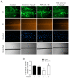
Fasudil prevents the TNF-α/IL-1β-induced decrease in primary mesangial cell coupling. (A) Representative fluorescence micrographs of SL/DT with Lucifer yellow (LY) by primary MCs under control conditions, 72 h after exposure to TNF-α/IL-1β (10 ng/mL) or treatment with Fasudil (15 µM). For each experiment are shown representative images of communicating cells stained with LY, dead cells along the cut stained with propidium iodide (PI), the total cells stained with Hoechst. Bar = 200 μm. (B) Averaged data normalized to control (black bars) of SL/DT with LY by primary MCs after 72 h of treatment with TNF-α/IL-1β (white bars) or in combination with Fasudil. Each bar represents the mean value ± SEM of 3 independent experiments. Statistical significance, * p < 0.05 vs. Ctrl; † p < 0.05 vs. Control + Fasudil; # p < 0.05 vs. TNF-α/IL-1β.
Figure 4
Figure 4
Blockade of ROCK prevents the increase in connexin43 and phosphorylated MYPT in mesangial cells induced by TNF-α/IL-1β. (B) Graphs show the relative amount of Cx43 and (E) phosphorylation of MYPT-1 determined by Western blotting analysis in MES-13 cells under control conditions (black bars) or exposed to TNF-α/IL-1β (white bars) for 72 h. (C) Fluorescence images depicting PDGFβR (green), Cx43 (red) and DAPI (blue) staining of primary MCs stimulated for 72 h with 10 ng/mL TNF-α/IL-1β. White arrows indicate GJ plaques at cell–cell interfaces, and white arrowheads indicate Cx43 HCs. Calibration bar: white = 50 μm. Fasudil (15 µM) or Y-27632 (15 µM) was added 24 h before harvesting the cells. Each bar represents the mean value ± SEM of ≥3 independent experiments. Statistical significance * p < 0.05 vs. Ctrl; † p < 0.05 vs. Control + Fasudil; # p < 0.05 vs. Control + Y-27632; ‡ p < 0.05 vs. TNF-α/IL-1β. Next to the graph, representative pictures of Cx43 (A), phosphorylated MYPT (p-MYPT), unphosphorylated MYPT positive bands (D), and its loading control (α-tubulin) are shown.
Figure 5
Figure 5
Blockade of ROCK prevents the increase in TBARS induced by TNF-α/IL-1β in primary mesangial cells. Graphs showing the amount of TBARS obtained from culture media of primary MCs under control conditions (black bars) or exposed to TNF-α/IL-1β (white bars) for 72 h. Fasudil (15 µM) was added 24 h before harvesting the cells. Each bar represents the mean value ± SEM of 4 independent experiments. Statistical significance, * p < 0.05 vs. Ctrl; † p < 0.05 vs. Control + Fasudil; # p < 0.05 vs. TNF-α/IL-1β.
Figure 6
Figure 6
Blockade of ROCK prevents apoptosis, preserving cell viability in primary mesangial cells stimulated with TNF-α/IL-1β. Graphs showing the measurement of (A) cell viability and (B) % apoptotic cells in MCs under control conditions (black bars) or exposed to TNF-α/IL-1β (white bars) for 72 h. (C) Fluorescence images depicting PDGFβR (green), Tunel (red) and DAPI (blue) staining by primary MCs stimulated for 72 h with 10 ng/mL TNF-α/IL-1β. Calibration bar: white = 50 μm. Fasudil (15 µM) was added 24 h before harvesting the cells. Each bar represents the mean value ± SEM of 3 independent experiments. Statistical significance * p < 0.05 vs. Ctrl; † p < 0.05 vs. Ctrl + Fasudil # p < 0.05 vs. TNF-α/IL-1β.
Figure 7
Figure 7
Schematic showing the possible signaling pathways involved in the TNF-α/IL-1β-mediated regulation of the functional state of Cx43 HCs and GJs in mesangial cells. High TNF-α/IL-1β concentrations could promote activation of a RhoA/ROCK-dependent pathway through TNF-α/IL-1β receptors (TNF-R and IL-1R) (continuous black arrows). Once the RhoA/ROCK-dependent pathway is activated, the formation of reactive oxygen species (ROS) takes place (generating TBARS) along with NF-κB-dependent production of proinflammatory cytokines such as TNF-α and IL-1β. The activated RhoA/ROCK-dependent pathway could increase the activity of Cx43 HCs, since Fasudil or Y-27632 (ROCK blockers) inhibit this response. The resulting increase in [Ca2+]i will also activate Cx43 HCs, allowing the increase in Ca2+ influx. The latter results in the rise of [Ca2+]i and further generation of ROS. The influx of Ca2+ establishes a positive feedback loop sensitive to different compounds such as the mimetic peptides Gap19 and TAT-L2, selective Cx43 HC blockers. This increase in the cellular activity caused by TNF-α/IL-1β, where the RhoA/ROCK pathway could be involved, also reduces cell–cell communication through GJs. Of note, alterations in [Ca2+]i homeostasis mediated by Cx43 HCs or Panx1 HCs might affect diverse aspects of MC functions (e.g., morphology and proinflammatory profile). Discontinuous black arrows indicate cell responses identified in other systems, whereas straight black arrows denote responses identified in the present work. Elaborated in biorender.com.

Similar articles

Cited by

References

    1. Yang Z.-J., Wang H.-R., Wang Y.-I., Zhai Z.-H., Wang L.-W., Li L., Zhang C., Tang L. Myricetin Attenuated Diabetes-Associated Kidney Injuries and Dysfunction via Regulating Nuclear Factor (Erythroid Derived 2)-Like 2 and Nuclear Factor-κB Signaling. Front. Pharmacol. 2019;10:647. doi: 10.3389/fphar.2019.00647. - DOI - PMC - PubMed
    1. Zhang X., Yang J., Yu X., Cheng S., Gan H., Xia Y. Angiotensin II-Induced Early and Late Inflammatory Responses Through NOXs and MAPK Pathways. Inflammation. 2017;40:154–165. doi: 10.1007/s10753-016-0464-6. - DOI - PubMed
    1. Romagnani P., Remuzzi G., Glassock R., Levin A., Jager K.J., Tonelli M., Massy Z., Wanner C., Anders H.-J. Chronic kidney disease. Nat. Rev. Dis. Primers. 2017;3:17088. doi: 10.1038/nrdp.2017.88. - DOI - PubMed
    1. Zhao J.-H. Renal Fibrosis: Mechanisms and Therapies. Advances in Experimental Medicine and Biology. Springer; Singapore: 2019. Mesangial Cells and Renal Fibrosis; pp. 165–194. - PubMed
    1. Kao M.P.C., Ang D.S.C., Pall A., Struthers A.D. Oxidative stress in renal dysfunction: Mechanisms, clinical sequelae and therapeutic options. J. Hum. Hypertens. 2010;24:1–8. doi: 10.1038/jhh.2009.70. - DOI - PubMed

MeSH terms

LinkOut - more resources